Studying Algae Byproducts using SERDS

Laura Ruvuna is guest web writer for Analyst. She is currently a PhD student at Northwestern University, Evanston, Illinois, USA.

SERDS combined with signal regression analysis to monitorthe phototrophic microorganisms

As the demand for non-renewable energy sources continues to soar, alternative renewable and environmentally friendly alternatives continue to be explored. The biomass conversion by algae has emerged as promising avenue. Additionally, algae produce other compounds relevant in pharmaceutical research. A bioreactor houses these phototropic organisms, but direct measurement of these compounds within the reactor can be costly, inefficient, and require complex modeling to understand.

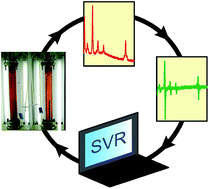
Researchers at University of Erlangen-Nuremberg in Germany used shifted-excitation Raman difference spectroscopy (SERDS) in a model algae system as a new way to probe these reactions. In SERDS, two spectra are acquired at slightly different wavelengths, and subtraction of the two removes the fluorescence background, a common problem in normal Raman spectroscopy. The red algae, Porphyridium purpureum produces a number of relevant compounds, such as sulfated polysaccharides, which have important anti-viral properties.

With the help of an algorithm to process the spectra, they tracked the separation of this promising pharmaceutical compound, which can be applied to other algae systems.

To know more about the study, please access the link below. This paper will be free to read for the next three weeks.

Combined shifted-excitation Raman difference spectroscopy and support vector regression for monitoring the algal production of complex polysaccharides
Kristina Noack, Björn Eskofier, Johannes Kiefer, Christina Dilk, Georg Bilow, Matthias Schirmer, Rainer Buchholz and   Alfred Leipertz
Analyst, 2013, Advance Article
DOI: 10.1039/C3AN01158E

Digg This
Reddit This
Stumble Now!
Share on Facebook
Bookmark this on Delicious
Share on LinkedIn
Bookmark this on Technorati
Post on Twitter
Google Buzz (aka. Google Reader)

Analytical Sciences in Brazil Web Themed Issue: Submissions Open!

Analyst and Analytical Methods invite you to submit your next article to a web-themed issue on

 “Analytical Sciences in Brazil”  

Interdisciplinary analytical science is particularly welcome

Submission deadline: 31st Jan 2014
Articles published online in a web collection

Guest Editors: Boris Mizaikoff, University of Ulm
                     Ivo M. Raimundo Jr., UNICAMP


Analyst: The home of premier fundamental discoveries, inventions and applications in the analytical and bioanalytical sciences with an Impact Factor of 4.0

Analytical Methods: Publishing early applied demonstrations of new analytical methods with clear societal impact and the fastest growing general analytical journal.

Digg This
Reddit This
Stumble Now!
Share on Facebook
Bookmark this on Delicious
Share on LinkedIn
Bookmark this on Technorati
Post on Twitter
Google Buzz (aka. Google Reader)

Analytical Toxicology of Nanoparticles Themed issue: Submissions Open!

Soon we will be publishing a themed issue of Analyst on Analytical Toxicology of Nanoparticles featuring research on new nanotechnologies and their potential impact on safety and environment.

The aim is to highlight analytical techniques developed to detect and characterise nanoparticles and determine their influence on biological and ecological systems.

Guest Edited by Christy Haynes, University of Minnesota

Do you work in the area of nanoparticle toxicology? Submit your work today!

All articles will be peer-reviewed, and to be suitable for publication, must meet the usual quality and significance standards of the journal. Please indicate in your covering letter that your article is to be considered for the Analytical Toxicology of Nanoparticle themed issue.

Email us at ANALYST-RSC@rsc.org for more information.

Digg This
Reddit This
Stumble Now!
Share on Facebook
Bookmark this on Delicious
Share on LinkedIn
Bookmark this on Technorati
Post on Twitter
Google Buzz (aka. Google Reader)

CMOS Imaging as a Potential Solution for Diabetic Patients

Pui Sai Lau is a guest web writer for Analyst and Analytical Methods. She is currently a PhD student at McMaster University, Hamilton, ON, Canada.

CMOS Image Sensor for Glucose Monitoring

CMOS Image Sensor for Glucose Monitoring

Millions of people suffer from diabetes, and the disease is quickly becoming a global epidemic. To address patient need, many electrochemical- and biochemical-based technologies have been developed over the last decade.

While considerable progress has been made, many of these methods still do not possess the sensitivity, specificity, accuracy, and user friendliness required to meet patient demands.

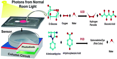
Researchers from Gachon University in the Republic of Korea, have developed the latest glucose sensor based on complementary metal oxide semiconductor (CMOS) imaging that provides enhanced efficiency for glucose monitoring. The novel method measures the concentration of oxidized glucose in blood plasma by the intensity of color produced, which leads to variations in photon count detected by the CMOS sensor.

Assess the link below for more details. This paper will be free to read for the next three weeks:

CMOS image sensors as an efficient platform for glucose monitoring
Jasmine Pramila Devadhasan, Sanghyo Kim and   Cheol Soo Choi
Analyst, 2013, Advance Article
DOI: 10.1039/C3AN00805C

What are your thoughts on this new discovery? Let us know by leaving a comment.

Digg This
Reddit This
Stumble Now!
Share on Facebook
Bookmark this on Delicious
Share on LinkedIn
Bookmark this on Technorati
Post on Twitter
Google Buzz (aka. Google Reader)

The second reason to attend HPLC 2013 – Hobart

Hear Prof. Justin Gooding present his plenary lecture on

Designing and Characterising Bioaffinity Surfaces for Sensing”,

Then treat your own senses to the sights,  sounds and aromas of Hobart’s historic Sullivans Cove and Salamanca Markets, location to some of Australia’s freshest and naturally delicious steak and seafood restaurants.

This is a unique opportunity for you to meet with scientists from throughout the world and to share your latest results!

Just over 1 week till the HPLC 2013 Hobart Abstract Deadline Closes!

Abstract Deadline – 31st July 2013

Early bird Registration Deadline – 16th August 2013

Don’t miss out and submit your abstract to be considered for the conference program
Click here to register and submit for the conference

http://www.hplc2013-hobart.org/

Digg This
Reddit This
Stumble Now!
Share on Facebook
Bookmark this on Delicious
Share on LinkedIn
Bookmark this on Technorati
Post on Twitter
Google Buzz (aka. Google Reader)

HOT Articles in Analyst

Take a look below at the new HOT Articles just published in Analyst. They will be free to read until August 16th. Enjoy!

Multiplexed DNA detection with a composite molecular beacon based on guanine-quenching
Dong-Shan Xiang, Kun Zhai and Lian-Zhi Wang  
Analyst, 2013, Advance Article
DOI: 10.1039/C3AN01060K 

In vivo characterization of protein uptake by yeast cell envelope: single cell AFM imaging and μ-tip-enhanced Raman scattering study
Denys Naumenko, Valentinas Snitka, Elena Serviene, Ingrida Bruzait and Boris Snopok  
Analyst, 2013, Advance Article
DOI: 10.1039/C3AN00362K 

Luminol-labeled gold nanoparticles for ultrasensitive chemiluminescence-based chemical analyses
Lateef U. Syed, Luxi Zhang Swisher, Hannah Huff, Caitlin Rochford, Fengli Wang, Jianwei Liu, Judy Wu, Mark Richter, Sivasai Balivada, Deryl Troyer and Jun Li   
Analyst, 2013, Advance Article
DOI: 10.1039/C3AN01005H

Toward optimal spatial and spectral quality in widefield infrared spectromicroscopy of IR labelled single cells
Eric C. Mattson, Miriam Unger, Sylvain Clède, François Lambert, Clotilde Policar, Asher Imtiaz, Roshan D’Souza and Carol J. Hirschmugl 
Analyst, 2013, Advance Article
DOI: 10.1039/C3AN00383C

Energy-selective neutron imaging for morphological and phase analysis of iron–nickel meteorites
S. Peetermans, F. Grazzi, F. Salvemini, E. H. Lehmann, S. Caporalid and G. Pratesie  
Analyst, 2013, Advance Article
DOI: 10.1039/C3AN00985H

Metal–tag labeling coupled with multiple reaction monitoring-mass spectrometry for absolute quantitation of proteins
Xueying Wang, Xin Wang, Weijie Qin, Hongjun Lin, Jifeng Wang, Junying Wei, Yangjun Zhang and Xiaohong Qian 
Analyst, 2013, Advance Article
DOI: 10.1039/C3AN00613A

A novel miniaturized radiofrequency potentiometer tag using ion-selective electrodes for wireless ion sensing
Marta Novell, Tomàs Guinovart, Ivana Murković Steinberg, Matthew Steinberg, F. Xavier Rius and Francisco J. Andrade  
Analyst, 2013, Advance Article
DOI: 10.1039/C3AN00727H

3D pulsed laser-triggered high-speed microfluidic fluorescence-activated cell sorter
Yue Chen, Ting-Hsiang Wu, Yu-Chun Kung, Michael A. Teitell and Pei-Yu Chiou  
Analyst, 2013, Advance Article
DOI: 10.1039/C3AN01266B

Digg This
Reddit This
Stumble Now!
Share on Facebook
Bookmark this on Delicious
Share on LinkedIn
Bookmark this on Technorati
Post on Twitter
Google Buzz (aka. Google Reader)

Forensic fingers

A team in the US has developed a detection system that crime scene investigators can wear on their fingertips to rapidly identify suspected traces of explosives and gunshot residue.

With the increase in crimes related to explosives and firearms, fast and accurate detection of possible traces of explosives and gunshot residue is of considerable importance. Forensic analysis is typically carried out in a central laboratory, requiring time-consuming sampling, transportation and storage. Joseph Wang and colleagues at the University California, San Diego, have built on their expertise with wearable sensors  to move forensic analysis away from the lab and to the crime scene.

Their new sensor is made up of two components: an electrode screen-printed onto a stretchable sheath worn on the index finger and a second sheath, worn on the thumb, coated with a solid-state ionogel electrolyte. ‘Initially we intended to use a hydrogel, but the lifetime of this media was finite due to evaporation of water,’ says Wang. The ionogel circumvents the need for carrying additional liquid reagents, making the device highly portable.

To read the full article please visit Chemistry World.

Solid-state Forensic Finger sensor for integrated sampling and detection of gunshot residue and explosives: towards ‘Lab-on-a-finger’
Amay J. Bandodkar, Aoife M. O’Mahony, Julian Ramírez, Izabela A. Samek, Sean M. Anderson, Joshua R. Windmiller and Joseph Wang   Analyst, 2013, Advance Article
DOI: 10.1039/C3AN01179H

Digg This
Reddit This
Stumble Now!
Share on Facebook
Bookmark this on Delicious
Share on LinkedIn
Bookmark this on Technorati
Post on Twitter
Google Buzz (aka. Google Reader)

Free to access Cancer Nanotechnology collection 2013

We are pleased to present a new Web Collection of articles from publications across the RSC journal portfolio demonstrating the use of (nano)technology in the diagnosis, imaging and treatment of cancer.

This web collection will be free to access until July 28th, so register for an RSC Publishing personal account and read this cutting edge research for free this week!

Here are just a few of the cancer nanotechnology articles:

Quantification of ovarian cancer markers with integrated microfluidic concentration gradient and imaging nanohole surface plasmon resonance
Carlos Escobedo, Yu-Wei Chou, Mohammad Rahman, Xiaobo Duan, Reuven Gordon, David Sinton, Alexandre G. Brolo and   Jacqueline Ferreira
Analyst, 2013,138, 1450-1458
DOI: 10.1039/C3AN36616B

Magnetic beads-based electrochemiluminescence immunosensor for determination of cancer markers using quantum dot functionalized PtRu alloys as labels
Yan Zhang, Shenguang Ge, Shaowei Wang, Mei Yan, Jinghua Yu, Xianrang Song and   Weiyan Liu
Analyst, 2012,137, 2176-2182
DOI: 10.1039/C2AN16170B

Magnetic quantitative immunoanalysis of carcinoembryonic antigen by ICP-MS with mercury labels
Hanyong Peng, Beibei Chen, Man He, Yuan Zhang and   Bin Hu
J. Anal. At. Spectrom., 2011,26, 1217-1223
DOI: 10.1039/C1JA00007A

A microfluidic system for introduction of nanolitre sample in inductively coupled plasma mass spectrometry using electrokinetic flow combined with hydrodynamic flow
Heyong Cheng, Zigang Xu, Jinhua Liu, Xiuzhong Wanga and   Xuefeng Yin
J. Anal. At. Spectrom., 2012,27, 346-353
DOI: 10.1039/C1JA10273G 

Visualising gold inside tumour cells following treatment with an antitumour gold(I) complex
Louise E. Wedlock, Matt R. Kilburn, John B. Cliff, Luis Filgueira, Martin Saunders and   Susan J. Berners-Price
Metallomics, 2011,3, 917-925
DOI: 10.1039/C1MT00053E

Digg This
Reddit This
Stumble Now!
Share on Facebook
Bookmark this on Delicious
Share on LinkedIn
Bookmark this on Technorati
Post on Twitter
Google Buzz (aka. Google Reader)

Protein analysis unlocks museum mysteries

Animal soft tissues feature regularly in cultural artefacts but it can be difficult to pinpoint their origin. Visual identification of these tissues relies on the skill of the examiner and the condition of the material. Analytical techniques, like gas chromatography-mass spectrometry, can detect and broadly classify proteins but give no answers as to their source. Precise identification of the sinews used for stitching is often impossible. Now, researchers in the US and UK have shown that peptide mass fingerprinting can be used to determine the animal species of collagen-based materials in a diverse range of museum objects.

Peptide mass fingerprinting (PMF) uses enzymes to digest proteins to produce a mixture of peptides. The mass spectrum of this mixture will have characteristic marker ions – called a peptide mass fingerprint – which are compared to a database of species-specific markers to identify the proteins.

Peabody Museum of Archaeology and Ethnology, #69-30-10/1619 © President and Fellows of Harvard College, US

 To read the full article please visit Chemistry World.

Identification of collagen-based materials in cultural heritage
Daniel P. Kirby, Michael Buckley, Ellen Promise, Sunia A. Traugerd and T. Rose Holdcraft  
Analyst, 2013, Advance Article
DOI: 10.1039/C3AN00925D

Digg This
Reddit This
Stumble Now!
Share on Facebook
Bookmark this on Delicious
Share on LinkedIn
Bookmark this on Technorati
Post on Twitter
Google Buzz (aka. Google Reader)

Top ten most accessed articles from April – June 2013

During the months April – June, the  following articles are in the Top Ten most accessed:-

Electrogenerated chemiluminescence of nanomaterials for bioanalysis 
Shengyuan Deng and Huangxian Ju 
Analyst, 2013,138, 43-61
DOI: 10.1039/C2AN36122A     

Electrogenerated chemiluminescence detection of trace level pentachlorophenol using carbon quantum dots 
Jiezhen Li, Niya Wang, ThanhThuy Tran.T, Chen’an Huang, Lan Chen, Lijuan Yuan, Liping Zhou, Rui Shen and Qingyun Cai 
Analyst, 2013,138, 2038-2043
DOI: 10.1039/C3AN36653G    

Metabolomics study on Fuzi and its processed products using ultra-performance liquid-chromatography/electrospray-ionization synapt high-definition mass spectrometry coupled with pattern recognition analysis 
Hui Sun, Bei Ni, Aihua Zhang, Mo Wang, Hui Dong and Xijun Wang 
Analyst, 2012,137, 170-185
DOI: 10.1039/C1AN15833C     

Upconversion nanoparticles in biological labeling, imaging, and therapy 
Feng Wang, Debapriya Banerjee, Yongsheng Liu, Xueyuan Chen and Xiaogang Liu 
Analyst, 2010,135, 1839-1854
DOI: 10.1039/C0AN00144A     

Quantum dots for fluorescent biosensing and bio-imaging applications 
Jingjing Lia and Jun-Jie Zhu 
Analyst, 2013,138, 2506-2515
DOI: 10.1039/C3AN36705C     

A real-time colorimetric and ratiometric fluorescent probe for sulfite 
Ming-Yu Wu, Ting He, Kun Li, Ming-Bo Wu, Zheng Huang and Xiao-Qi Yu 
Analyst, 2013,138, 3018-3025
DOI: 10.1039/C3AN00172E     

A turn-on fluorescent probe for detection of tyrosinase activity 
Changcheng Wang, Shengyong Yan, Rong Huang, Shuo Feng, Boshi Fu, Xiaocheng Weng and Xiang Zhou 
Analyst, 2013,138, 2825-2828
DOI: 10.1039/C3AN00272A     

A ratiometric fluorescent probe based on a BODIPY–DCDHF conjugate for the detection of hypochlorous acid in living cells 
Jeesook Park, Hyunjin Kim, Yongdoo Choi and Youngmi Kim 
Analyst, 2013,138, 3368-3371
DOI: 10.1039/C3AN36820C    

Electrochemical impedance spectroscopy versus cyclic voltammetry for the electroanalytical sensing of capsaicin utilising screen printed carbon nanotube electrodes 
Edward P. Randviir, Jonathan P. Metters, John Stainton and Craig E. Banks 
Analyst, 2013,138, 2970-2981 
DOI: 10.1039/C3AN00368J     

ATR-FTIR spectroscopic imaging: recent advances and applications to biological systems 
Sergei G. Kazarian and K. L. Andrew Chan  
Analyst, 2013,138, 1940-1951 
DOI: 10.1039/C3AN36865C     

Why not take a look at the articles today and blog your thoughts and comments below.

Fancy submitting an article to Analyst? Then why not submit to us today or alternatively email us your suggestions.

Digg This
Reddit This
Stumble Now!
Share on Facebook
Bookmark this on Delicious
Share on LinkedIn
Bookmark this on Technorati
Post on Twitter
Google Buzz (aka. Google Reader)