Archive for the ‘Hot Article’ Category

Label-free biosensing using photonic crystals and single nanoparticles

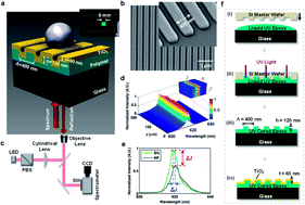
Photonic crystal enhanced microscopy (PCEM) is a relatively new technique that utilizes a grating like substrate composed of a high refractive index material coating a low refractive index one. When irradiated with light at a resonant wavelength, this photonic crystal reflects all of the light, and is very sensitive to any changes on the surface.

Photonic Crystal Enhanced Microscopy of a nanoparticle on a photonic crystal

Photonic Crystal Enhanced Microscopy of a nanoparticle on a photonic crystal

Researchers at the University of Illinois in the United States studied the effects of gold nanoparticles binding to  photonic crystals composed of a liquid epoxy polymer and TiO2.  Although PCEM has been used to detect biological analytes without labels, they observed how a nanoparticle tag can reduce  the signal of a photonic crystal. Furthermore, they determined the distance required between the nanoparticles for detection, and this could be used in future amplification schemes such as in single molecule sensing.

To read more about this topic, click the link below. It will be free to read until February 28.

Single nanoparticle detection using photonic crystal enhanced microscopy
Yue Zhuo,  Huan Hu, Weili Chen,  Meng Lu,  Limei Tian,  Hojeong Yu, Kenneth D. Long,  Edmond Chow,  William P.   King, Srikanth Singamaneni  and  Brian T. Cunningham
Analyst, 2014, Advance Article
DOI: 10.1039/C3AN02295A

Digg This
Reddit This
Stumble Now!
Share on Facebook
Bookmark this on Delicious
Share on LinkedIn
Bookmark this on Technorati
Post on Twitter
Google Buzz (aka. Google Reader)

HOT Articles in Analyst

Take a look at our new HOT articles just published in Analyst and free for you for the next couple of weeks:

Chemometric determination of lipidic parameters in serum using ATR measurements of dry films of solvent extracts
David Perez-Guaita, Angel Sanchez-Illana, Josep Ventura-Gayete, Salvador Garrigues and Miguel de la Guardia
Analyst, 2014,139, 170-178
DOI: 10.1039/C3AN01057K, Paper

Infrared imaging in breast cancer: automated tissue component recognition and spectral characterization of breast cancer cells as well as the tumor microenvironment
Audrey Benard, Christine Desmedt, Margarita Smolina, Philippe Szternfeld, Magali Verdonck, Ghizlane Rouas, Naima Kheddoumi, Françoise Rothé, Denis Larsimont, Christos Sotiriou and Erik Goormaghtigh
Analyst, 2014, Advance Article
DOI: 10.1039/C3AN01454A, Paper

SEDFIT–MSTAR: molecular weight and molecular weight distribution analysis of polymers by sedimentation equilibrium in the ultracentrifuge
Peter Schuck, Richard B. Gillis, Tabot M. D. Besong, Fahad Almutairi, Gary G. Adams, Arthur J. Rowe and Stephen E. Harding
Analyst, 2014,139, 79-92
DOI: 10.1039/C3AN01507F, Paper

Structured illumination for tomographic X-ray diffraction imaging
Joel A. Greenberg, Mehadi Hassan, Kalyani Krishnamurthy and David Brady
Analyst, 2014,139, 709-713
DOI: 10.1039/C3AN01641B, Communication

Molecular interactions of nanomaterials and organisms: defining biomarkers for toxicity and high-throughput screening using traditional and next-generation sequencing approaches
Rebecca Klaper, Devrah Arndt, Jared Bozich and Gustavo Dominguez
Analyst, 2014, Advance Article
DOI: 10.1039/C3AN01644G, Critical Review

Conformational and mechanical changes of DNA upon transcription factor binding detected by a QCM and transmission line model
Jorge de-Carvalho, Rogério M. M. Rodrigues, Brigitte Tomé, Sílvia F. Henriques, Nuno P. Mira, Isabel Sá-Correia and Guilherme N. M. Ferreira
Analyst, 2014, Advance Article
DOI: 10.1039/C3AN01682J, Paper

Real-time evaluation of aggregation using confocal imaging and image analysis tools
Zahra Hamrang, Egor Zindy, David Clarke and Alain Pluen
Analyst, 2014,139, 564-568
DOI: 10.1039/C3AN01693E, Communication

Enhancing the sensitivity of potential step voltammetry using chemometric resolution
Jiarun Tu, Wensheng Cai and Xueguang Shao
Analyst, 2014, Advance Article
DOI: 10.1039/C3AN01719B, Paper

Amplified plasmonic detection of DNA hybridization using doxorubicin-capped gold particles
Jolanda Spadavecchia, Ramesh Perumal, Alexandre Barras, Joel Lyskawa, Patrice Woisel, William Laure, Claire-Marie Pradier, Rabah Boukherroub and Sabine Szunerits
Analyst, 2014,139, 157-164
DOI: 10.1039/C3AN01794J, Paper

Quantitative dielectrophoretic tracking for characterization and separation of persistent subpopulations of Cryptosporidium parvum
Yi-Hsuan Su, Mikiyas Tsegaye, Walter Varhue, Kuo-Tang Liao, Lydia S. Abebe, James A. Smith, Richard L. Guerrant and Nathan S. Swami
Analyst, 2014,139, 66-73
DOI: 10.1039/C3AN01810E, Paper

A dual-plate ITO–ITO generator–collector microtrench sensor: surface activation, spatial separation and suppression of irreversible oxygen and ascorbate interference
Mohammad A. Hasnat, Andrew J. Gross, Sara E. C. Dale, Edward O. Barnes, Richard G. Compton and Frank Marken
Analyst, 2014,139, 569-575
DOI: 10.1039/C3AN01826A, Communication
Digg This
Reddit This
Stumble Now!
Share on Facebook
Bookmark this on Delicious
Share on LinkedIn
Bookmark this on Technorati
Post on Twitter
Google Buzz (aka. Google Reader)

Nanoparticles for platelet adhesion and aggregation

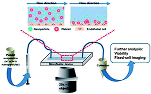
On-chip evaluation of platelet adhesion and aggregation

The use of nanoparticles in diagnostic and therapeutic roles requires that we understand their interactions and behaviour within complex biological systems. Where clinical applications would involve intravenous injection of nanoparticle-based therapeutics, it will be necessary to understand the interaction between nanoparticles and native blood components.

To address this issue, Christy Haynes and coworkers from the University of Minnesota, USA, have created a microfluidic device coated with endothelial cells to mimic the walls of the vascular system. This initial in vitro approach was carried out with both activated and unactivated platelets in the presence of various (therapeutically-relevant) concentrations of fluorescently-labelled, mesophorous silica nanoparticles. The impact of nanoparticles on the critical platelet functions of adhesion and aggregation was assessed.

Microfluidic platforms are readily customised and future work would be expected to expand upon these initial conditions to advance our understanding of nanotoxicology and interaction studies.

To read more about this study and the impact of nanoparticles on platelet interactions you can access this Analyst HOT Article free until 6 January 2014:

On-chip evaluation of platelet adhesion and aggregation upon exposure to mesoporous silica nanoparticles
Donghyuk Kim, Solaire Finkenstaedt-Quinn, Katie R. Hurley, Joseph T. Buchman and Christy L. Haynes 
Analyst, 2014, Advance Article
DOI: 10.1039/C3AN01679J

Digg This
Reddit This
Stumble Now!
Share on Facebook
Bookmark this on Delicious
Share on LinkedIn
Bookmark this on Technorati
Post on Twitter
Google Buzz (aka. Google Reader)

Enhancing transmission Raman spectroscopy (TRS) for pharmaceutical analysis

Data sub-selection in transmission Raman spectroscopy

Data sub-selection in transmission Raman spectroscopy

The analysis of compound mixtures in powder and tablet form has a range of purposes, from monitoring the stability of a formulation over time and quality control of a product, to the forensic analysis of illicit substances. Transmission Raman spectroscopy is a promising candidate for this type of analysis. TRS is fast and non-destructive, it produces data that is easy to interpret, and has good penetration depth for opaque samples such as powders.

Researchers led by Jonathan Burley at the University of Nottingham (UK) have investigated ways to improve the accuracy of TRS for quantitative analysis. In this HOT Analyst paper, they report the first detailed analysis of data sub-selection for a set of transmission Raman data obtained from a model pharmaceutical formulation. Burley and co-workers also focus on the utility of low-wavenumber data, which has only become accessible in recent years. The authors anticipate that their findings may shape the future development of Raman instrumentation.

To read more about this work, please access the link below. This paper will be free to read until 6 January 2014.

Quantification of pharmaceuticals via transmission Raman spectroscopy: data sub-selection
Jonathan C. Burley, Adeyinka Aina, Pavel Matousek and Christopher Brignell
Analyst, 2014,139, 74-78
DOI: 10.1039/C3AN01293J

Digg This
Reddit This
Stumble Now!
Share on Facebook
Bookmark this on Delicious
Share on LinkedIn
Bookmark this on Technorati
Post on Twitter
Google Buzz (aka. Google Reader)

Immobilized phage proteins for detection of staphylococci

The rapid and specific detection of pathogenic bacteria, such as staphylococci, is an important worldwide concern. Staphylococci cause serious infections in humans and animals and certain species such as S. aureus and methicillin resistant S. aureus (MRSA) are developing antibiotic resistance that is quickly becoming a global crisis. The ability to detect these bacteria rapidly and in situ would allow for early treatment even in countries with poor access to healthcare.

Immobilized phage proteins for specific detection of staphylococci

Canadian researchers from McGill University and Polytechnique Montreal have recently shown the use of immobilized bacteriophage proteins to develop a biosensor for the specific detection of 8 clinical isolates of staphylococciHicham Chibli and coworkers examined several purified phage proteins from the Endolysin class of enzymes and exploited their ability to specifically bind staphylococci. Indeed these immobilized phage proteins did not bind to other closely related bacteria.

Bacteriophages have been used as recognition elements for biosensors before, but this research shows the use of specific phage proteins instead of whole bacteriophages. “Compared with whole phages, these single proteins are smaller, easier to purify, and easier to assemble in a reproducible manner of a surface” – says Jay Nadeau of McGill University – “The small size is key for development of some types of biosensors where sensitivity is dependent upon thickness of the functionalizing layer, for example, microresonators” – he adds. The ability to develop microresonators with specific detection can provide small, portable and specific biosensors for in field use.

To know more about this study, please access the link below. This paper will be free to read for the next three weeks.

Immobilized phage proteins for specific detection of staphylococci
Hicham Chibli, Hala Ghali, Soonhyang Park, Yves-Alain Peter and Jay L. Nadeau  
Analyst, 2014, 139, 179-186
DOI: 10.1039/C3AN01608K

Digg This
Reddit This
Stumble Now!
Share on Facebook
Bookmark this on Delicious
Share on LinkedIn
Bookmark this on Technorati
Post on Twitter
Google Buzz (aka. Google Reader)

HOT Articles in Analyst

Take a look at our new HOT articles just published in Analyst and free for you for the next couple of weeks:

Development of coated-wire silver ion selective electrodes on paper using conductive films of silver nanoparticles
Wanwisa Janrungroatsakul, Chutiparn Lertvachirapaiboon, Wittaya Ngeontae, Wanlapa Aeungmaitrepirom, Orawon Chailapakul, Sanong Ekgasitb and Thawatchai Tuntulani
Analyst, 2013,138, 6786-6792
DOI: 10.1039/C3AN01385E

A visible light photoelectrochemical sensor for tumor marker detection using tin dioxide quantum dot–graphene as labels
Yanhu Wang, Meng Li, Yuanna Zhu, Shenguang Ge, Jinghua Yu, Mei Yan and Xianrang Song
Analyst, 2013,138, 7112-7118
DOI: 10.1039/C3AN01410J

Retention in continuous two-dimensional thermal field-flow fractionation: comparison of experimental results with theory
Pertti Vastamäki, P. Stephen Williams, Matti Jussila, Michel Martin and Marja-Liisa Riekkola
Analyst, 2014, Advance Article
DOI: 10.1039/C3AN01047C

Cascade signal amplification for ultrasensitive electrochemical DNA detection
Jin Xu, Qiong Wang, Yun Xiang, Ruo Yuan and Yaqin Chai
Analyst, 2014, Advance Article
DOI: 10.1039/C3AN01673K

Detection and characterization of silver nanoparticles and dissolved species of silver in culture medium and cells by AsFlFFF-UV-Vis-ICPMS: application to nanotoxicity tests
E. Bolea, J. Jiménez-Lamana, F. Laborda, I. Abad-Álvaro, C. Bladé, L. Arola and J. R. Castillo
Analyst, 2014, Advance Article
DOI: 10.1039/C3AN01443F

MALDI mechanisms: wavelength and matrix dependence of the coupled photophysical and chemical dynamics model
Richard Knochenmuss
Analyst, 2014, Advance Article
DOI: 10.1039/C3AN01446K

Target-induced quenching for highly sensitive detection of nucleic acids based on label-free luminescent supersandwich DNA/silver nanoclusters
Guangfeng Wang, Yanhong Zhu, Ling Chen, Lun Wang and Xiaojun Zhang
Analyst, 2014, Advance Article
DOI: 10.1039/C3AN01702H

LED-based interferometric reflectance imaging sensor for the detection of amyloid-β aggregation
Xin R. Cheng, George G. Daaboul, M. Selim Ünlü and Kagan Kerman
Analyst, 2014, Advance Article
DOI: 10.1039/C3AN01307C

Improved accuracy for label-free absolute quantification of proteome by combining the absolute protein expression profiling algorithm and summed tandem mass spectrometric total ion current
Qi Wu, Yichu Shan, Yanyan Qu, Hao Jiang, Huiming Yuan, Jianxi Liu, Shen Zhang, Zhen Liang, Lihua Zhang and Yukui Zhang
Analyst, 2014, Advance Article
DOI: 10.1039/C3AN01738A

Focussed ion beam serial sectioning and imaging of monolithic materials for 3D reconstruction and morphological parameter evaluation
Mercedes Vázquez, David Moore, Xiaoyun He, Aymen Ben Azouz, Ekaterina Nesterenko, Pavel Nesterenko, Brett Paull and Dermot Brabazon
Analyst, 2014, Advance Article
DOI: 10.1039/C3AN01827J

Digg This
Reddit This
Stumble Now!
Share on Facebook
Bookmark this on Delicious
Share on LinkedIn
Bookmark this on Technorati
Post on Twitter
Google Buzz (aka. Google Reader)

Analysis of thyroid tissues by FT-IR microscopectroscopy


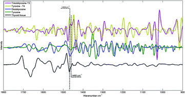
FTIR spectra of hormones triiodothyronine, thyroxine, diiodotyrosine, tyrosine and thyroid tissue

The combination of vibrational spectroscopy with mapping and imaging techniques to address important biochemical questions is an area of active and expanding research.  By subjecting adjacent tissue sections to standard histopathological screening and spectroscopic imaging, a greater understanding of the biochemical processes underlying tissue form and function can be achieved.

Researchers from Northeastern University, USA, and the Institute of Energy and Nuclear Research (IPEN), Sao Paolo, have used Fourier transform infrared (FTIR) microspectroscopy to examine healthy thyroid tissues. FTIR allows spatially resolved images, or maps, to be obtained by use of focal plane array detectors and spectral processing of the data then reveals chemical images.  In this HOT Analyst paper, Denise Zezell and coworkers present an example from their investigation of 80 different patient samples, which were analysed by transflection-mode FTIR mapping.  The approach was  applied to the study of healthy thyroid tissue and, with reference to thyroid specific hormones, iodination state. 

To know more about this research, click on the links below. This paper will be free to read for the next three weeks:

The characterization of normal thyroid tissue by micro-FTIR spectroscopy
Thiago M. Pereira, Denise M. Zezell, Benjamin Bird, Milos Miljković and Max Diem
Analyst, 2013,138, 7094-7100
DOI: 10.1039/C3AN00296A

Digg This
Reddit This
Stumble Now!
Share on Facebook
Bookmark this on Delicious
Share on LinkedIn
Bookmark this on Technorati
Post on Twitter
Google Buzz (aka. Google Reader)

Classifying myobacterial strains using surface enhanced Raman spectroscopy

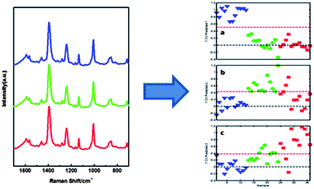
Spectroscopic analysis of mycolic acid profiles

In bacterial-based diagnostics, the bacteria can evolve over time and it is important to be able to differentiate between multiple strains. Mycobacterium tuberculosis, which is responsible for causing tuberculosis (TB), has multiple drug-resistant strains. The most accurate method to identify toxic strains of TB, high performance liquid chromatography (HPLC), requires fluorescent labeling and extraction of the mycolic acids, from the outer membrane of the bacteria.

Researchers at the University of Georgia in the United States wanted a more direct detection method and approached this problem in two important aspects. They began by identifying the high molecular weight lipids within the bacteria using HPLC, gas chromatography (GC), and nuclear magnetic resonance (NMR) for several TB bacterial strains. NMR in particular was very important in identifying the structures of mycolic acids. Using the information from these techniques and surface enhanced Raman spectroscopy (SERS), they chemically classified the mycolic acid from both TB and non-TB strains. The SERS spectra underwent further statistical analysis and accurately identified different strains of TB label-free and with high sensitivity.

To know more about this research, click on the link below. This paper will be free to read for the next three weeks.

Identification of mycobacteria based on spectroscopic analyses of mycolic acid profiles
Omar E. Rivera-Betancourt, Russell Karls, Benjamin Grosse-Siestrup, Shelly Helms, Frederick Quinn and Richard A. Dluhy
Analyst, 2013,138, 6774-6785
DOI: 10.1039/C3AN01157G

Digg This
Reddit This
Stumble Now!
Share on Facebook
Bookmark this on Delicious
Share on LinkedIn
Bookmark this on Technorati
Post on Twitter
Google Buzz (aka. Google Reader)

More HOT Articles in Analyst

Take a look at our new HOT articles just published in Analyst! These papers are all free to read for the next three weeks. Enjoy!

In vitro detection of calcium in bone by modified carbon dots
A. Shanti Krishna, C. Radhakumary and K. Sreenivasan
Analyst, 2013,138, 7107-7111
DOI: 10.1039/C3AN01239E, Paper

Measuring and modelling cell-to-cell variation in uptake of gold nanoparticles
J. Charles. G. Jeynes, Christopher Jeynes, Michael J. Merchant and Karen J. Kirkby
Analyst, 2013,138, 7070-7074
DOI: 10.1039/C3AN01406A, Communication

Adenosine capped QDs based fluorescent sensor for detection of dopamine with high selectivity and sensitivity
Qin Mu, Hu Xu, Yan Li, Shijian Ma and Xinhua Zhong
Analyst, 2013, Advance Article
DOI: 10.1039/C3AN01592K, Paper

A pyrene-based simple but highly selective fluorescence sensor for Cu2+ ions via a static excimer mechanism
Soma Sarkar, Swapnadip Roy, Anindita Sikdar, R. N. Saha and Sujit S. Panja
Analyst, 2013,138, 7119-7126
DOI: 10.1039/C3AN00928A, Paper

Whole organ cross-section chemical imaging using label-free mega-mosaic FTIR microscopy
Paul Bassan, Ashwin Sachdeva, Jonathan H. Shanks, Mick D. Brown, Noel W. Clarke and Peter Gardner
Analyst, 2013,138, 7066-7069
DOI: 10.1039/C3AN01674A, Communication

A highly sensitive LED-induced chemiluminescence platform for aptasensing of platelet-derived growth factor
Xinfeng Zhang, Hui Zhang, Shuxia Xu and Yonghua Sun
Analyst, 2013, Advance Article
DOI: 10.1039/C3AN01566A, Paper

Effects of surface charges of graphene oxide on neuronal outgrowth and branching
Qin Tu, Long Pang, Yun Chen, Yanrong Zhang, Rui Zhang, Bingzhang Lu and Jinyi Wang
Analyst, 2013, Advance Article
DOI: 10.1039/C3AN01796F, Paper

Paracrine signalling of inflammatory cytokines from an in vitro blood brain barrier model upon exposure to polymeric nanoparticles
Michelle Nic Raghnaill, Mattia Bramini, Dong Ye, Pierre-Olivier Couraud, Ignacio A. Romero, Babette Weksler, Christoffer Åberg, Anna Salvati, Iseult Lynch and Kenneth A Dawson
Analyst, 2013, Accepted Manuscript
DOI: 10.1039/C3AN01621H, Paper

Adenosine capped QDs based fluorescent sensor for detection of dopamine with high selectivity and sensitivity
Qin Mu, Hu Xu, Yan Li, Shijian Ma and Xinhua Zhong
Analyst, 2013, Advance Article
DOI: 10.1039/C3AN01592K, Paper

Digg This
Reddit This
Stumble Now!
Share on Facebook
Bookmark this on Delicious
Share on LinkedIn
Bookmark this on Technorati
Post on Twitter
Google Buzz (aka. Google Reader)

HOT Articles in Analyst

Take a look at our new HOT articles just published in Analyst and free for you for the next couple of weeks:

Combined in situ atomic force microscopy and infrared attenuated total reflection spectroelectrochemistry
Daniel Neubauer, Jochen Scharpf, Alberto Pasquarelli, Boris Mizaikoff and Christine Kranz
Analyst, 2013,138, 6746-6752
DOI: 10.1039/C3AN01169K, Paper

An iridium(III)-based lab-on-a-molecule for cysteine/homocysteine and tryptophan using triple-channel interrogation Kun Chen and Michael Schmittel
Analyst, 2013,138, 6742-6745
DOI: 10.1039/C3AN01530K, Communication

Determination of aerosol oxidative activity using silver nanoparticle aggregation on paper-based analytical devices
Wijitar Dungchai, Yupaporn Sameenoi, Orawon Chailapakul, John Volckens and Charles S. Henry
Analyst, 2013,138, 6766-6773
DOI: 10.1039/C3AN01235B, Paper

Biocatalytic analysis of biomarkers for forensic identification of ethnicity between Caucasian and African American groups
Friederike Kramer, Lenka Halámková, Arshak Poghossian, Michael J. Schöning, Evgeny Katz and Jan Halámek
Analyst, 2013,138, 6251-6257
DOI: 10.1039/C3AN01062G, Communication

Biocatalytic analysis of biomarkers for forensic identification of ethnicity between Caucasian and African American groups
Friederike Kramer, Lenka Halámková, Arshak Poghossian, Michael J. Schöning, Evgeny Katz and Jan Halámek
Analyst, 2013,138, 6251-6257
DOI: 10.1039/C3AN01062G, Communication

Analysis of free fractions for chiral drugs using ultrafast extraction and multi-dimensional high-performance affinity chromatography
Xiwei Zheng, Michelle J. Yoo and David S. Hage
Analyst, 2013,138, 6262-6265
DOI: 10.1039/C3AN01315D, Communication

Radical cascades in electron transfer dissociation (ETD) – implications for characterizing peptide disulfide regio-isomers
Lei Tan, Kirt L. Durand, Xiaoxiao Ma and Yu Xia
Analyst, 2013,138, 6759-6765
DOI: 10.1039/C3AN01333B, Paper

Enhanced peroxydisulfate electrochemiluminescence for dopamine biosensing based on Au nanoparticle decorated reduced graphene oxide
Yuting Yan, Qian Liu, Kun Wang, Ling Jiang, Xingwang Yang, Jing Qian, Xiaoya Dong and Baijing Qiu
Analyst, 2013,138, 7101-7106
DOI: 10.1039/C3AN01533E, Paper

Digg This
Reddit This
Stumble Now!
Share on Facebook
Bookmark this on Delicious
Share on LinkedIn
Bookmark this on Technorati
Post on Twitter
Google Buzz (aka. Google Reader)