Archive for the ‘Hot Article 2013’ Category

Enhancing transmission Raman spectroscopy (TRS) for pharmaceutical analysis

Data sub-selection in transmission Raman spectroscopy

Data sub-selection in transmission Raman spectroscopy

The analysis of compound mixtures in powder and tablet form has a range of purposes, from monitoring the stability of a formulation over time and quality control of a product, to the forensic analysis of illicit substances. Transmission Raman spectroscopy is a promising candidate for this type of analysis. TRS is fast and non-destructive, it produces data that is easy to interpret, and has good penetration depth for opaque samples such as powders.

Researchers led by Jonathan Burley at the University of Nottingham (UK) have investigated ways to improve the accuracy of TRS for quantitative analysis. In this HOT Analyst paper, they report the first detailed analysis of data sub-selection for a set of transmission Raman data obtained from a model pharmaceutical formulation. Burley and co-workers also focus on the utility of low-wavenumber data, which has only become accessible in recent years. The authors anticipate that their findings may shape the future development of Raman instrumentation.

To read more about this work, please access the link below. This paper will be free to read until 6 January 2014.

Quantification of pharmaceuticals via transmission Raman spectroscopy: data sub-selection
Jonathan C. Burley, Adeyinka Aina, Pavel Matousek and Christopher Brignell
Analyst, 2014,139, 74-78
DOI: 10.1039/C3AN01293J

Digg This
Reddit This
Stumble Now!
Share on Facebook
Bookmark this on Delicious
Share on LinkedIn
Bookmark this on Technorati
Post on Twitter
Google Buzz (aka. Google Reader)

Immobilized phage proteins for detection of staphylococci

The rapid and specific detection of pathogenic bacteria, such as staphylococci, is an important worldwide concern. Staphylococci cause serious infections in humans and animals and certain species such as S. aureus and methicillin resistant S. aureus (MRSA) are developing antibiotic resistance that is quickly becoming a global crisis. The ability to detect these bacteria rapidly and in situ would allow for early treatment even in countries with poor access to healthcare.

Immobilized phage proteins for specific detection of staphylococci

Canadian researchers from McGill University and Polytechnique Montreal have recently shown the use of immobilized bacteriophage proteins to develop a biosensor for the specific detection of 8 clinical isolates of staphylococciHicham Chibli and coworkers examined several purified phage proteins from the Endolysin class of enzymes and exploited their ability to specifically bind staphylococci. Indeed these immobilized phage proteins did not bind to other closely related bacteria.

Bacteriophages have been used as recognition elements for biosensors before, but this research shows the use of specific phage proteins instead of whole bacteriophages. “Compared with whole phages, these single proteins are smaller, easier to purify, and easier to assemble in a reproducible manner of a surface” – says Jay Nadeau of McGill University – “The small size is key for development of some types of biosensors where sensitivity is dependent upon thickness of the functionalizing layer, for example, microresonators” – he adds. The ability to develop microresonators with specific detection can provide small, portable and specific biosensors for in field use.

To know more about this study, please access the link below. This paper will be free to read for the next three weeks.

Immobilized phage proteins for specific detection of staphylococci
Hicham Chibli, Hala Ghali, Soonhyang Park, Yves-Alain Peter and Jay L. Nadeau  
Analyst, 2014, 139, 179-186
DOI: 10.1039/C3AN01608K

Digg This
Reddit This
Stumble Now!
Share on Facebook
Bookmark this on Delicious
Share on LinkedIn
Bookmark this on Technorati
Post on Twitter
Google Buzz (aka. Google Reader)

HOT Articles in Analyst

Take a look at our new HOT articles just published in Analyst and free for you for the next couple of weeks:

Development of coated-wire silver ion selective electrodes on paper using conductive films of silver nanoparticles
Wanwisa Janrungroatsakul, Chutiparn Lertvachirapaiboon, Wittaya Ngeontae, Wanlapa Aeungmaitrepirom, Orawon Chailapakul, Sanong Ekgasitb and Thawatchai Tuntulani
Analyst, 2013,138, 6786-6792
DOI: 10.1039/C3AN01385E

A visible light photoelectrochemical sensor for tumor marker detection using tin dioxide quantum dot–graphene as labels
Yanhu Wang, Meng Li, Yuanna Zhu, Shenguang Ge, Jinghua Yu, Mei Yan and Xianrang Song
Analyst, 2013,138, 7112-7118
DOI: 10.1039/C3AN01410J

Retention in continuous two-dimensional thermal field-flow fractionation: comparison of experimental results with theory
Pertti Vastamäki, P. Stephen Williams, Matti Jussila, Michel Martin and Marja-Liisa Riekkola
Analyst, 2014, Advance Article
DOI: 10.1039/C3AN01047C

Cascade signal amplification for ultrasensitive electrochemical DNA detection
Jin Xu, Qiong Wang, Yun Xiang, Ruo Yuan and Yaqin Chai
Analyst, 2014, Advance Article
DOI: 10.1039/C3AN01673K

Detection and characterization of silver nanoparticles and dissolved species of silver in culture medium and cells by AsFlFFF-UV-Vis-ICPMS: application to nanotoxicity tests
E. Bolea, J. Jiménez-Lamana, F. Laborda, I. Abad-Álvaro, C. Bladé, L. Arola and J. R. Castillo
Analyst, 2014, Advance Article
DOI: 10.1039/C3AN01443F

MALDI mechanisms: wavelength and matrix dependence of the coupled photophysical and chemical dynamics model
Richard Knochenmuss
Analyst, 2014, Advance Article
DOI: 10.1039/C3AN01446K

Target-induced quenching for highly sensitive detection of nucleic acids based on label-free luminescent supersandwich DNA/silver nanoclusters
Guangfeng Wang, Yanhong Zhu, Ling Chen, Lun Wang and Xiaojun Zhang
Analyst, 2014, Advance Article
DOI: 10.1039/C3AN01702H

LED-based interferometric reflectance imaging sensor for the detection of amyloid-β aggregation
Xin R. Cheng, George G. Daaboul, M. Selim Ünlü and Kagan Kerman
Analyst, 2014, Advance Article
DOI: 10.1039/C3AN01307C

Improved accuracy for label-free absolute quantification of proteome by combining the absolute protein expression profiling algorithm and summed tandem mass spectrometric total ion current
Qi Wu, Yichu Shan, Yanyan Qu, Hao Jiang, Huiming Yuan, Jianxi Liu, Shen Zhang, Zhen Liang, Lihua Zhang and Yukui Zhang
Analyst, 2014, Advance Article
DOI: 10.1039/C3AN01738A

Focussed ion beam serial sectioning and imaging of monolithic materials for 3D reconstruction and morphological parameter evaluation
Mercedes Vázquez, David Moore, Xiaoyun He, Aymen Ben Azouz, Ekaterina Nesterenko, Pavel Nesterenko, Brett Paull and Dermot Brabazon
Analyst, 2014, Advance Article
DOI: 10.1039/C3AN01827J

Digg This
Reddit This
Stumble Now!
Share on Facebook
Bookmark this on Delicious
Share on LinkedIn
Bookmark this on Technorati
Post on Twitter
Google Buzz (aka. Google Reader)

Analysis of thyroid tissues by FT-IR microscopectroscopy


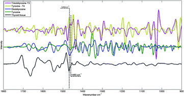
FTIR spectra of hormones triiodothyronine, thyroxine, diiodotyrosine, tyrosine and thyroid tissue

The combination of vibrational spectroscopy with mapping and imaging techniques to address important biochemical questions is an area of active and expanding research.  By subjecting adjacent tissue sections to standard histopathological screening and spectroscopic imaging, a greater understanding of the biochemical processes underlying tissue form and function can be achieved.

Researchers from Northeastern University, USA, and the Institute of Energy and Nuclear Research (IPEN), Sao Paolo, have used Fourier transform infrared (FTIR) microspectroscopy to examine healthy thyroid tissues. FTIR allows spatially resolved images, or maps, to be obtained by use of focal plane array detectors and spectral processing of the data then reveals chemical images.  In this HOT Analyst paper, Denise Zezell and coworkers present an example from their investigation of 80 different patient samples, which were analysed by transflection-mode FTIR mapping.  The approach was  applied to the study of healthy thyroid tissue and, with reference to thyroid specific hormones, iodination state. 

To know more about this research, click on the links below. This paper will be free to read for the next three weeks:

The characterization of normal thyroid tissue by micro-FTIR spectroscopy
Thiago M. Pereira, Denise M. Zezell, Benjamin Bird, Milos Miljković and Max Diem
Analyst, 2013,138, 7094-7100
DOI: 10.1039/C3AN00296A

Digg This
Reddit This
Stumble Now!
Share on Facebook
Bookmark this on Delicious
Share on LinkedIn
Bookmark this on Technorati
Post on Twitter
Google Buzz (aka. Google Reader)

Classifying myobacterial strains using surface enhanced Raman spectroscopy

Spectroscopic analysis of mycolic acid profiles

In bacterial-based diagnostics, the bacteria can evolve over time and it is important to be able to differentiate between multiple strains. Mycobacterium tuberculosis, which is responsible for causing tuberculosis (TB), has multiple drug-resistant strains. The most accurate method to identify toxic strains of TB, high performance liquid chromatography (HPLC), requires fluorescent labeling and extraction of the mycolic acids, from the outer membrane of the bacteria.

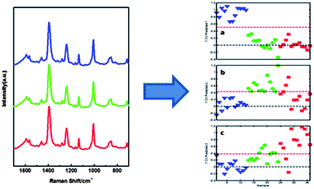
Researchers at the University of Georgia in the United States wanted a more direct detection method and approached this problem in two important aspects. They began by identifying the high molecular weight lipids within the bacteria using HPLC, gas chromatography (GC), and nuclear magnetic resonance (NMR) for several TB bacterial strains. NMR in particular was very important in identifying the structures of mycolic acids. Using the information from these techniques and surface enhanced Raman spectroscopy (SERS), they chemically classified the mycolic acid from both TB and non-TB strains. The SERS spectra underwent further statistical analysis and accurately identified different strains of TB label-free and with high sensitivity.

To know more about this research, click on the link below. This paper will be free to read for the next three weeks.

Identification of mycobacteria based on spectroscopic analyses of mycolic acid profiles
Omar E. Rivera-Betancourt, Russell Karls, Benjamin Grosse-Siestrup, Shelly Helms, Frederick Quinn and Richard A. Dluhy
Analyst, 2013,138, 6774-6785
DOI: 10.1039/C3AN01157G

Digg This
Reddit This
Stumble Now!
Share on Facebook
Bookmark this on Delicious
Share on LinkedIn
Bookmark this on Technorati
Post on Twitter
Google Buzz (aka. Google Reader)

More HOT Articles in Analyst

Take a look at our new HOT articles just published in Analyst! These papers are all free to read for the next three weeks. Enjoy!

In vitro detection of calcium in bone by modified carbon dots
A. Shanti Krishna, C. Radhakumary and K. Sreenivasan
Analyst, 2013,138, 7107-7111
DOI: 10.1039/C3AN01239E, Paper

Measuring and modelling cell-to-cell variation in uptake of gold nanoparticles
J. Charles. G. Jeynes, Christopher Jeynes, Michael J. Merchant and Karen J. Kirkby
Analyst, 2013,138, 7070-7074
DOI: 10.1039/C3AN01406A, Communication

Adenosine capped QDs based fluorescent sensor for detection of dopamine with high selectivity and sensitivity
Qin Mu, Hu Xu, Yan Li, Shijian Ma and Xinhua Zhong
Analyst, 2013, Advance Article
DOI: 10.1039/C3AN01592K, Paper

A pyrene-based simple but highly selective fluorescence sensor for Cu2+ ions via a static excimer mechanism
Soma Sarkar, Swapnadip Roy, Anindita Sikdar, R. N. Saha and Sujit S. Panja
Analyst, 2013,138, 7119-7126
DOI: 10.1039/C3AN00928A, Paper

Whole organ cross-section chemical imaging using label-free mega-mosaic FTIR microscopy
Paul Bassan, Ashwin Sachdeva, Jonathan H. Shanks, Mick D. Brown, Noel W. Clarke and Peter Gardner
Analyst, 2013,138, 7066-7069
DOI: 10.1039/C3AN01674A, Communication

A highly sensitive LED-induced chemiluminescence platform for aptasensing of platelet-derived growth factor
Xinfeng Zhang, Hui Zhang, Shuxia Xu and Yonghua Sun
Analyst, 2013, Advance Article
DOI: 10.1039/C3AN01566A, Paper

Effects of surface charges of graphene oxide on neuronal outgrowth and branching
Qin Tu, Long Pang, Yun Chen, Yanrong Zhang, Rui Zhang, Bingzhang Lu and Jinyi Wang
Analyst, 2013, Advance Article
DOI: 10.1039/C3AN01796F, Paper

Paracrine signalling of inflammatory cytokines from an in vitro blood brain barrier model upon exposure to polymeric nanoparticles
Michelle Nic Raghnaill, Mattia Bramini, Dong Ye, Pierre-Olivier Couraud, Ignacio A. Romero, Babette Weksler, Christoffer Åberg, Anna Salvati, Iseult Lynch and Kenneth A Dawson
Analyst, 2013, Accepted Manuscript
DOI: 10.1039/C3AN01621H, Paper

Adenosine capped QDs based fluorescent sensor for detection of dopamine with high selectivity and sensitivity
Qin Mu, Hu Xu, Yan Li, Shijian Ma and Xinhua Zhong
Analyst, 2013, Advance Article
DOI: 10.1039/C3AN01592K, Paper

Digg This
Reddit This
Stumble Now!
Share on Facebook
Bookmark this on Delicious
Share on LinkedIn
Bookmark this on Technorati
Post on Twitter
Google Buzz (aka. Google Reader)

LEDs Overcome the “Enemy” of Raman Spectroscopy

LED-SERDS in Raman Spectroscopy

LED-SERDS in Raman Spectroscopy

Raman spectroscopy finds a range of uses in analytical chemistry. Like infra-red (IR) spectroscopy it can provide a vibrational fingerprint by which compounds may be identified. Unlike IR, Raman is a scattering technique that does not require complex sample preparation, and water can be used as a solvent. In some samples, however, the Raman spectrum is rendered useless by intense fluorescence signals.

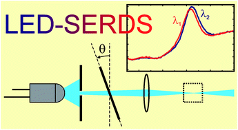
Chemical engineers Renata Adami (University of Salerno, Italy) and Johannes Kiefer (University of Aberdeen, UK) have developed a new approach to shifted-excitation Raman difference spectroscopy (SERDS) for suppression of fluorescence. SERDS experiments conventionally require expensive diode lasers, but in this Analyst paper Kiefer and Adami demonstrate the use of light-emitting diodes (LEDs) as an inexpensive light source. A dielectric bandpass filter narrows and stabilises the broad LED spectrum to make it usable for SERDS. While the resulting spectral resolution is low, different compounds and functional groups can be clearly distinguished using this method.

To read the full access, please click on the link below. This paper will be free to read for the next three weeks:

Light-emitting diode based shifted-excitation Raman difference spectroscopy (LED-SERDS)
Renata Adami and Johannes Kiefer
Analyst, 2013,138, 6258-6261
DOI: 10.1039/C3AN01367G
Digg This
Reddit This
Stumble Now!
Share on Facebook
Bookmark this on Delicious
Share on LinkedIn
Bookmark this on Technorati
Post on Twitter
Google Buzz (aka. Google Reader)

High Sensitivity Using Low Cost Equipment

Low cost webcam based prism spectrometer

Techniques such as cavity ring-down spectroscopy (CRDS) have greatly enhanced the sensitivity in traditional absorption spectroscopy methods. Although CRDS can detect analytes in very small concentrations in solution, it requires expensive and complex instrumentation, which has restricted its widespread implementation. Alternatively, broadband cavity enhanced absorption spectroscopy (BBCEAS) has emerged to overcome some of these limitations. These adjustments allow for multiplexed data collection and improved resolution.

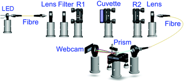
Researchers at the University of Cambridge, UK,  have constructed a BBCEAS apparatus using relatively simple components and achieved sensitivity levels similar to much more expensive systems.  Using a well-studied process, the Griess Assay, they measured different concentrations of nitrite and the dye Rhodamine 6 G with a system that contained a low cost prism spectrometer and a webcam detector. The limits of detection were found to be 3.7 nM (nitrite) and 850 pM (Rhodamine), and by adding a better detector, these can further be improved upon.

To read the full article, please access the link below. This paper will be free to read for the next three weeks:

High sensitivity liquid phase measurements using broadband cavity enhanced absorption spectroscopy (BBCEAS) featuring a low cost webcam based prism spectrometer
Zhechao Qu, Julia Engstrom, Donald Wong, Meez Islam and   Clemens F. Kaminski
Analyst, 2013,138, 6372-6379
DOI: 10.1039/C3AN01441J

Digg This
Reddit This
Stumble Now!
Share on Facebook
Bookmark this on Delicious
Share on LinkedIn
Bookmark this on Technorati
Post on Twitter
Google Buzz (aka. Google Reader)

Intracellular enzymatic activity by SERRS

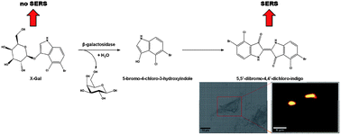
Analysis of intracellular enzyme activity by SERS

Enzymatic activity in cells has been revealed by surface enhanced Raman scattering (SERRS) spectroscopic studies, according to new research led by Duncan Graham at the University of Strathclyde.

X-Gal, a SERRS inactive precursor and a common marker for galactosidase enzymes, was introduced to macrophage cells along with metal nanoparticles. On incubation, enzymatic activity of galactosidase on X-Gal produces a SERRS active trans-alkene dimerised product. This product adsorbs onto the delivered citrate capped gold nanoparticles producing a distinctive SERRS signal. Using SERRS mapping to evaluate the formation of this product, enzyme activity was assessed, not only across a cell population, but also at sub-cellular levels. The authors note that the SERRS activity in this study may not reflect the only sites of galactosidase activity, but rather the sites where both galactosidase and the nanoparticles are co-located. Nevertheless, approaches towards sub-cellular analysis of enzyme activity are important.

Advances in targeted nanoparticle uptake combined with the multiplexing capability of SERRS make this an interesting approach to sub-cellular studies of biochemical activity.

To read the full article, please click on the link below. This paper will be free to read until October 18th.

Analysis of intracellular enzyme activity by surface enhanced Raman scattering
Ross Stevenson, Sarah McAughtrie, Laura Senior, Robert J. Stokes, Helen McGachy, Laurence Tetley, Paola Nativo, James M. Brewer, James Alexander, Karen Faulds and   Duncan Graham
Analyst, 2013, Advance Article
DOI: 10.1039/C3AN00729D

Digg This
Reddit This
Stumble Now!
Share on Facebook
Bookmark this on Delicious
Share on LinkedIn
Bookmark this on Technorati
Post on Twitter
Google Buzz (aka. Google Reader)

HOT Articles in Analyst

Take a look at these new HOT articles just published in Analyst! These papers will be free to read for the next three weeks. Download them today!

The global identification of tRNA isoacceptors by targeted tandem mass spectrometry
Collin Wetzel and Patrick A. Limbach  
Analyst, 2013,138, 6063-6072
DOI: 10.1039/C3AN01224G

Extraction and separation of proteins by ionic liquid aqueous two-phase system
Xiao Lin, Yuzhi Wang, Qun Zeng, Xueqin Ding and Jing Chen  
Analyst, 2013, Advance Article
DOI: 10.1039/C3AN01301D

A smart T1-weighted MRI contrast agent for uranyl cations based on a DNAzyme–gadolinium conjugate
Weichen Xu, Hang Xing and Yi Lu  
Analyst, 2013, Advance Article
DOI: 10.1039/C3AN01182H

Improved analysis of oligosaccharides for matrix-assisted laser desorption/ionization time-of-flight mass spectrometry using aminopyrazine as a derivatization reagent and a co-matrix
Yan Cai, Ying Zhang, Pengyuan Yang and Haojie Lu
Analyst, 2013, Advance Article
DOI: 10.1039/C3AN01228J

A chemical quenching- and physical blocking-based method to minimize process-mediated aggregation of antibody-crosslinked nanoparticles for imaging application
Chandra K. Dixit, Shibsekhar Roy, Conor Byrne, Richard O’Kennedy and Colette McDonagh  
Analyst, 2013, Advance Article
DOI: 10.1039/C3AN01294H

Exploring the electrochemical performance of graphitic paste electrodes: graphene vs. graphite
Luiz C. S. Figueiredo-Filho, Dale A. C. Brownson, Maria Gómez-Mingot, Jesús Iniesta, Orlando Fatibello-Filho and Craig E. Banks  
Analyst, 2013, Advance Article
DOI: 10.1039/C3AN00950E

An investigation into the simultaneous enzymatic and SERRS properties of silver nanoparticles
Kristy S. McKeating, Sian Sloan-Dennison, Duncan Graham and Karen Faulds  
Analyst, 2013, Advance Article
DOI: 10.1039/C3AN01451G

Electrochemical piezoelectric-excited millimeter-sized cantilever (ePEMC) for simultaneous dual transduction biosensing
Blake N. Johnson and Raj Mutharasan  
Analyst, 2013, Advance Article
DOI: 10.1039/C3AN01353G

Digg This
Reddit This
Stumble Now!
Share on Facebook
Bookmark this on Delicious
Share on LinkedIn
Bookmark this on Technorati
Post on Twitter
Google Buzz (aka. Google Reader)