Author Archive

Biocatalytic Conversion of Glucose on an Enzyme Electrode

Enzyme electrodes for glucose biosensing

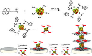
Glucose oxidase (GOx) is responsible for breaking down glucose and produces a detectable secondary signal used in a variety of biological assays. The primary electrical response from this catalytic process is difficult to detect because of the distance of the signal from within the enzyme to the electrode. To overcome these limitations, Jingquan Liu and researchers at the Qingdao University, China, have developed an enzyme-based electrode with pyrene functionalized GOx, which self assembles onto graphene sheets. The large surface area and high conductivity of graphene sheets enhance the electron transfer from the GOx via interaction with the pyrene, down to the electrode. In addition, increasing alternating layers of the pyrene-GOx and graphene enhances the biocatalytic activity in glucose solutions.

To know more about this research, please access the link below. This paper will be free to read until April 12th.

Graphene bridged enzyme electrodes for glucose biosensing application
Jingquan Liu ,  Na Kong ,  Aihua Li ,  Xiong Luo ,  Liang Cui ,  Rui Wang and Shengyu Feng
Analyst, 2013, Advance Article
DOI: 10.1039/C3AN36929C

Digg This
Reddit This
Stumble Now!
Share on Facebook
Bookmark this on Delicious
Share on LinkedIn
Bookmark this on Technorati
Post on Twitter
Google Buzz (aka. Google Reader)

miRNA and Palladium Nanoparticles for Label-Free Cancer Testing

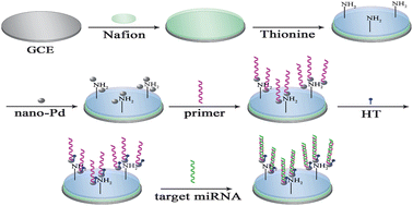
Schematic steps for biosensor detecting mRNAs

Short non-coding single stranded nucleotides known as microRNA (miRNA) have become important biomarkers due to their up- and down-regulated expression in certain cancers.

Jing Han and colleagues at Southwest University, China, targeted miRNAs using an electrochemical-based biological sensor containing palladium nanoparticles. A multilayer film coated the glass electrodes in order to bind the palladium particles, and then catalyzed hydrogen peroxide to enhance the signal of the miRNA binding. Using a model of miRNA overexpressed in several diseases, miRNA-155, they detected even low concentrations in blood serum samples previously prepared and demonstrated the high reproducibility and adaptability of this assay for a variety of other biomarkers.

To read more about this study, access to the link below. This paper will be free to read until January 24th.

A novel label-free electrochemical microRNA biosensor using Pd nanoparticles as enhancer and linker
Xiaoyan Wu ,  Yaqin Chai ,  Ruo Yuan ,  Huilan Su and Jing Han
Analyst, 2013, Advance Article
DOI: 10.1039/C2AN36506E

Digg This
Reddit This
Stumble Now!
Share on Facebook
Bookmark this on Delicious
Share on LinkedIn
Bookmark this on Technorati
Post on Twitter
Google Buzz (aka. Google Reader)

Next Generation Carbohydrate Assays

Carbohydrate microarrays have become an important tool for studying glycan-cell interactions, carbohydrate-binding proteins, and in detecting dangerous pathogens. The current sensor methods require complex labeling or indirect analyte detection.

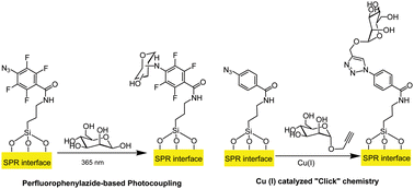
C–H insertion photocoupling reaction and copper “click” methods

Using surface plasmon resonance spectroscopy (SPR), Sabine Szunerits and coworkers at  Lille 1 University detected carbohydrate-binding protein complexes directly and without labels. In order to use SPR, they developed a unique substrate of Ti, Au, and silicon dioxide and tested two carbohydrate surface attachment chemistries. The azide-functionalized surfaces were linked to a glycan using a copper “click” chemistry or photocoupling. Both methods showed comparable binding efficiency and sensitivity of the carbohydrate binding protein.

To read the full article, click on the link below. It will be free to read until January 9th.

Comparison of photo- and Cu(I)-catalyzed “click” chemistries for the formation of carbohydrate SPR interfaces
Nazek Maalouli, Alexandre Barras, Aloysius Siriwardena, Mohamed Bouazaoui, Rabah Boukherroub and Sabine Szunerits
Analyst, 2013, Advance Article
DOI: 10.1039/C2AN36272D

Digg This
Reddit This
Stumble Now!
Share on Facebook
Bookmark this on Delicious
Share on LinkedIn
Bookmark this on Technorati
Post on Twitter
Google Buzz (aka. Google Reader)

Detecting Trace Levels of Pesticide in Fruit Juices

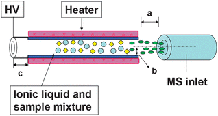
Pesticides are necessary for effective crop production but their toxicity can pass onto consumers and cause dangerous health effects. Mass spectrometry continues to be the gold standard for pesticide residue testing by providing highly sensitive and specific tests for food safety. Although many versions of mass spectrometry have been applied to this field, there are still problems such as loss of sample, requiring high voltages, complex ionization preparation, and difficulty in heterogeneous solutions. To overcome these limitations, researchers at East China Institute of Technology and Nanchang University have coupled thermal dissociation atmospheric chemical ionization (TDCI) to linear trap mass spectrometry (LTQ-MS), for a more environmentally friendly pesticide detection technique. TDCI uses ionic liquids to create ions, instead of an external source, and the resulting solution exchanges charge with the thermally evaporated analytes. In order to test the capabilities of TDCI, a common pesticide, dimethoate, was measured in highly viscous solutions, fruit juices. TDCI rapidly detected trace levels of dimethoate in a variety of fruit juices below the EU standardized levels for fruit and with high specificity.

Dissociation atmospheric chemical ionization (TDCI) source coupled with LTQ-MS

To know more about the study, click on the link below:

Thermal dissociation atmospheric chemical ionization ion trap mass spectrometry with a miniature source for selective trace detection of dimethoate in fruit juices
Yongzhong Ouyang,  Xinglei Zhang,  Jing Han,  Xiali Guo,  Zhiqiang Zhu,  Huanwen Chen and Liping Luo
Analyst, 2013, Advance Article
DOI: 10.1039/C2AN36244A

Digg This
Reddit This
Stumble Now!
Share on Facebook
Bookmark this on Delicious
Share on LinkedIn
Bookmark this on Technorati
Post on Twitter
Google Buzz (aka. Google Reader)

DNA Rotaxanes for Cancer Detection

Bi et al., Analyst, 2013, Advance Article

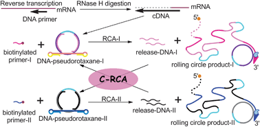
Interlocking molecular structures known as rotaxanes have been expanded to DNA to create a new class of nanomachines for studying biological systems. Li Li and researchers at Qingdao University used DNA psuedorotaxnes as an alternative method for detecting low concentrations of mRNA in cancerous cell lines. Instead of the traditional polymerase chain reaction, a cross-rolling circular amplification synthesizes and links two DNA psuedorotaxnes into a single DNA rotaxane, which contains a biotinylated primer detected by a streptavidin coated magnetic nanoparticle. To read more about the zeptomole sensitivity and high selectivity accomplished by this assay, check out the article below.

Ultrasensitive detection of mRNA extracted from cancerous cells achieved by DNA rotaxane-based cross-rolling circle amplification
Sai Bi, Yangyang Cui and Li Li
Analyst, 2013, Advance Article
DOI: 10.1039/C2AN36118C

Digg This
Reddit This
Stumble Now!
Share on Facebook
Bookmark this on Delicious
Share on LinkedIn
Bookmark this on Technorati
Post on Twitter
Google Buzz (aka. Google Reader)

Carbon Nanodot Aptamer Assay

Communication Previous Article | Next Article

Qu et al., Analyst, 2012, 137, 5483

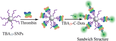
Trying to incorporate quantum dots into biological systems has proven difficult due to their lack of biocompatibility and the toxicity of heavy metals inside cells. Recently developed carbon nanodots retain the advantages of quantum dots, but can function in biological media. Xianogang Qu and researchers at the Chinese Academy of Sciences incorporated carbon nanodots in a thrombin detection assay using DNA aptamers. Thrombin contains two binding sites that are recognized by different aptamers on both a silica nanoparticle and carbon nanodot. The multi-binding site capabilities of aptamers allow for greater sensitivity when compared to single site antibodies, and the fluorescent signal of the carbon nanodot is only detected when bound to thrombin on the silica nanoparticle. Click on the paper below to read more, it will be free to read until November 16th.

Aptamer carbon nanodot sandwich used for fluorescent detection of protein
Bailu Xu, Chuanqi Zhao, Weili Wei, Jinsong Ren, Daisuke Miyoshi, Naoki Sugimoto and Xiaogang Qu
Analyst, 2012, 137, 5483-5486
DOI: 10.1039/C2AN36174D

Digg This
Reddit This
Stumble Now!
Share on Facebook
Bookmark this on Delicious
Share on LinkedIn
Bookmark this on Technorati
Post on Twitter
Google Buzz (aka. Google Reader)

Quick Testing for Lead Poisoning in Blood

Peroxidase mimicking DNA–gold nanoparticles for fluorescence detection of the lead ions in blood

Chang et al., Analyst, 2012, 137, 5222

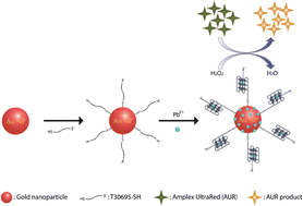
Lead has no biological function in the human body, and high concentrations can cause detrimental health effects, especially for children. The large instruments used for detecting lead mostly test contaminated water, and cannot be transported to remote areas or test blood samples. Huan-Tsung Chang and researchers at National Taiwan University created a unique binding complex composed of gold nanoparticles, DNA, and Amplex UltraRed to combat this problem. In this system, lead ions form an alloy with the gold nanoparticles increasing catalytic activity, which results in oxidation of the DNA-conjugated Amplex UltraRed and production of a detectable fluorescent signal. This simple yet effective sensor detected lead concentrations down to 0.1 nM in blood samples.

This paper will be free to read until the end of the month.

Peroxidase mimicking DNA–gold nanoparticles for fluorescence detection of the lead ions in blood
Chi-Lin Li, Chih-Ching Huang, Wei-Hsi Chen, Cheng-Kang Chiang and Huan-Tsung Chang
Analyst, 2012, 137, 5222-5228
DOI: 10.1039/C2AN35599J

Digg This
Reddit This
Stumble Now!
Share on Facebook
Bookmark this on Delicious
Share on LinkedIn
Bookmark this on Technorati
Post on Twitter
Google Buzz (aka. Google Reader)