Author Archive

Identifying DNA stucture using SERS

Detecting G-quadruplexes using SERS of silver nanoparticles

Differentiating quadruplexes from duplexes using silver nanoparticles and SERS

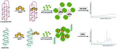
A promising drug target and ligand-binding site, G-quadruplexes form via Hoogsteen hydrogen bonding in guanine bases. These quartets are rich in guanine and are stabilized through π-π stacking interactions and a cation such as potassium or sodium. As researchers explore new compounds to target this region to treat diseases such as cancer, the structure of these quadruplexes and their response to ligands compared to standard DNA remains understudied. 

Researchers at the University of Strathclyde have used the sensitivity of surface-enhanced Raman spectroscopy (SERS) to detect the formation of G-quadruplexes in the presence of stabilizing ligands and silver nanoparticles. They successfully differentiated the quadruplex DNA structure from the duplex based on the SERS signal, and demonstrated the capability of SERS in identifying higher order DNA structures. To read more about the qualitative detection of these structures, click the link below. It will be free to read until June 30. 

Qualitative SERS analysis of G-quadruplex DNAs using selective stabilizing ligands
K. Gracie, V. Dhamodharan, P. I. Pradeepkumar, K. Faulds and   D. Graham
Analyst
, 2014, Advance Article
DOI: 10.1039/C4AN00551A
<!–[if gte mso 9]> <![endif]–>
Digg This
Reddit This
Stumble Now!
Share on Facebook
Bookmark this on Delicious
Share on LinkedIn
Bookmark this on Technorati
Post on Twitter
Google Buzz (aka. Google Reader)

Label-free biosensing using photonic crystals and single nanoparticles

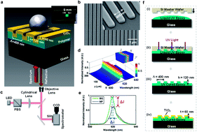
Photonic crystal enhanced microscopy (PCEM) is a relatively new technique that utilizes a grating like substrate composed of a high refractive index material coating a low refractive index one. When irradiated with light at a resonant wavelength, this photonic crystal reflects all of the light, and is very sensitive to any changes on the surface.

Photonic Crystal Enhanced Microscopy of a nanoparticle on a photonic crystal

Photonic Crystal Enhanced Microscopy of a nanoparticle on a photonic crystal

Researchers at the University of Illinois in the United States studied the effects of gold nanoparticles binding to  photonic crystals composed of a liquid epoxy polymer and TiO2.  Although PCEM has been used to detect biological analytes without labels, they observed how a nanoparticle tag can reduce  the signal of a photonic crystal. Furthermore, they determined the distance required between the nanoparticles for detection, and this could be used in future amplification schemes such as in single molecule sensing.

To read more about this topic, click the link below. It will be free to read until February 28.

Single nanoparticle detection using photonic crystal enhanced microscopy
Yue Zhuo,  Huan Hu, Weili Chen,  Meng Lu,  Limei Tian,  Hojeong Yu, Kenneth D. Long,  Edmond Chow,  William P.   King, Srikanth Singamaneni  and  Brian T. Cunningham
Analyst, 2014, Advance Article
DOI: 10.1039/C3AN02295A

Digg This
Reddit This
Stumble Now!
Share on Facebook
Bookmark this on Delicious
Share on LinkedIn
Bookmark this on Technorati
Post on Twitter
Google Buzz (aka. Google Reader)

Classifying myobacterial strains using surface enhanced Raman spectroscopy

Spectroscopic analysis of mycolic acid profiles

In bacterial-based diagnostics, the bacteria can evolve over time and it is important to be able to differentiate between multiple strains. Mycobacterium tuberculosis, which is responsible for causing tuberculosis (TB), has multiple drug-resistant strains. The most accurate method to identify toxic strains of TB, high performance liquid chromatography (HPLC), requires fluorescent labeling and extraction of the mycolic acids, from the outer membrane of the bacteria.

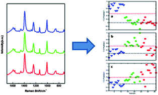
Researchers at the University of Georgia in the United States wanted a more direct detection method and approached this problem in two important aspects. They began by identifying the high molecular weight lipids within the bacteria using HPLC, gas chromatography (GC), and nuclear magnetic resonance (NMR) for several TB bacterial strains. NMR in particular was very important in identifying the structures of mycolic acids. Using the information from these techniques and surface enhanced Raman spectroscopy (SERS), they chemically classified the mycolic acid from both TB and non-TB strains. The SERS spectra underwent further statistical analysis and accurately identified different strains of TB label-free and with high sensitivity.

To know more about this research, click on the link below. This paper will be free to read for the next three weeks.

Identification of mycobacteria based on spectroscopic analyses of mycolic acid profiles
Omar E. Rivera-Betancourt, Russell Karls, Benjamin Grosse-Siestrup, Shelly Helms, Frederick Quinn and Richard A. Dluhy
Analyst, 2013,138, 6774-6785
DOI: 10.1039/C3AN01157G

Digg This
Reddit This
Stumble Now!
Share on Facebook
Bookmark this on Delicious
Share on LinkedIn
Bookmark this on Technorati
Post on Twitter
Google Buzz (aka. Google Reader)

High Sensitivity Using Low Cost Equipment

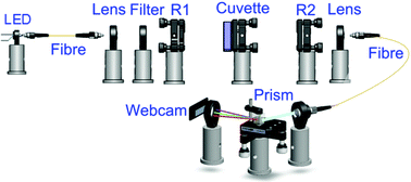
Low cost webcam based prism spectrometer

Techniques such as cavity ring-down spectroscopy (CRDS) have greatly enhanced the sensitivity in traditional absorption spectroscopy methods. Although CRDS can detect analytes in very small concentrations in solution, it requires expensive and complex instrumentation, which has restricted its widespread implementation. Alternatively, broadband cavity enhanced absorption spectroscopy (BBCEAS) has emerged to overcome some of these limitations. These adjustments allow for multiplexed data collection and improved resolution.

Researchers at the University of Cambridge, UK,  have constructed a BBCEAS apparatus using relatively simple components and achieved sensitivity levels similar to much more expensive systems.  Using a well-studied process, the Griess Assay, they measured different concentrations of nitrite and the dye Rhodamine 6 G with a system that contained a low cost prism spectrometer and a webcam detector. The limits of detection were found to be 3.7 nM (nitrite) and 850 pM (Rhodamine), and by adding a better detector, these can further be improved upon.

To read the full article, please access the link below. This paper will be free to read for the next three weeks:

High sensitivity liquid phase measurements using broadband cavity enhanced absorption spectroscopy (BBCEAS) featuring a low cost webcam based prism spectrometer
Zhechao Qu, Julia Engstrom, Donald Wong, Meez Islam and   Clemens F. Kaminski
Analyst, 2013,138, 6372-6379
DOI: 10.1039/C3AN01441J

Digg This
Reddit This
Stumble Now!
Share on Facebook
Bookmark this on Delicious
Share on LinkedIn
Bookmark this on Technorati
Post on Twitter
Google Buzz (aka. Google Reader)

A Competitive Binding Scheme for Cancer Cells

Ultrasensitive electrochemical detection of leukemia cells

Detecting cancerous cells, such as those of leukemia, in early stages can greatly affect patient treatment and survival. Current diagnostic tools such as immunohistochemistry require large concentrations of cells, which occur in advanced stages of the disease.

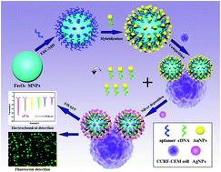
Jun-Jie Zhu and co-workers at Nanjing University , China, developed an assay for leukemia cells using aptamers in a unique, competition-based electrochemical sensor. This involves the use of magnetic (Fe3O4) and gold nanoparticles and the detection of electrochemical signals from deposited silver nanoparticles using square wave anodic stripping voltammetery. The gold nanoparticles bind to the much larger magnetic particles through an apatmer, but the apatmer has a higher specificity for the leukemia cells. Upon introduction of the cells, some of the gold nanoparticles are released from the magnetic surface affecting the amount of detectable signal from the silver. The researchers successfully detected as few as 10 cells and demonstrated a high specificity of the aptamers for only one type of leukemia cells. Although they chose leukemia cells, the only limitation is the specificity of aptamers available and ideally, any cancer could be tested for in this system.

To read the full article, please access the link below. This paper will be free to read for the next three weeks.

A novel aptamer-based competition strategy for ultrasensitive electrochemical detection of leukemia cells
Kui Zhang, Tingting Tan, Jia-Ju Fu, Tingting Zheng and   Jun-Jie Zhu
Analyst, 2013, Advance Article
DOI: 10.1039/C3AN01255G

Digg This
Reddit This
Stumble Now!
Share on Facebook
Bookmark this on Delicious
Share on LinkedIn
Bookmark this on Technorati
Post on Twitter
Google Buzz (aka. Google Reader)

Studying Algae Byproducts using SERDS

SERDS combined with signal regression analysis to monitorthe phototrophic microorganisms

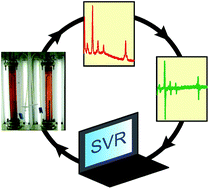
As the demand for non-renewable energy sources continues to soar, alternative renewable and environmentally friendly alternatives continue to be explored. The biomass conversion by algae has emerged as promising avenue. Additionally, algae produce other compounds relevant in pharmaceutical research. A bioreactor houses these phototropic organisms, but direct measurement of these compounds within the reactor can be costly, inefficient, and require complex modeling to understand.

Researchers at University of Erlangen-Nuremberg in Germany used shifted-excitation Raman difference spectroscopy (SERDS) in a model algae system as a new way to probe these reactions. In SERDS, two spectra are acquired at slightly different wavelengths, and subtraction of the two removes the fluorescence background, a common problem in normal Raman spectroscopy. The red algae, Porphyridium purpureum produces a number of relevant compounds, such as sulfated polysaccharides, which have important anti-viral properties.

With the help of an algorithm to process the spectra, they tracked the separation of this promising pharmaceutical compound, which can be applied to other algae systems.

To know more about the study, please access the link below. This paper will be free to read for the next three weeks.

Combined shifted-excitation Raman difference spectroscopy and support vector regression for monitoring the algal production of complex polysaccharides
Kristina Noack, Björn Eskofier, Johannes Kiefer, Christina Dilk, Georg Bilow, Matthias Schirmer, Rainer Buchholz and   Alfred Leipertz
Analyst, 2013, Advance Article
DOI: 10.1039/C3AN01158E

Digg This
Reddit This
Stumble Now!
Share on Facebook
Bookmark this on Delicious
Share on LinkedIn
Bookmark this on Technorati
Post on Twitter
Google Buzz (aka. Google Reader)

Analyzing Century Old Blood Samples with DART-MS

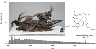
The Detroit Institute of Arts' Komo mask. Image courtesy of the DIA.

Typically dried or degraded blood samples at crime scenes are positively identified via a catalytic reaction with the haem protein, which produces a colored signal or interacts uniquely with light. In addition to blood, other proteins can undergo the same reaction, yielding inaccurate results. This high failure rate makes these tests unreliable and requires further analysis in a laboratory.

Conservators at the Detroit Art Institute in collaboration with chemistry researchers in the US used direct real time mass spectrometry (DART-MS) for the detection of degraded blood samples. This required little sample preparation, when compared to other standard conservation techniques such as HPLC and GC-MS, and identified the haem protein in the coating of an African Komo mask. In addition to cultural artifacts, this methodology can potentially be applied in forensic investigations. The authors detected haem peaks from both myoglobin and haemoglobin quickly and with greater specificity than the simple techniques described previously.

To know more, please access the link below. This paper will be free to read until July 25th.

Characterization of blood in an encrustation on an African mask: spectroscopic and direct analysis in real time mass spectrometric identification of haem
Daniel Fraser, Cathy Selvius DeRoo, Robert B. Cody and   Ruth Ann Armitage  
Analyst, 2013, Advance Article
DOI: 10.1039/C3AN00633F

Digg This
Reddit This
Stumble Now!
Share on Facebook
Bookmark this on Delicious
Share on LinkedIn
Bookmark this on Technorati
Post on Twitter
Google Buzz (aka. Google Reader)

A Combination Probe for Characterizing Cancerous Tissue

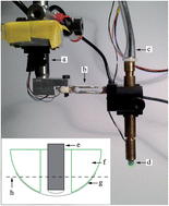
Instrument with a thin fibre optic Raman probe mounted inside a hollow tactile resonance sensor

In some types of cancers, such as in prostate cancers, surgical removal of the entire organ provides the most effective treatment option. Unfortunately, after removal of the prostate a few tumor cells may remain and cause a recurrence of the disease in the patient. If the surgical area could be tested shortly after removal, it would determine if any cancerous cells remain and improve patient mortality.

Morgan Nyberg and researchers at Umea University in Sweden have harnessed the power of two techniques in a single probe to differentiate healthy and cancerous cells: Raman spectroscopy and tactile resonance method (TRM). Although an inherently weak effect, Raman spectroscopy can identify tissues based on their unique vibrational spectra. TRM measures tissue stiffness and successfully detects cancerous tissues in a prostate. However, it fails at the cellular level in differentiating between other benign growth tissues from cancerous ones. The combination of these two techniques removes the drawbacks of implementing Raman spectroscopy in surgery such as interfering ambient light and increases the specificity lacking in TRM. The researchers have successfully identified muscle and fat tissues from an animal sample and plan to move onto prostate tissue samples in the near future.

To know more about this ressearch, please access the full article below. This paper will be free to read for the next three weeks.

Optical fibre probe NIR Raman measurements in ambient light and in combination with a tactile resonance sensor for possible cancer detection
Morgan Nyberg, Kerstin Ramser and   Olof A. Lindahl
Analyst, 2013, Advance Article
DOI: 10.1039/C3AN00243H

Digg This
Reddit This
Stumble Now!
Share on Facebook
Bookmark this on Delicious
Share on LinkedIn
Bookmark this on Technorati
Post on Twitter
Google Buzz (aka. Google Reader)

Enhancing Dopamine Sensitivity by Reducing Surface Fouling

Researchers have adapted a gold electrode to enhance electrochemical dopamine measurements and overcome the fouling problems that typically occur on the surface when using this technique.

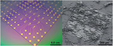
Cells on doped overoxidised PPy modified electrodes

Many diseases such as Parkinson’s and schizophrenia are caused by irregularities in the neurotransmitter dopamine. Each disease affects dopamine production differently, however in all cases studying the process both inside and outside the body has presented many challenges.

Jenny Emnéus at the Technical University of Denmark and collaborators in Italy improved the detection of dopamine by placing a doped overoxidised polypyrrole (PPy) film on the electrode surface. The film was doped with different counter ions to inhibit dopamine polymerisation and the binding of negatively charged species. Although the overoxidation of PPy did affect the conductivity of the film, it also became more sensitive to dopamine, suggesting that doped overoxidised PPy can be used as sensors for dopamine.

To learn more about the techniques the authors used to reduce surface fouling and detect dopamine release from live cells, check out the article below. It will be free to read until May 28th .

Doped overoxidized polypyrrole microelectrodes as sensors for the detection of dopamine released from cell populations
Luigi Sasso, Arto Heiskanen, Francesco Diazzi, Maria Dimaki, Jaime Castillo-León, Marco Vergani, Ettore Landini, Roberto Raiteri, Giorgio Ferrari, Marco Carminati, Marco Sampietro, Winnie E. Svendsen and   Jenny Emnéus  
Analyst, 2013, Advance Article
DOI: 10.1039/C3AN00085K

Digg This
Reddit This
Stumble Now!
Share on Facebook
Bookmark this on Delicious
Share on LinkedIn
Bookmark this on Technorati
Post on Twitter
Google Buzz (aka. Google Reader)

Probing Alzheimer’s disease Plaques using Synchrotron FTIR

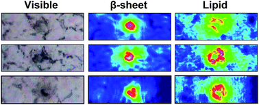
Visible and FTIR images showing plaques and lipids in mice tissues

Alzheimer’s disease (AD) affects millions of patients worldwide, and with much still unknown, few treatments options are available for the long duration of this disease. Although opinions differ on the cause of AD, histological staining shows the presence of neurotoxic plaques and tangles in deceased patients and serves as the primary diagnostic method.

Researchers at the University of Manitoba, Canada, used high resolution Fourier Transform Infrared Spectroscopy (FTIR) to image brain tissue in mice and human samples in order to study plaque formation at the sub cellular level. Different components in the cell such as DNA, proteins or sugars, produce unique chemical signatures which correspond to vibrational bands. The researchers discovered infiltration of lipid membrane components surrounding the plaques, which increase the signal as the plaques become larger. The spatial resolution of the system used here enables detection of lipids that a staining would miss, but it is important to understand disease progression. Although the relationship between plaque formation and lipid concentration remains unclear, the authors have developed a new method towards understanding plaque formation in AD.

To read the full article, please access the link below. This paper will be free to read until May 14th .

Synchrotron FTIR reveals lipid around and within amyloid plaques in transgenic mice and Alzheimer’s disease brain
Catherine R. Liao, Margaret Rak, Jillian Lund, Miriam Unger, Eric Platt, Benedict C. Albensi, Carol J. Hirschmugl and   Kathleen M. Gough
Analyst, 2013, Advance Article
DOI: 10.1039/C3AN00295K

Digg This
Reddit This
Stumble Now!
Share on Facebook
Bookmark this on Delicious
Share on LinkedIn
Bookmark this on Technorati
Post on Twitter
Google Buzz (aka. Google Reader)