Author Archive

Understanding the “How” of Disease Biomarker Discovery

An Overview of Biomarker Discovery

An Overview of Biomarker Discovery

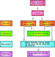
A disease biomarker is a key indicator that when detected from the body can help determine whether an individual is at risk to develop a certain disease. Thus, the discovery of suitable biomarkers is very important to enable earlier diagnosis of disease, better treatment options, and lower overall healthcare costs. Over the years, substantial progress has been made in the field of proteomics to study proteins as disease biomarkers. However, while most effort has been made towards identifying new biomarkers, less focus has been made towards understanding and optimizing the methodologies, which is required in order to find effective biomarkers.

Nina Bergman and Jonas Bergquist from Uppsala University in Sweden have written a review article to outline the proteomic methods that are currently used for biomarker identification and analysis, assess some of the advantages and disadvantages of each method and provide insight into what the future may hold for biomarker discovery. Click on the link below to learn more (free access until September 6th):

Recent developments in proteomic methods and disease biomarkers
Nina Bergman and Jonas Bergquist
Analyst, 2014, Advance Article
DOI: 10.1039/C4AN00627E

Digg This
Reddit This
Stumble Now!
Share on Facebook
Bookmark this on Delicious
Share on LinkedIn
Bookmark this on Technorati
Post on Twitter
Google Buzz (aka. Google Reader)

Lights! Camera! ePetri!

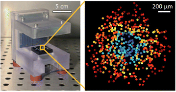
Tracking the activity of viruses has never been so easy thanks to the latest development from Chao Han and Changhuei Yang, researchers at the California Institute of Technology, USA.  The duo has developed ePetri, a high performance imaging platform capable of monitoring the formation of viral plaques, an event that occurs when a virus infects a host cell. Unlike conventional plaque assays, ePetri provides high resolution, time-lapse imaging for precise monitoring of the dynamics of viral plaque formation and growth. In addition to enabling better analysis of viral behaviour, the capabilities of ePetri may also facilitate future applications in antiviral drug discovery. Learn more by accessing the link below. 

ePetri Instrument for Viral Plaque Analysis

ePetri Instrument for Viral Plaque Analysis


Viral plaque analysis on a wide field-of-view, time-lapse, on-chip imaging platform 

Chao Han and Changhuei Yang 

Analyst, 2014, Advance Article

DOI: 10.1039/C3AN02323K, Paper

Digg This
Reddit This
Stumble Now!
Share on Facebook
Bookmark this on Delicious
Share on LinkedIn
Bookmark this on Technorati
Post on Twitter
Google Buzz (aka. Google Reader)

The Ongoing Hunt for Deadly Microbes – A New Lateral Flow Assay for Pathogen Detection

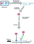
Early and accurate monitoring of pathogens such as Escherichia coli is critical for food and water safety, clinical diagnosis, and biosecurity. However there are a number of limitations to current detection methods such as cell culturing, and immunoassays. These drawbacks include non-specificity, insensitivity, as well as the requirement of long analysis time, intensive labor, and extensive sample purification. To address some of these limitations, Christopher Pöhlmann and his colleagues from the University of Bayreuth in Germany have developed a lateral flow device based on a sandwich assay design using gold nanoparticle-oligonucleotides to detect ribosomal RNA of E. coli. The new device is able to provide specific detection of E. coli within 25 minutes and requires no signal amplification step.

A Lateral Flow Assay for Detection of E. coli Ribosomal RNA

A Lateral Flow Assay for Detection of E. coli Ribosomal RNA

Find out more about this discovery by reading the full paper below, which is free to download until March 7th:

A lateral flow assay for identification of Escherichia coli by ribosomal RNA hybridization
Christopher Pöhlmann, Irina Dieser and Mathias Sprinzl
Analyst, 2014, 139, 1063-1071
DOI: 10.1039/C3AN02059B

Let us know what your thoughts are by leaving a comment!

Digg This
Reddit This
Stumble Now!
Share on Facebook
Bookmark this on Delicious
Share on LinkedIn
Bookmark this on Technorati
Post on Twitter
Google Buzz (aka. Google Reader)

BODIPY-Based Probe for HClO Imaging: Resolving the Paradox of Detection?

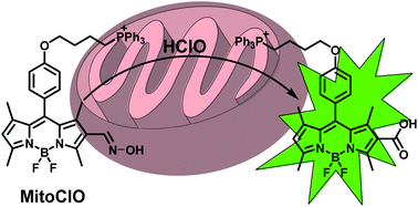
BODIPY-Based Probe for HClO Imaging

BODIPY-Based Probe for HClO Imaging

Detection of suitable biomarkers of disease is critical for medical diagnosis. However, detection becomes a paradoxical challenge when the biomarker is naturally found in healthy individuals. This is the case for the biomarker hypochlorous acid (HClO), a reactive oxygen species that plays an essential role in maintaining a healthy immune system under normal conditions. Yet, when optimal levels of HClO cannot be maintained by the body, cellular functions become disrupted and this can result in diseases such as atherosclerosis, arthritis, and cancer. Consequently, HClO requires medical monitoring, particularly in the mitochondria where a high concentration tends to reside. To help distinguish between healthy and diseased states, the ideal biosensor must provide fast response, high selectivity, high sensitivity and mitochondrial permeability.

To address this challenge, Xiaojun Peng and colleagues from the Dalian University of Technology in China have created a fluorescent imaging probe to detect HClO by localizing to mitochondria in live cells. Their probe makes use of boron dipyrromethene (BODIPY) dye for its outstanding fluorescent properties. Incorporation of an oxime group onto the BODIPY scaffold allows the tuning of fluorescence to an on/off state depending on the presence/absence of HClO, respectively. Lastly, incorporation of a triphenylphosphine group onto BODIPY provides the localization signal to the mitochondria. Learn all the details of this exciting new discovery by accessing the link below:


A highly specific BODIPY-based probe localized in mitochondria for HClO imaging
Guanghui Cheng, Jiangli Fan, Wen Sun, Kun Sui, Xin Jin, Jingyun Wang and   Xiaojun Peng  
Analyst, 2013, Advance Article
DOI: 10.1039/C3AN01152F

This pap[er will be free to read for the next three weeks. Let us know what your thoughts are by leaving a comment!

Digg This
Reddit This
Stumble Now!
Share on Facebook
Bookmark this on Delicious
Share on LinkedIn
Bookmark this on Technorati
Post on Twitter
Google Buzz (aka. Google Reader)

CMOS Imaging as a Potential Solution for Diabetic Patients

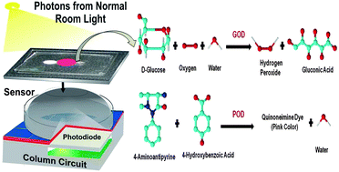
CMOS Image Sensor for Glucose Monitoring

CMOS Image Sensor for Glucose Monitoring

Millions of people suffer from diabetes, and the disease is quickly becoming a global epidemic. To address patient need, many electrochemical- and biochemical-based technologies have been developed over the last decade.

While considerable progress has been made, many of these methods still do not possess the sensitivity, specificity, accuracy, and user friendliness required to meet patient demands.

Researchers from Gachon University in the Republic of Korea, have developed the latest glucose sensor based on complementary metal oxide semiconductor (CMOS) imaging that provides enhanced efficiency for glucose monitoring. The novel method measures the concentration of oxidized glucose in blood plasma by the intensity of color produced, which leads to variations in photon count detected by the CMOS sensor.

Assess the link below for more details. This paper will be free to read for the next three weeks:

CMOS image sensors as an efficient platform for glucose monitoring
Jasmine Pramila Devadhasan, Sanghyo Kim and   Cheol Soo Choi
Analyst, 2013, Advance Article
DOI: 10.1039/C3AN00805C

What are your thoughts on this new discovery? Let us know by leaving a comment.

Digg This
Reddit This
Stumble Now!
Share on Facebook
Bookmark this on Delicious
Share on LinkedIn
Bookmark this on Technorati
Post on Twitter
Google Buzz (aka. Google Reader)

What does it mean to be an “Analyst”?

The Thinker

The Thinker (from The Bubble Chamber blog)

There appears to be many journal titles that contain the word “analyst” in some form. Examples include Analytical Biochemistry, Analytical Chemistry, Analytica Chimica Acta, Analytica, Journal of Analytical Toxicology, Analytical Methods, and of course, Analyst.

With so many variations of analytical journals available, some effort is needed to determine the best fit when submitting a manuscript for publication. This process requires classification of your own area(s) of research, which can be surprisingly challenging.

Take for instance, the areas of biochemistry, chemical biology, and biological chemistry. The prefix of each word appears to form the suffix of the next, and vice versa. In fact, if all of these words are to be read one after another, the whole phrase sounds more like a tongue-twister! While each of these respective scientific fields is specialized in its own right, some ambiguity still remains upon deeper contemplation of what exactly each field encompasses and what it does not. Differences among the fields can become more difficult to discern, the boundaries that separate them become less defined, and these multi-disciplinary approaches begin to converge into common research goals.

An underlying factor that unifies various scientific areas is the requirement of high quality analysis.

So, what does it mean exactly to be an analyst? Perhaps it is someone who can decipher the fine details of chemical reactions, molecular structures, computational algorithms, and biochemical mechanisms; then piece together these components into an overall composite for scientific understanding. Perhaps it is someone who is well-attuned with his/her surroundings, and inspired to find answers to everyday peculiarities. It may be someone who foresees the raw potential in new discoveries, even before direct applications are demonstrated. Or simply someone who loves scientific questioning and appreciates the sake of learning for what it is worth in and of itself. Whatever the precise definition is, frequent publications and updates on the latest scientific breakthroughs by journals like Analyst continue to ignite the passion of those who are motivated to discover and to know more.

So what does being an Analyst mean to you? Share your thoughts with us by commenting below.

From discovery to recovery – Analyst and Analytical Methods working together for the analytical community
Analyst, 2011,136, 429-430
DOI: 10.1039/C0AN90013C
Digg This
Reddit This
Stumble Now!
Share on Facebook
Bookmark this on Delicious
Share on LinkedIn
Bookmark this on Technorati
Post on Twitter
Google Buzz (aka. Google Reader)

Using Infrared Spectroscopy to Predict Cancer Origin

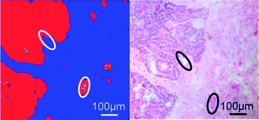
FTIR Based Classification of Carcinoma Regions in Brain Tissue

FTIR based classification of carcinoma regions in brain tissue

Cancer is one of the top causes of death in the world, particularly for developed countries. Regardless of the cancer type, up to 40% of all cases metastasize to the brain during disease progression. Indeed, better survival rates are possible with early and accurate cancer detection.

While histopathologic evaluation involving staining of brain tissue is the current gold standard method for diagnosis, major drawbacks include the complexity of analysis and the non-specific staining of some dyes for non-cancerous cells. Moreover, histopathological staining, and other screening methods often cannot identify the primary tumor of brain metastasis. Without knowledge of the cancer origin, determination of the optimal treatment strategy can be difficult.

Christoph Krafft and colleagues from the Institute of Photonic Technology of Jena, Germany, have developed a strategy to help identify the primary tumor by using Fourier transform infrared (FTIR) and associated software. By analysing brain metastasis tissue, the “molecular fingerprint”, or vibrational spectra characteristic of the primary tumor can be found to deduce the cancer source.

Learn more about this exciting discovery by accessing the link below. This paper will be free to read until April 29th:

Tumor margin identification and prediction of the primary tumor from brain metastases using FTIR imaging and support vector machines
Norbert Bergner ,  Bernd F. M. Romeike ,  Rupert Reichart ,  Rolf Kalff ,  Christoph Krafft and Jürgen Popp
Analyst, 2013, Advance Article
DOI: 10.1039/C3AN00326D

Digg This
Reddit This
Stumble Now!
Share on Facebook
Bookmark this on Delicious
Share on LinkedIn
Bookmark this on Technorati
Post on Twitter
Google Buzz (aka. Google Reader)

DNA Detection Using Novel Fiber-Optic Biosensing Strategy

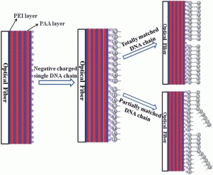
DNA Detection Using Fiber Optic Biosensor

Overview of DNA Detection Using Fiber Optic Biosensor

Ming-jie Yin and others from The Hong Kong Polytechnic University in China have made the latest stride towards development of label-free fiber-optic biosensors for DNA detection.

Their new design involves fabrication of a DNA sensor as part of a thin sensing film on the surface of a thin-core fiber modal interferometer (TCFMI) to measure differences in refractive-index upon detection of target DNA sequences. The research team makes use of a layer-by-layer self-assembly approach, which enables controlled tunability in preparation of the sensing film.

Unlike many other reported fiber-optic biosensing methods, this current strategy confers the advantages of high detection sensitivity, lower cost and ease in preparation.

Read more about the latest discovery by accessing the link below. This article will be free to read until March 6th.

Label-free, disposable fiber-optic biosensors for DNA hybridization detection
Ming-jie Yin ,  Chuang Wu ,  Li-yang Shao ,  Wing Kin Edward Chan ,  A. Ping Zhang ,  Chao Lu and Hwa-yaw Tam
Analyst, 2013, Advance Article
DOI: 10.1039/C3AN36791F

Digg This
Reddit This
Stumble Now!
Share on Facebook
Bookmark this on Delicious
Share on LinkedIn
Bookmark this on Technorati
Post on Twitter
Google Buzz (aka. Google Reader)

Using Chemosensors to Monitor pH Dynamics

The cell effectively uses the mechanism of pH fluctuation as a form of regulatory control for diverse physiological functions. Over the years, optical based methods have been used to develop chemosensors to measure the dynamics of intracellular pH, some of which have demonstrated considerable potential for biological applications.

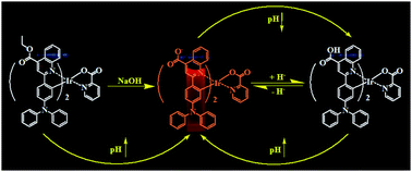
Wei Huang and colleagues from the Nanjing University of Posts and Telecommunications, China, have developed the latest optical pH probes by making use of cyclometalated iridium (III) complexes, a well known phosphorescent emitter. Huang’s group has expanded the repertoire of useful chemosensors by synthesizing and characterizing the photophysical properties of two iridium (III) complexes using a novel substituent, carboxylic-acid.

Find out more about their discovery by accessing the link below. This paper will be free to read for the next two weeks.

Effect of pH on the photophysical properties of two new carboxylic-substituted iridium(III) complexes
Jiena Weng ,  Qunbo Mei ,  Weiwei Jiang ,  Quli Fan ,  Bihai Tong ,  Qidan Ling and Wei Huang
Analyst, 2013, Advance Article
DOI: 10.1039/C2AN36298H

pH effect on carboxylic-substituted iridium(III) complex

pH effect on carboxylic-substituted iridium(III) complex

Digg This
Reddit This
Stumble Now!
Share on Facebook
Bookmark this on Delicious
Share on LinkedIn
Bookmark this on Technorati
Post on Twitter
Google Buzz (aka. Google Reader)

Drug Resistance in Cancer Cells

For cancer patients, the course of treatment is a critical factor that determines their rate of survival. The use of imatinib-mesylate (IMA) for instance, has successfully induced remission in most patients suffering from chronic myelogenous leukemia (CML). The IMA drug and its derivatives effectively inhibit abnormal tyrosine kinase activity of key oncoproteins. However, many of these patients develop resistance to IMA-based drugs due to mutations that arise in the oncoproteins. While new drug development is currently underway, methods that can predict drug response are invaluable in order to determine the degree of drug-resistance.

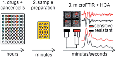
Giuseppe Bellisola and colleagues from the University of Verona, Italy, combined the ability of Fourier Transform Infrared microspectroscopy (microFTIR) and unsupervised Hierarchical Cluster Analysis (HCA) to assess drug-resistance in cancer cells.

Overview of microFTIR and HCA

Overview of microFTIR and HCA

To find out more about this study, check out the link below:

Rapid recognition of drug-resistance/sensitivity in leukemic cells by Fourier transform infrared microspectroscopy and unsupervised hierarchical cluster analysis
Giuseppe Bellisola ,  Gianfelice Cinque ,  Marzia Vezzalini ,  Elisabetta Moratti ,  Giovannino Silvestri ,  Sara Redaelli ,  Carlo Gambacorti Passerini ,  Katia Wehbe and Claudio Sorio
Analyst, 2013, Advance Article
DOI: 10.1039/C2AN36393C

Digg This
Reddit This
Stumble Now!
Share on Facebook
Bookmark this on Delicious
Share on LinkedIn
Bookmark this on Technorati
Post on Twitter
Google Buzz (aka. Google Reader)