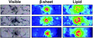
Visible and FTIR images showing plaques and lipids in mice tissues
Alzheimer’s disease (AD) affects millions of patients worldwide, and with much still unknown, few treatments options are available for the long duration of this disease. Although opinions differ on the cause of AD, histological staining shows the presence of neurotoxic plaques and tangles in deceased patients and serves as the primary diagnostic method.
Researchers at the University of Manitoba, Canada, used high resolution Fourier Transform Infrared Spectroscopy (FTIR) to image brain tissue in mice and human samples in order to study plaque formation at the sub cellular level. Different components in the cell such as DNA, proteins or sugars, produce unique chemical signatures which correspond to vibrational bands. The researchers discovered infiltration of lipid membrane components surrounding the plaques, which increase the signal as the plaques become larger. The spatial resolution of the system used here enables detection of lipids that a staining would miss, but it is important to understand disease progression. Although the relationship between plaque formation and lipid concentration remains unclear, the authors have developed a new method towards understanding plaque formation in AD.
To read the full article, please access the link below. This paper will be free to read until May 14th .
Synchrotron FTIR reveals lipid around and within amyloid plaques in transgenic mice and Alzheimer’s disease brain
Catherine R. Liao, Margaret Rak, Jillian Lund, Miriam Unger, Eric Platt, Benedict C. Albensi, Carol J. Hirschmugl and Kathleen M. Gough
Analyst, 2013, Advance Article
DOI: 10.1039/C3AN00295K


