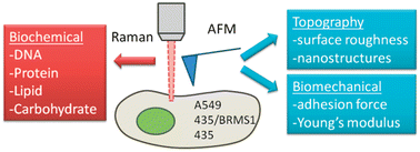
In 2004 the World Health Organisation estimated that over half a million women died from breast cancer. A great deal of research is now conducted to improve the diagnostics and prognosis of cancers. One avenue of research is to improve our understanding of cancers at a cellular level. Anhong Zhou and coworkers from Utah State University have recently used atomic force microscopy (AFM) and Raman spectroscopy to attempt just that.

Combined Raman spectroscopy and AFM to detect differences in cancer cells
Their preliminary study, in this new Analyst HOT paper, examines the use of atomic force microscopy (AFM) and Raman spectroscopy to study a breast cancer cell line and the effect of the presence or absence of a metastasis suppressor gene on cell behaviour. They have also compared various cancer cell lines to examine the differences in behaviour at the cellular level between cancer types. The authors provide a comprehensive analysis of both the practical and data processing techniques required to differentiate the cell types. The use of both AFM and Raman reveals information about the biochemical and biomechanical attributes of the cell lines and is an approach that could increase our understanding of cancer cell behaviour and tumour development.
Subcellular spectroscopic markers, topography and nanomechanics of human lung cancer and breast cancer cells examined by combined confocal Raman microspectroscopy and atomic force microscopy
Gerald D. McEwen, Yangzhe Wu, Mingjie Tang, Xiaojun Qi, Zhongmiao Xiao, Sherry M. Baker, Tian Yu, Timothy A. Gilbertson, Daryll B. DeWald and Anhong Zhou
Analyst, 2013, Advance Article
DOI: 10.1039/C2AN36359C
This article will be free to read for the next two weeks.


