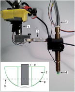
Instrument with a thin fibre optic Raman probe mounted inside a hollow tactile resonance sensor
In some types of cancers, such as in prostate cancers, surgical removal of the entire organ provides the most effective treatment option. Unfortunately, after removal of the prostate a few tumor cells may remain and cause a recurrence of the disease in the patient. If the surgical area could be tested shortly after removal, it would determine if any cancerous cells remain and improve patient mortality.
Morgan Nyberg and researchers at Umea University in Sweden have harnessed the power of two techniques in a single probe to differentiate healthy and cancerous cells: Raman spectroscopy and tactile resonance method (TRM). Although an inherently weak effect, Raman spectroscopy can identify tissues based on their unique vibrational spectra. TRM measures tissue stiffness and successfully detects cancerous tissues in a prostate. However, it fails at the cellular level in differentiating between other benign growth tissues from cancerous ones. The combination of these two techniques removes the drawbacks of implementing Raman spectroscopy in surgery such as interfering ambient light and increases the specificity lacking in TRM. The researchers have successfully identified muscle and fat tissues from an animal sample and plan to move onto prostate tissue samples in the near future.
To know more about this ressearch, please access the full article below. This paper will be free to read for the next three weeks.
Optical fibre probe NIR Raman measurements in ambient light and in combination with a tactile resonance sensor for possible cancer detection
Morgan Nyberg, Kerstin Ramser and Olof A. Lindahl
Analyst, 2013, Advance Article
DOI: 10.1039/C3AN00243H


