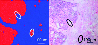
FTIR based classification of carcinoma regions in brain tissue
Cancer is one of the top causes of death in the world, particularly for developed countries. Regardless of the cancer type, up to 40% of all cases metastasize to the brain during disease progression. Indeed, better survival rates are possible with early and accurate cancer detection.
While histopathologic evaluation involving staining of brain tissue is the current gold standard method for diagnosis, major drawbacks include the complexity of analysis and the non-specific staining of some dyes for non-cancerous cells. Moreover, histopathological staining, and other screening methods often cannot identify the primary tumor of brain metastasis. Without knowledge of the cancer origin, determination of the optimal treatment strategy can be difficult.
Christoph Krafft and colleagues from the Institute of Photonic Technology of Jena, Germany, have developed a strategy to help identify the primary tumor by using Fourier transform infrared (FTIR) and associated software. By analysing brain metastasis tissue, the “molecular fingerprint”, or vibrational spectra characteristic of the primary tumor can be found to deduce the cancer source.
Learn more about this exciting discovery by accessing the link below. This paper will be free to read until April 29th:
Tumor margin identification and prediction of the primary tumor from brain metastases using FTIR imaging and support vector machines
Norbert Bergner , Bernd F. M. Romeike , Rupert Reichart , Rolf Kalff , Christoph Krafft and Jürgen Popp
Analyst, 2013, Advance Article
DOI: 10.1039/C3AN00326D


