Archive for December, 2012

Next Generation Carbohydrate Assays

Carbohydrate microarrays have become an important tool for studying glycan-cell interactions, carbohydrate-binding proteins, and in detecting dangerous pathogens. The current sensor methods require complex labeling or indirect analyte detection.

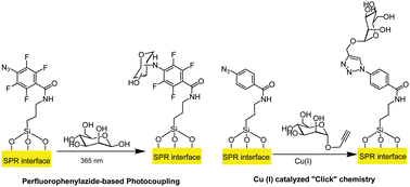
C–H insertion photocoupling reaction and copper “click” methods

Using surface plasmon resonance spectroscopy (SPR), Sabine Szunerits and coworkers at  Lille 1 University detected carbohydrate-binding protein complexes directly and without labels. In order to use SPR, they developed a unique substrate of Ti, Au, and silicon dioxide and tested two carbohydrate surface attachment chemistries. The azide-functionalized surfaces were linked to a glycan using a copper “click” chemistry or photocoupling. Both methods showed comparable binding efficiency and sensitivity of the carbohydrate binding protein.

To read the full article, click on the link below. It will be free to read until January 9th.

Comparison of photo- and Cu(I)-catalyzed “click” chemistries for the formation of carbohydrate SPR interfaces
Nazek Maalouli, Alexandre Barras, Aloysius Siriwardena, Mohamed Bouazaoui, Rabah Boukherroub and Sabine Szunerits
Analyst, 2013, Advance Article
DOI: 10.1039/C2AN36272D

Digg This
Reddit This
Stumble Now!
Share on Facebook
Bookmark this on Delicious
Share on LinkedIn
Bookmark this on Technorati
Post on Twitter
Google Buzz (aka. Google Reader)

Electrochemical Detection of Lead Using Target-Induced Strand Release Strategy

Electrochemical Sensing Using Target-Induced Strand Release

Electrochemical Sensing Using Target-Induced Strand Release

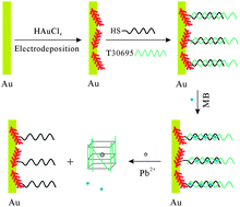
The toxic, bioaccumulative, and non-biodegradable effects of heavy metals pose a severe environmental concern. Hence, monitoring toxic metals such as lead requires effective detection methods. Bo Tang and colleagues from the Shandong Normal University, China, have developed the latest electrochemical detection strategy, which is based on the target-induced strand release method. In their design, the lead-binding DNA aptamer forms a duplex with a complementary DNA sequence that is immobilized to an electrode surface. In this state, methylene blue redox indicators are able to intercalate along the duplex structure, and provide a readable electrochemical signal. However, in the presence of lead, the metal ions bind specifically to the aptamer, which allows release of the lead-aptamer complex into solution. The subsequent release of methylene blue molecules away from the electrode surface leads to a measurable decrease in signal to signify lead detection.

Find out more details by accessing the link below:

A selective amperometric sensing platform for lead based on target-induced strand release
Feng Li, Limin Yang, Mingqin Chen, Peng Li and Bo Tang
Analyst, 2013, 138, 461-466
DOI: 10.1039/C2AN36227A

Digg This
Reddit This
Stumble Now!
Share on Facebook
Bookmark this on Delicious
Share on LinkedIn
Bookmark this on Technorati
Post on Twitter
Google Buzz (aka. Google Reader)

HOT Articles in Analyst

Take a look at these new articles published in Analyst free for you to enjoy until January 9th.

Analyst Issue 2Topics include aptamer based spectrofluorometry, the development of enzyme-free colorimetric bioassays based on gold nanoparticle catalyzed dye decolorization, new ways to identify DNA G-quadruplex structures, and a new set-up to monitor biphasic switchable solvent synthesis. We also have an interesting review on coupled isothachophoresis and capillary electrophoresis which describes the features, advantages and limitations of the methodologies currently in use.

To have a read now, click on the links below:

Coupling isotachophoresis and capillary electrophoresis: a review and comparison of methods
Supreet S. Bahga and Juan G. Santiago
Analyst, 2013, Advance Article
DOI: 10.1039/C2AN36150G

A novel signal-amplified strategy based on assembly reactivation for highly specific and sensitive detection of chair-like antiparallel G-quadruplex
Wei Gai ,  Qianfan Yang ,  Junfeng Xiang ,  Wei Jiang ,  Qian Li ,  Hongxia Sun ,  Lijia Yu ,  Qian Shang ,  Aijiao Guan ,  Hong Zhang and Yalin Tang
Analyst, 2013, Advance Article
DOI: 10.1039/C2AN36557J

Aptamer-based spectrofluorometry for cellular prion protein using N,N′-bis[3,3′-(dimethylamino)propylamine]-3,4,9,10-perylenetetracarboxylic diimide
Lei Zhan ,  Li Jiao Liang ,  Shu Jun Zhen ,  Chun Mei Li and Cheng Zhi Huang
Analyst, 2013, Advance Article
DOI: 10.1039/C2AN36322D

Comprehensive monitoring of a biphasic switchable solvent synthesis
Sonja Hardy ,  Irene M. de Wispelaere ,  Walter Leitner and Marcel A. Liauw
Analyst, 2013, Advance Article
DOI: 10.1039/C2AN36044F

Enzyme-free colorimetric bioassay based on gold nanoparticle-catalyzed dye decolorization
Wei Li ,  Jie Li ,  Weibing Qiang ,  Jingjuan Xu and Danke Xu
Analyst, 2013, Advance Article
DOI: 10.1039/C2AN36374G

Digg This
Reddit This
Stumble Now!
Share on Facebook
Bookmark this on Delicious
Share on LinkedIn
Bookmark this on Technorati
Post on Twitter
Google Buzz (aka. Google Reader)

Graphene-DNA biosensors to detect DNA polymorphisms

The affordable and rapid testing of DNA hybridization and polymorphisms is crucial for the diagnosis of genetic and infectious diseases. DNA biosensors based on nucleic recognition represent one of the most recent technologies currently employed. Electrochemical transducers are commonly used for sensing of DNA hybridization because they are inexpensive, small and highly sensitive. However, this kind of device sees the attachment of a single DNA probe onto the electrode surface to act as recognition element for the complementary target, and an external electro-active label that recognises this complex structure is required. To avoid the use of an external electro-active label, researchers have started to use new materials.

Huiling Loo et al., Analyst, 2013

Differential pulse voltammetry to examine the oxidation of guanine on target DNA

Adeline Huiling Loo and co-workers, from the Nanyang Technological University of Singapore, used graphene, a low cost substance made of a single layer of carbon atoms densely packed. Graphene has high electron conductivity, large specific surface area and fast electron transfer, and is considered a material with a great potential for biosening applications.

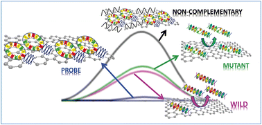
The authors analysed the properties of graphene oxide, electrochemically reduced graphene oxide and thermally reduced graphene oxide and investigated for the first time the mechanisms behind physically immobilized hairpin DNA hybridization. For the label free detection of DNA hybridization and polymorphism, they employed differential pulse voltammetry and examined the oxidation of guanine on target DNA molecules hybridized with an inosine-substituted hairpin DNA probe. According to the study, graphene oxides show the best performance in terms of recognition of complementary and non-complementary DNA sequences.

To know more about this research, click on the link below. This paper will be free to read until January 9th.

An insight into the hybridization mechanism of hairpin DNA physically immobilized on chemically modified graphenes
Adeline Huiling Loo ,  Alessandra Bonanni and Martin Pumera
Analyst, 2013, 138, 467-471
DOI: 10.1039/C2AN36199J

Digg This
Reddit This
Stumble Now!
Share on Facebook
Bookmark this on Delicious
Share on LinkedIn
Bookmark this on Technorati
Post on Twitter
Google Buzz (aka. Google Reader)

Analyst Issue 2 now online!

The latest issue of Analyst is now online: take a look at these beautiful covers and read all about the research behind them.

Analyst Issue 2

Front cover: Liu and Boyd, Analyst, 2013,138, 391-409

Our outside front cover features the work of Qingtao Liu and Ben J. Boyd from the Monash University, Parkville, Australia. In their critical review, the authors offer a detailed overview on the recent use of liposomes in the field of biosensors and bioanalysis.

Liposome surface structures can be modified in a way that they recognise a wide range of analytes. Thus, the possibility to translate liposomes into commercial devices for biosensing is discussed.

Liposomes in biosensors
Qingtao Liu and Ben J. Boyd
Analyst, 2013, 138, 391-409
DOI: 10.1039/C2AN36140J

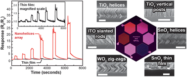
The inside front cover gives a snapshot of a study from the Republic of Korea. Jong Kyu Kim and colleagues present a high performance gas sensor based on near single crystalline TiO2 array nanohelices fabricated by rotating oblique angle deposition (OAD). This new OAD method can be used to fabricate a functional electronic nose and multifunctional smart chips for in situ environmental monitoring.

Analyst Issue 2

Inside front cover: Hwang et al., Analyst, 2013,138, 443-450

A near single crystalline TiO2 nanohelix array: enhanced gas sensing performance and its application as a monolithically integrated electronic nose
Sunyong Hwang, Hyunah Kwon, Sameer Chhajed, Ji Won Byon, Jeong Min Baik, Jiseong Im, Sang Ho Oh, Ho Won Jang, Seok Jin Yoon and Jong Kyu Kim
Analyst, 2013, 138, 443-450
DOI: 10.1039/C2AN35932D

Analyst Issue 2

Back cover: Chen et al., Analyst 2013, 138, 451-460

Finally, the back cover of this issue shows wonderful pictures taken using a dual-beam focused ion beam/scanning electron microscope (FIB/SEM) system developed by Alexander Laskin and his group, from the Pacific Northwest Laboratories, USA. The researchers suggest a novel approach for particle microanalysis, including inorganic refractive materials like fly ash and mineral dust.

Chemical imaging analysis of environmental particles using the focused ion beam/scanning electron microscopy technique: microanalysis insights into atmospheric chemistry of fly ash
Haihan Chen, Vicki H. Grassian, Laxmikant V. Saraf and Alexander Laskin
Analyst, 2013, 138, 451-460
DOI: 10.1039/C2AN36318F

Along with the covers of this issue, here are some interesting HOT articles for you to take a look at:

A selective amperometric sensing platform for lead based on target-induced strand release
Feng Li, Limin Yang, Mingqin Chen, Peng Li and Bo Tang
Analyst, 2013, 138, 461-466
DOI: 10.1039/C2AN36227A

An insight into the hybridization mechanism of hairpin DNA physically immobilized on chemically modified graphenes
Adeline Huiling Loo, Alessandra Bonanni and Martin Pumera
Analyst, 2013, 138, 467-471
DOI: 10.1039/C2AN36199J

Thermal dissociation atmospheric chemical ionization ion trap mass spectrometry with a miniature source for selective trace detection of dimethoate in fruit juices
Yongzhong Ouyang, Xinglei Zhang, Jing Han, Xiali Guo, Zhiqiang Zhu, Huanwen Chen and Liping Luo
Analyst, 2013, 138, 472-479
DOI: 10.1039/C2AN36244A

These papers will be free until January 9th.

Digg This
Reddit This
Stumble Now!
Share on Facebook
Bookmark this on Delicious
Share on LinkedIn
Bookmark this on Technorati
Post on Twitter
Google Buzz (aka. Google Reader)

AFM and Raman to understand cancer cell behaviour

In 2004 the World Health Organisation estimated that over half a million women died from breast cancer.  A great deal of research is now conducted to improve the diagnostics and prognosis of cancers.  One avenue of research is to improve our understanding of cancers at a cellular level. Anhong Zhou and coworkers from Utah State University have recently used atomic force microscopy (AFM) and Raman spectroscopy to attempt just that.

McEwen et al., Analyst 2013

Combined Raman spectroscopy and AFM to detect differences in cancer cells

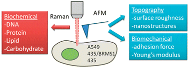
Their preliminary study, in this new Analyst HOT paper, examines the use of atomic force microscopy (AFM) and Raman spectroscopy to study a breast cancer cell line and the effect of the presence or absence of a metastasis suppressor gene on cell behaviour.  They have also compared various cancer cell lines to examine the differences in behaviour at the cellular level between cancer types. The authors provide a comprehensive analysis of both the practical and data processing techniques required to differentiate the cell types.  The use of both AFM and Raman reveals information about the biochemical and biomechanical attributes of the cell lines and is an approach that could increase our understanding of cancer cell behaviour and tumour development.

Subcellular spectroscopic markers, topography and nanomechanics of human lung cancer and breast cancer cells examined by combined confocal Raman microspectroscopy and atomic force microscopy
Gerald D. McEwen, Yangzhe Wu, Mingjie Tang, Xiaojun Qi, Zhongmiao Xiao, Sherry M. Baker, Tian Yu, Timothy A. Gilbertson, Daryll B. DeWald and Anhong Zhou
Analyst, 2013, Advance Article
DOI: 10.1039/C2AN36359C

This article will be free to read for the next two weeks.

Digg This
Reddit This
Stumble Now!
Share on Facebook
Bookmark this on Delicious
Share on LinkedIn
Bookmark this on Technorati
Post on Twitter
Google Buzz (aka. Google Reader)

Detecting Trace Levels of Pesticide in Fruit Juices

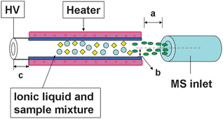
Pesticides are necessary for effective crop production but their toxicity can pass onto consumers and cause dangerous health effects. Mass spectrometry continues to be the gold standard for pesticide residue testing by providing highly sensitive and specific tests for food safety. Although many versions of mass spectrometry have been applied to this field, there are still problems such as loss of sample, requiring high voltages, complex ionization preparation, and difficulty in heterogeneous solutions. To overcome these limitations, researchers at East China Institute of Technology and Nanchang University have coupled thermal dissociation atmospheric chemical ionization (TDCI) to linear trap mass spectrometry (LTQ-MS), for a more environmentally friendly pesticide detection technique. TDCI uses ionic liquids to create ions, instead of an external source, and the resulting solution exchanges charge with the thermally evaporated analytes. In order to test the capabilities of TDCI, a common pesticide, dimethoate, was measured in highly viscous solutions, fruit juices. TDCI rapidly detected trace levels of dimethoate in a variety of fruit juices below the EU standardized levels for fruit and with high specificity.

Dissociation atmospheric chemical ionization (TDCI) source coupled with LTQ-MS

To know more about the study, click on the link below:

Thermal dissociation atmospheric chemical ionization ion trap mass spectrometry with a miniature source for selective trace detection of dimethoate in fruit juices
Yongzhong Ouyang,  Xinglei Zhang,  Jing Han,  Xiali Guo,  Zhiqiang Zhu,  Huanwen Chen and Liping Luo
Analyst, 2013, Advance Article
DOI: 10.1039/C2AN36244A

Digg This
Reddit This
Stumble Now!
Share on Facebook
Bookmark this on Delicious
Share on LinkedIn
Bookmark this on Technorati
Post on Twitter
Google Buzz (aka. Google Reader)

An Electronic Nose for Detection of Toxic Air Pollutants

Our health and well-being is deeply impacted by the air that we breathe. Maintaining good quality of air is highly dependent on the ability of chemical gas sensors to monitor toxic air pollutants in the environment. While many sensors are currently available, there is still a need for a detection system that is highly sensitive and selective, robust, accurate, fast, adaptable to mass production, and capable of monitoring many different chemical gases. Jong Kyu Kim and colleagues from the Republic of Korea have addressed some of these challenges by developing an “electronic nose” – a series of high performance gas sensors based on an array of TiO2 nanohelices fabricated by the method of rotating oblique angle deposition (OAD).

Electronic Nose

Gas sensors based on an array of near single crystalline TiO2

Find out more by clicking on the link below:

A near single crystalline TiO2 nanohelix array: enhanced gas sensing performance and its application as a monolithically integrated electronic nose
Sunyong Hwang,  Hyunah Kwon,  Sameer Chhajed,  Ji Won Byon,  Jeong Min Baik,  Jiseong Im,  Sang Ho Oh,  Ho Won Jang,  Seok Jin Yoon and Jong Kyu Kim
Analyst, 2013, Advance Article
DOI: 10.1039/C2AN35932D

Digg This
Reddit This
Stumble Now!
Share on Facebook
Bookmark this on Delicious
Share on LinkedIn
Bookmark this on Technorati
Post on Twitter
Google Buzz (aka. Google Reader)

2012 FACSS Innovation Award winner announced

FACCS logoThe Federation of Analytical Chemistry & Spectroscopy Societies (FACSS), of which the RSC is a member society, has announced the winner of the 2012 FACSS Innovation Award as Rohit Bhargava, from the University of Illinois, Beckman Institute for Advanced Science and Technology.

This is a juried prize awarded to the most innovative, creative, and outstanding work making its world debut at the FACSS-organized SciX conference. The winner is selected in a special oral session featuring five candidates selected from the many talks contributed to the program.

This year’s award was given for “Advancing Infrared Microscopy Instrumentation by Theory and Computation,” Rohit Bhargava, P. Scott Carney, Rohith Reddy, Kevin Yeh, Thomas van Dijk, Matthew Gelber, Matthew V. Schulmerich; University of Illinois, Beckman Institute for Advanced Science and Technology.

Dr Rohit Bhargava, recipient of the 2012 FACSS Innovation Award

Rohit Bhargava completed his PhD at Case Western Reserve University in 2000 after first completing his undergraduate degree at the Indian Institute of Technology. Since then he has worked at the UIUC Department of Bioengineering and is a full time faculty member with the Beckman Institute Bioimaging Science and Technology group. His research interests include infrared spectroscopy imaging, molecular imaging and cancer pathology research.

Take a look at a selection of papers published by Bhargava in Analyst. They will be free to read until December 24th.

Accurate histopathology from low signal-to-noise ratio spectroscopic imaging data
Rohith K. Reddy and Rohit Bhargava
Analyst, 2010, 135, 2818-2825
DOI: 10.1039/C0AN00350F

Characterization of porcine skin as a model for human skin studies using infrared spectroscopic imaging
Rong Kong and Rohit Bhargava
Analyst, 2011, 136, 2359-2366
DOI: 10.1039/C1AN15111H

Subcellular localization of early biochemical transformations in cancer-activated fibroblasts using infrared spectroscopic imaging
Sarah E. Holton, Michael J. Walsh and Rohit Bhargava
Analyst, 2011, 136, 2953-2958
DOI: 10.1039/C1AN15112F

Characterization of tumor progression in engineered tissue using infrared spectroscopic imaging
Rong Kong, Rohith K. Reddy and Rohit Bhargava
Analyst, 2010, 135, 1569-1578
DOI: 10.1039/C0AN00112K

Digg This
Reddit This
Stumble Now!
Share on Facebook
Bookmark this on Delicious
Share on LinkedIn
Bookmark this on Technorati
Post on Twitter
Google Buzz (aka. Google Reader)

How clean are your contact lenses?

Although silicone hydrogel contact lenses have been available since 1999, the accumulation of biological material as solid deposit on the surface of lens polymer during lenses wear is still a reason of discomfort.
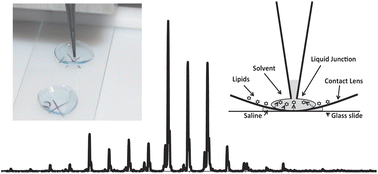
With this work, Simon Brown and colleagues from the University of Wollongong, Australia, describe a method to analyse lipids directly from a contact lens using liquid extraction surface analysis coupled to mass spectrometry (LESA-MS/MS). To demonstrate the efficiency of LESA-MS/MS, the researchers analysed the lipids on 25 worn contact lenses and extracted lipids with limited degradation of the lens material in only 15 minutes.

Brown et al., Analyst 2013

Photograph and schematic diagram of the LESA process from a contact lens

Liquid extraction surface analysis allows the extraction of analytes from a surface by creating a micro-junction between a small volume of solvent at the surface and a pipette tip. The solvent is then aspirated into the pipette tip and infused onto a sensitive mass analyser.
According to the authors, this improved technique will determine the molecular composition of biological deposits and identify the lipid molecular species representing all major classes present in human tear films.

To know more about this study click on the link below.

Communication Automated surface sampling of lipids from worn contact lenses coupled with tandem mass spectrometry
Simon H. J. Brown ,  Liam H. Huxtable ,  Mark D. P. Willcox ,  Stephen J. Blanksby and Todd W. Mitchell
Analyst, 2013, Advance Article
DOI: 10.1039/C2AN36189B

Digg This
Reddit This
Stumble Now!
Share on Facebook
Bookmark this on Delicious
Share on LinkedIn
Bookmark this on Technorati
Post on Twitter
Google Buzz (aka. Google Reader)