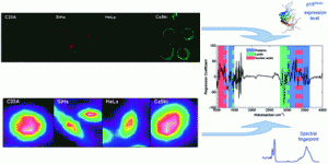
 In this HOT Article a team from Dublin present an impressive study that links imunohistochemistry and fluorescence with FTIR to demonstrate the effectiveness of spectroscopic techniques in the diagnosis of cervical cancer.
Fluorescence microscopy and flow cytometry are used to deomonstrate that expression levels of the biomarker protein p16INK4A in cervical cancer cell lines correlates with HPV invection levels. This confirms p16INK4A as a potential diagnostic marker of cervical cancer. FTIR imaging is then used to identify the specific spectral features of nuclear and cytoplasmic regions of the cervical cancer cells. By correlating all the findings it was possible to construct a model which can predict the p16INK4A expression level based on a spectral fingerprint of a cell line, demonstrating the diagnostic potential of spectroscopic techniques.
Interested in knowing more? Read the article here, free until March 11th.
Correlation of p16INK4A expression and HPV copy number with cellular FTIR spectroscopic signatures of cervical cancer cells
Kamila M. Ostrowska, Amaya Garcia, Aidan D. Meade, Alison Malkin, Ifeoluwapo Okewumi, John J. O’Leary, Cara Martin, Hugh J. Byrne and Fiona M. Lyng
Analyst, 2011, Advance Article
DOI: 10.1039/C0AN00910E


