Archive for the ‘Hot Articles’ Category

Keeping a remote eye on the microworld

Creating a lab-on-chip device with multiple sensing capabilities has been a long-desired goal in the biotechnology field. A sensor-rich chip would pack the power of a full-scale laboratory as initially envisioned in the lab on a chip concept, yet it remained challenging for decades. Many researchers suggest that real-time monitoring of the culture conditions can provide higher-quality drug screening testing. Although many platforms have been proposed up till now, none of them has yet taken off. A recent work from Andreas Weltin’s laboratory at IMTEK published in the Lab on a Chip, however, sounds very promising.

Embedding the cells in synthetic or natural 3D matrices makes it possible to mimic an in vivo cellular environment, but also frequently takes away control over the culturing conditions. The present microfluidic platform achieves the spheroid growth of tumor cells using Matrigel for several days. However, without continuous monitoring, large amounts of independent cultures would be necessary, as each must be sacrificed to conduct separate assays at separate time points, raising the degree of uncertainty in statistical analysis. The authors addressed this problem by integrating several sensors which can perform continuous readout of different molecules.

Creating hypoxia (low oxygen levels) or hyperoxia (high oxygen levels) conditions in cell culture is often needed to mimic disease conditions for fundamental studies. On the other hand, monitoring the local oxygen levels in a culture is not straightforward. In the present study, the authors fabricated the reference electrodes by electroplating silver/silver chloride. The oxygen sensors were modified using a platinum-coated surface located at multiple spots in close vicinity of the culture chamber, allowing for a comparison of the initial and waste streams (Figure 1).

Cell metabolism underpins cell activity, as well as its viability. Some of the key parameters to monitor cell metabolism include glucose and lactate concentrations in the culture media. While high uptake of glucose is indicative of faster cell respiration and replication, high production of lactate is indicative of an increased anaerobic respiration rate. The authors embedded enzymatic sensors in hydrogel to measure glucose and lactate levels. Regions with lactate oxidase and glucose oxidase enzymes are strategically located in close vicinity to the culture chamber. Such locations were ideal because the cell metabolites released into the media are not yet diffused to a lower concentration at this spot, while it is far enough from the chamber so the by-products of the enzymatic reaction cannot interfere with the cell culture (Figure 1).

As a proof-of-concept, Weltin’s team applied this multifunctional sensing platform to the growth of patient-derived triple-negative breast cancer stem cells, which are highly metastatic and less responsive to treatment. The developed platform unveiled the difference in temporal and concentration-dependent drug response of the cells in spheroids. The authors state that this work underlines the importance of in situ, real-time metabolite monitoring in 3D cell cultures as a future standard in cancer research-related studies.

chip with multiple sensors

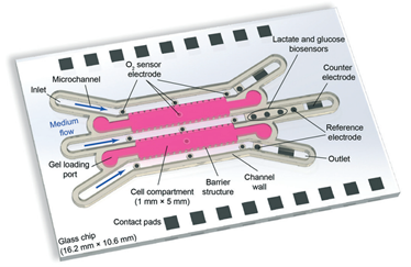
Figure 1. The layout of the microfluidic chip. The authors created a compartmentalized matrix-embedded cell culture with medium perfusion and rigorous control of the liquid and gas composition in one platform but also incorporated continuous sensors that are capable to deliver real-time readouts of the oxygen concentrations and cell metabolism by-products.


About the Web writers

Burcu Gumuscu is an assistant professor at Eindhoven University of Technology in the Netherlands, and the chair of the Biosensors and Devices Laboratory. She strives for the development, fabrication, and application of smart biomaterials to realize high-precision processing in high-throughput microfluidic settings. She specifically focuses on the design and development of lab-on-a-chip devices containing hydrogels for diversified life sciences applications.

OksanaSavchak


Oksana Savchak is a Ph.D. student in Biosensors and Devices Laboratory at the Eindhoven University of Technology in the Netherlands. She focuses on the development of microfluidic screening platforms to investigate cell-material interactions.


Original publication

Dornhof, J., Kieninger, J., Muralidharan, H., Maurer, J., Urban, G. A., Weltin, A. (2022): Microfluidic organ-on-chip system for multi-analyte monitoring of metabolites in 3D cell cultures. In: Lab on a Chip, 22 (2). DOI: 10.1039/d1lc00689d

Digg This
Reddit This
Stumble Now!
Share on Facebook
Bookmark this on Delicious
Share on LinkedIn
Bookmark this on Technorati
Post on Twitter
Google Buzz (aka. Google Reader)

Digital microfluidics for handling picoliters

Just another day at the chemistry lab. Get in, make sure to wear PPE thoroughly, grab a pipette set, start reading, and applying the steps of the experiment. Pipette hundreds of microliters of a solution, done. Pipette tens of microliters of another solution, no problems. Pipette several picoliters of …, what? Wait!  Can we pipet sub-microliter size liquids? The answer will be yes at this time, but we will twist it a bit. As the name suggests, microfluidics is the art of handling tiny amounts of liquids. Can it be helpful when it comes to handling sub-microliter size droplets?

Since the early 2000s, digital microfluidics (DMF) has been proposed as a more versatile candidate in sample processing applications. Most DMF devices use electric field application to control the displacement of droplets, which are separated from the underlying electrodes via a dielectric layer and an nm-thick hydrophobic layer. Once we actuate an electrode, the contact angle, and in turn, the hydrophobicity, of a droplet sitting on top of the electrode decreases due to surface energy changes. This would be a golden moment because if we actuate a neighboring electrode now, the droplet will displace towards the activated electrode. Based on this principle, we can transport, dispense, merge, and split droplets in a digital microfluidic platform.

One constraint of current digital microfluidic systems is that droplet volume is limited by electrode area. Droplets can only be reliably split and displaced above a certain volume as they will not be capable of displacing towards another electrode if their contact area is too small. Working with smaller scale electrodes does not solve the problem either, but it makes evaporation-related problems more pronounced.

In the recent Lab on a Chip article of researchers from the University of Macau (CN) and the University of Lisboa (PT) demonstrated the controllable ejection of satellite droplets to create a digital microfluidic platform that is capable of transporting picoliter-volume droplets. Precise control of ejection position and volume was made possible using a narrow electrode, or a jetting bar, which focused the area of high voltage AC actuation. During a rapid change in electric field intensity beyond the contact angle saturation threshold voltage, the excess energy is released by the ejection of satellite droplets. The dispensing droplet would release picoliter sized volumes onto the jetting bar which were then collected by another droplet moving over the jetting bar (Fig. 1). Volumes between 5 pL to 20 nL can be produced by repeatedly dispensing and collecting picoliter-size droplets at the jetting bar. The dispensed volume could also be controlled by changing increasing the width of the jetting bar for greater volumes. Dispensing volume can also be carefully controlled by the strength of the electric field, the actuation frequency, and actuation time.

The authors showed that the picoliter- volume dosing system can be used in a dequenching assay. Fluorescent DNA probes specific to S. aureus were delivered to droplets containing S. aureus and K. pneumonia using this assay. The dispensed satellite droplets successfully delivered the DNA probes to the droplets. The authors built a quantitative relationship between the degree of fluorescence and volume delivered in the S. aureus droplet accordingly. The authors think that this new technique may make microfluidics more accessible in sample preparation for clinical and lab studies in the future.

Figure 1. The dispensing drop releases satellite droplets under high voltage AC actuation of the jetting bar. The satellite droplets are then taken up by another droplet, facilitating volume transfer without the merging of either the dispensing or pickup-up droplet.


To download the full article for free* click the link below:

Turning on/off satellite droplet ejection for flexible sample delivery on digital microfluidics

Haoran Li, Ren Shen, Cheng Dong, Tianlan ChenYanwei Jia, Pui-In Mak and Rui P. Martins, Lab Chip, 2020, Lab on a Chip Hot Articles

DOI: 10.1039/D0LC00701C


About the Webwriters

Burcu Gumuscu is an assistant professor in BioInterface Science Group at Eindhoven University of Technology in the Netherlands. She strives for the development, fabrication, and application of smart biomaterials to realize high-precision processing in high-throughput microfluidic settings. She specifically focuses on the design and development of lab-on-a-chip devices containing hydrogels for diversified life sciences applications. She is also interested in combining data-mining and machine learning techniques with hypothesis-driven experimental research for future research.


Miguel FaaseMiguel Faase is a Ph.D. student in BioInterface Science Group at Eindhoven University of Technology in the Netherlands. He focuses on the development of high-throughput screening platforms for biomaterial research under the supervision of Burcu Gumuscu.

Digg This
Reddit This
Stumble Now!
Share on Facebook
Bookmark this on Delicious
Share on LinkedIn
Bookmark this on Technorati
Post on Twitter
Google Buzz (aka. Google Reader)

Modelling arthritis

The function of tissues degrade during the course of human life, whether the cause is genetics, accidents, or wear and tear. Frequently experienced medical conditions associated with aging include blocked arteries, cataracts, and arthritis. The former two can be thoroughly treated via surgeries, but arthritis remains a bane to millions of sufferers because its treatment is rather palliative than intensive.

Let’s take the example of rheumatoid arthritis. It occurs when macrophage cells (a main component of the immune system) attack to the membrane (synovium) that surrounds the joints as a result of an auto-immune response. The damaged synovium thickens significantly, its anatomy undergoes striking changes such as metabolic activation of synoviocytes and continuous mass of cells invading into the cartilage and bone. If unchecked this inflammation can destroy the cartilage and the bone within the joint. What does this mean for the patients? A relatively short answer is painful knees, reduced physical activities, continuous uptake of anti-inflammatory medication, and painkillers. A cure does not yet loom on the horizon, but new tools to model rheumatoid arthritis could definitely make it easier to predict the onset of the disorder.

Peter Ertl and his colleagues at Vienna University of Technology, Austria, decided to tackle this problem in the article they recently published in Lab on a Chip. They researched the mechanisms governing the destructive inflammatory reaction in rheumatoid arthritis by designing a first-ever 3D synovium-on-chip tool, which can monitor the onset and progression of the tissue responses. The researchers were focused on monitoring the behavior of the inflated fibroblast-like synoviocytes, which makes the synovium membrane thicker. They used tumor necrosis factor-alpha (TNF-α) to trigger synoviocytes to demonstrate inflammation response. The chips contained circular microchambers, where surface coatings are applied and Matrigel is filled for obtaining 3D organoids. Different hydrogel types were also examined to observe cell response. For the monitoring, the researchers used collimated laser beams, and the scattered light was collected using embedded organic photodiodes. This powerful optical measurement setup allowed for adjusting the detection range (50 nm to 10 µm) and sensitivity to any tissue construct (Figure 1). The researchers implemented a PDMS waveguide structure to the optical measurement setup to turn the light scattering measurements into reproducible ones. The measurement setup also enabled continuous monitoring of hydrogel polymerization, which was also controlled in this work since the polymerization time influences hydrogel stiffness, which in turn affects the fate of cell behavior. A typical measurement took four days, where the researchers obtained cultures accurately mimicking in-vivo rheumatoid arthritis conditions. A diseased phenotype becomes distinguishable within 2-3 days in the organ-on-chip platform, as this takes at least 14 days in conventional cell culture platforms. The developed tool can very well serve as a new modeling system for inflammatory arthritis and joint-related disease models.


Figure 1. Overview of the synovium-on-a-chip system with integrated time-resolved light scatter biosensing.


To download the full article for free* click the link below:

Monitoring tissue-level remodelling during inflammatory arthritis using a three-dimensional synovium-on-a-chip with non-invasive light scattering biosensing

Mario Rothbauer, Gregor Höll, Christoph Eilenberger, Sebastian R. A. Kratz, Bilal Farooq, Patrick Schuller, Isabel Olmos Calvo, Ruth A. Byrne, Brigitte Meyer, Birgit Niederreiter, Seta Küpcü, Florian Sevelda, Johannes Holinka, Oliver Hayden, Sandro F. Tedde, Hans P. Kiener and Peter Ertl, Lab Chip, 2020, Lab on a Chip Hot Articles

DOI: 10.1039/c9lc01097a


About the Webwriter

Burcu Gumuscu is an assistant professor in BioInterface Science Group at Eindhoven University of Technology in the Netherlands. She strives for the development, fabrication, and application of smart biomaterials to realize high-precision processing in high-throughput microfluidic settings. She specifically focuses on the design and development of lab-on-a-chip devices containing hydrogels for diversified life sciences applications. She is also interested in combining data-mining and machine learning techniques with hypothesis-driven experimental research for future research.

Digg This
Reddit This
Stumble Now!
Share on Facebook
Bookmark this on Delicious
Share on LinkedIn
Bookmark this on Technorati
Post on Twitter
Google Buzz (aka. Google Reader)

Alcohol testing on skin

Our skin perspires every day by about 2.5 million sweat glands under the dermis layer. Not all the sweat glands have the same function. While some glands release sweat through the opening of hair follicles−aka apocrine glands−, some others open directly onto the skin surface−aka eccrine glands−. The most prevalent type in the body is the eccrine glands, which produce interesting signaling subtleties for monitoring health. Actually, the sweat produced at different times contains the time-stamps of noteworthy information such as electrolytes, metabolites, micronutrients, hormones exogenous agents, each of which can change in concentration in the content of sweat with diet, stress level, hydration status, and physiologic or metabolic state. Monitoring these cues using appropriate sensors makes possible to track an individual’s health in real-time. Sweat analysis potentially complements or even obviates the need for approaches relying on puncturing the skin with needles.

We encounter two major types of skin-interfaced measurement units in the literature: (1) equipped with electronics to power measurement units or run electrochemical measurements (2) colorimetric detection relying on no electronic sensors. Each type has different application areas. The colorimetric detection is particularly interesting as it relies on uniquely designed rapid chemical reactions between the sweat and the unit, in many cases with improved repeatability and accuracy. As a recent example of this kind, a research team from Northwestern University and the University of Illinois at Urbana-Champaign have introduced a skin sensor to detect signaling subtleties emanating from the skin. The promise of this work lies in the ability to control the reaction kinetics and the mixing of different reagents and samples in a user-operated device. This achievement was made possible by introducing a multi-layered microfluidic device platform containing stop valves and a super absorbent polymer to initiate colorimetric reactions for microliter volumes of ammonia and ethanol in microliter volumes of sweat.

The patch absorbs sweat via super absorbent polymer layers located subjacent to individual wells (Figure 1). The super absorbent polymer layer expands upon soaking sweat, activating a microfluidic mechanical pump that releases pre-loaded reaction buffers into the wells. The colorimetric reaction subsequently takes place (Figure 1). This pad was equipped for running ammonia and ethanol (alcohol) assays. But why two metabolites only? The patch is dedicated to monitoring alcohol testing in daily living. As mentioned in the paper, ammonia levels could serve as an index for hepatic encephalopathy diagnosis in subjects who are experiencing alcohol abuse. Hepatic encephalopathy refers to liver failure mostly caused by alcohol uptake. The researchers also tested the patch on volunteers resting in a warm bath after consuming alcoholic beverages to highlight the operational advantages of such patches. The authors note that this work has direct implications for sweat biomarker research, health monitoring in daily life, and simultaneous drug/alcohol testing.

Figure 1. The skin-interfaced measurement patch, its measuring units, and working mechanism.

To download the full article for free* click the link below:

Soft, skin-interfaced microfluidic systems with integrated enzymatic assays for measuring the concentration of ammonia and ethanol in sweat

Sung Bong Kim, Jahyun Koo, Jangryeol Yoon, Aurélie Hourlier-Fargette, Boram Lee,f Shulin Chen, Seongbin Jo, Jungil Choi, Yong Suk Oh, Geumbee Lee, Sang Min Won, Alexander J. Aranyosi, Stephen P. Lee, Jeffrey B. Model, Paul V. Braun, Roozbeh Ghaffari, Chulwhan Park and John A. Rogers, Lab Chip, 2020, Lab on a Chip Hot Articles

DOI: 10.1039/c9lc01045a


About the Webwriter

Burcu Gumuscu is a researcher in Mesoscale Chemical Systems Group at the University of Twente in the Netherlands. Her research interests include the development of microfluidic devices for quantitative analysis of proteins from single-cells, next-generation sequencing, compartmentalized organ-on-chip studies, and desalination of water on the microscale.

Digg This
Reddit This
Stumble Now!
Share on Facebook
Bookmark this on Delicious
Share on LinkedIn
Bookmark this on Technorati
Post on Twitter
Google Buzz (aka. Google Reader)

Processing one-trillionth the size of a liter

A picoliter of a droplet is approximately 1 µm. It is so small that a raindrop can contain thousands of picoliters. Despite the small quantity, precise control of picoliter size liquids spawns a growing number of applications including in point-of-care tools and drug synthesis. Such a task is not easy to achieve for conventional liquid-manipulation methods due to precision, evaporation and the ease of post-processing problems.

When it comes to small volume liquid handling, no other platform would be better than microfluidic devices. As such, a recent Lab Chip paper from Prof. Bai Yang’s Lab at Jilin University in China demonstrates the fabrication of a microfluidic platform to handle sub-picoliter size droplets. The authors use glass as the core material of the microchips because glass is mechanically strong and it enables higher precision in droplet manipulation (Figure 1). In the paper, pressure-controlled syringes are used to dispense down to 0,5 picoliters in a series of trapezoidal-shaped microchambers, each composed of 10 subunits. The boundaries of the subunits act as pressure barriers, preventing the liquid from flowing through the entire microchamber and allowing for fine-tuning of the volume to be handled. The authors also showed that the working range, stability, and precision of the device could be increased by adjusting the boundary properties and microchamber dimensions (e.g., height). However, the magnitude of the minimum volume will still be bound to two main factors. (1) The resolution of the fabrication technology determines the microchannel and boundary dimensions. (2) Laplace pressure difference between the channel and the boundaries determines the accuracy of the device. The smaller channels yield decreased accuracy due to higher pressure differences between the boundary-microchannel interfaces. Relying on these design criteria, the authors fabricated up to three interconnected picoliter-volume injection units. Combined units work together to synthesize gold nanoparticles and nanorods with a wide size range. The authors believe that the platform can be further used in several different applications including lab-on-chip platforms, analytical chemistry for medicine approaches, quantitative cell cultures, and even drug injection into single cells.

Figure 1. The microfluidic device for sub-picoliter volume liquid handling

To download the full article for free* click the link below:

Pressure-controlled microfluidic sub-picoliter ultramicro-volume syringes based on integrated micro-nanostructure arrays

Nianzuo Yu, Yongshun Liu, Shuli Wang, Xiaoduo Tang, Peng Ge, Jingjie Nan, Junhu Zhang, and Bai Yang, Lab Chip, 2019, Lab on a Chip Hot Articles

DOI: 10.1039/C9LC00730J


About the Webwriter

Burcu Gumuscu is a researcher in Mesoscale Chemical Systems Group at the University of Twente in the Netherlands. Her research interests include the development of microfluidic devices for quantitative analysis of proteins from single-cells, next-generation sequencing, compartmentalized organ-on-chip studies, and desalination of water on the microscale.

Digg This
Reddit This
Stumble Now!
Share on Facebook
Bookmark this on Delicious
Share on LinkedIn
Bookmark this on Technorati
Post on Twitter
Google Buzz (aka. Google Reader)

What are the challenges to sample-in-answer-out technologies in clinical settings?

The boom in microfluidic total analysis systems is spurred by the use of microscale fabrication techniques. In 1999, Agilent Technologies introduced a coin-size device called “LabChip” to the market for the analysis of DNA. The timing of the market release of this device was remarkable as it benefited from the hype of the Human Genome Project. The LabChip has drawn considerable attention from both academia and industry. As a result, the shrink of chemistry labs into coin-sized microfluidic devices has evolved more from technological push than from market pull. Technology-driven development pathway brought challenges, especially for the clinical integration of microfluidic devices due to the lack of standards, focus, and communication between academia and clinics.

A recent Lab on a Chip paper from Martyn Boutelle’s Lab addresses this issue. The authors identify the problem from a well-addressed microdialysis perspective. They state that “taking a microfluidic system into clinical environment brings lots of challenges, not least that during setups and developments the very low the flow rates used in combination with microdialysis means that leaks and misdirection of flows are very hard to see.” The authors define the most significant challenge as the development of a device that’s robust enough to be used by and provide enough information to, the clinical team without micromanagement by experts.

The authors attack the problem by creating a sensor-based online system associated with electrochemical measurement, which would be able to analyze the sample in a miniaturized platform continuously. They developed the microfluidic sensors and chip, but they wanted to increase the use of their technology in real-world scenarios by non-experts, so they looked into ways to introduce more precision and control to the platform. The authors combined their technology with LabSmith microfluidic components and constructed breadboard like layouts for typical lab protocols. The authors add that “the main surprise was the ability to bring the rigor of an analytical laboratory into unusual places such as abattoirs, surgical theatres and public transport!”

In this work and the previous work of the authors, the performance of 3-D printed chips was compared to the PDMS ones. The authors found that PDMS material is much more vulnerable than the 3D print outs when frequently handled. The sensor attached to the chip is programmed to calibrate in regular intervals (e.g., every three hours) in single or multiphase flow conditions. Authors describe the advantage of the system that ”remote access to the scripts allows interaction with the system without the requirement of a highly skilled person being right next to it, which in the context of surgical theatres and hospital wards is a distinct advantage. If the codes and scripts are available to less skilled personnel, they are still able to interact and use the system and by making the system more user-friendly a wider audience and more enthusiasm is generated for the product, increasing interest, uptake, and use.”

The authors would like to improve the platform further by making it wearable since it already has grounds for such an operation with wireless sensors. The next thing to be improved in the system is the feed. At this moment, the syringes have to be regularly refilled. This might not be a problem in the laboratory; however, monitoring can last for days in a clinical setting, and periodically refilling the syringes may lead to noise artifacts. Another improvement could be the ease of operation and troubleshooting when, e.g., a tubing becomes blocked in the middle of the measurement when the user is short on space and time.

Lastly, the authors think that this pioneering platform can help shape the future in the market by giving more people access to an area of science that was previously highly skilled, whilst maintaining analytical robustness. This will be the start to break the barrier between academia-made devices and clinical settings.


To download the full article for free* click the link below:

Clinical translation of microfluidic sensor devices: focus on calibration and analytical robustness

Sally A. N. Gowers, Michelle L. Rogers, Marsilea A. Booth, Chi L. Leong, Isabelle C. Samper, Tonghathai Phairatana, Sharon L. Jewell, Clemens Pahl, Anthony J. Strong and Martyn G. Boutelle, Lab Chip, 2019, Lab on a Chip Hot Articles

DOI: 10.1039/C9LC00400A

*access is free with an RSC account (free to register)


About the Webwriter

Burcu Gumuscu is a researcher in Mesoscale Chemical Systems Group at the University of Twente in the Netherlands. Her research interests include the development of microfluidic devices for quantitative analysis of proteins from single-cells, next-generation sequencing, compartmentalized organ-on-chip studies, and desalination of water on the microscale.

Digg This
Reddit This
Stumble Now!
Share on Facebook
Bookmark this on Delicious
Share on LinkedIn
Bookmark this on Technorati
Post on Twitter
Google Buzz (aka. Google Reader)

Through a cheaper ‘book of life’

Mapping the human genome project has been one of the world’s largest scientific collaborations. Completing the full genome sequencing for “the book of life” took more than 10 years with the efforts of 1000’s scientists and a budget of $3 billion. About 20 years after the finalization of this enormous project, it is now possible to complete a full human genome sequencing within 8 days for about $1,000 thanks to more advanced sequencing tools. Further improvements in genome sequencing tools are still warranted today because the genome sequencing field has been embraced by many more applications, including forensics, disease modeling & identification, and personalized medicine (e.g., identifying the genes that cause a medicine to work in one patient but not in another).

Initial sequencing technologies relied on standard DNA electrophoresis techniques such as slab gels and capillaries, allowing for the preparation of only small numbers of samples at a time. The sample preparation limitation was the primary reason for the increased costs and processing duration during the human genome project. Many efforts have been directed towards improving sample preparation techniques in the last decades. As the first step, electrophoresis techniques have been optimized to boost the sample throughput with user-friendly, smaller, and functional platforms. Traditional DNA separation gels, which have been used as the golden standard for many decades, have been replaced by microfabricated post arrays and nanometer-scale deterministic lateral displacement arrays.

A nice example of nanometer-scale deterministic lateral displacement arrays has been demonstrated recently by researchers from IBM T.J. Watson Research Center and Icahn School of Medicine at Mount Sinai in New York, USA. In this work, the researchers fractioned DNA in the range of 100-10.000 base pairs with a size-selective resolution of 200 base pairs. To achieve that, four different microchip configurations were fabricated on silicon wafers, where the array and nanopillar sizes were tuned for each configuration to obtain the optimum separation performance for the selected range of DNA fragments. Each configuration contained several separate arrays to conduct independent runs in a single chip. Instead of applying an electric field, the researchers applied pressure-driven force to separate the fragments. This strategy is particularly useful for separating non-charged species without being affected by buffer conditions (e.g., ionic strength).

The separation mechanism in the nanometer-scale deterministic lateral displacement array is simple: If the size of a DNA fragment is larger than the diameter of the pillars, the fragment is deflected towards the collection wall at a large angle, also called the bump mode. If the size of a DNA fragment is smaller than the diameter of the pillars, the fragment migrates at an angle nominally zero, termed as the zig zag mode. In such a system, diffusion of DNA fragments lead to intermediate migration angles, termed as the partial-bump mode. Different sizes of DNA fragments could be separated in the array since the fragments will follow distinct trajectories thanks to the existence of different modes. Figure 1 summarizes the separation mechanisms and gives an outline for the nanometer-scale deterministic lateral displacement array.

In the nanometer-scale deterministic lateral displacement array, the gap sizes were tuned from microscale to nanoscale only, without application of any other molecules that could change the DNA diffusion behavior, ionic strength (changing the effective gap distances). In such a setting, the researchers identified, for the first time, the flow velocity-dependence of different fragment lengths. Mainly, changing flow velocity caused a transition between bump and zig zag modes for the given size range of different DNA fragments: Slow speeds lead to partial-bump mode, and high speeds lead to the collapse of all DNA fragments to zigzag mode. The nanometer-scale deterministic lateral displacement array could also be used as a purification tool with 75% recovery and 3-fold concentration enhancement of DNA fragments. This tool could be used effectively for preparing next-generation sequencing libraries, on-chip DNA characterization, and circulating DNA characterization applications.


Figure 1. The nanometer-scale deterministic lateral displacement array and DNA separation mechanism at different flow velocities.

To download the full article for free* click the link below:

Gel-on-a-chip: continuous, velocity-dependent DNA separation using nanoscale lateral displacement

Benjamin H. Wunsch, Sung-Cheol Kim, Stacey M. Gifford, Yann Astier, Chao Wang, Robert L. Bruce, Jyotica V. Patel, Elizabeth A. Duch, Simon Dawes, Gustavo Stolovitzky and  Joshua T. Smith, Lab Chip, 2018, Lab on a Chip Articles

DOI: 10.1039/c8lc01053f


About the Webwriter

Burcu Gumuscu is a researcher in Mesoscale Chemical Systems Group at the University of Twente in the Netherlands. Her research interests include the development of microfluidic devices for quantitative analysis of proteins from single-cells, next-generation sequencing, compartmentalized organ-on-chip studies, and desalination of water on the microscale.


Digg This
Reddit This
Stumble Now!
Share on Facebook
Bookmark this on Delicious
Share on LinkedIn
Bookmark this on Technorati
Post on Twitter
Google Buzz (aka. Google Reader)

Microfluidics for improving the natural gas extraction process


shale rock

Figure 1. Natural gas extraction from shale rock.

Shale is a type of fine-grained rock that contains silt, clay, mineral particles, and pores ranging from meter to nanometer scales. The high organic material content in shale rock is used in natural gas extraction, for which shale reservoirs are mechanically stimulated to create permeability in the pores. A preferred stimulation method is called hydraulic fracturing, where a pressurized fluid fractures shale stone and keeps the fractures open for gas extraction (Figure 1). Natural gas extraction from shale rock is a relatively new process compared to existing energy sources. It has attracted growing interest in America and Asia especially after the 2000s because of being an environmentally friendly alternative to other consumable energy sources. On the other hand, the gas industry currently struggles with optimizing the use of pore space and fractions for efficient extraction of the gas. In a newly opened shale rock reservoir, volatile components vaporize from meter to micrometer-scale pores first, leaving heavier components in hard-to-access nanometer-scale pores. Extraction of the remaining components is necessary for full utilization of the reservoirs but poses a hard-to-solve problem for the industry.


In a recent study published in Lab on Chip, David Sinton and co-authors review the current state of the technology and demonstrate a nano-scale physical model of shale with pores. The authors also study the dynamics of gas production in nanopores via imaging the system optically and developing an analytical model for gas vaporization. They first created a microchip model matching shale nanoporous matrix properties (e.g., dominant pore sizes and permeability) (Figure 2). The microchip model contained approximately 5800 pores connected via 23000 throats, where a hydrocarbon mixture was injected. In the model, the number of the small pores (≤10 nm) is designed to be greater than the number of the larger pores (∼100 nm) to store most of the accessible hydrocarbons. This pore size distribution captures the influence of nanoscale throats connecting the larger pores and is relevant to shale production. High, medium, and low superheat was applied to the filled microchip to investigate the spatiotemporal dynamics of vaporization via optical imaging. An analytical model and experimental results showed that phase change (liquid to vapor) in a pore is largely independent of phase change in neighboring pores.

This work supports the hypothesis that the rapid decline in production rates is due to a shift from the large connected features to the nanoporous matrix, as over time the smallest pores become enriched with heavier fractions. The authors reveal that vaporization rate slows down 3000 times thanks to the nanoscale throat bottlenecks at high temperatures, while the rates reduce further with vaporization of light components in large pores at low temperatures. Even the pores with 10 nm and fewer diameters can significantly influence the production from larger pores by severely gating transport. The authors found that this problem can be solved by applying very low pressures, although currently not available in the field, during the later stages of hydraulic fracturing. This finding seems to open a new avenue in the field of shale rock processing for energy.

Figure 2. Close up view of shale rock, the description of how the evaporation works, and the description of the microchip operation.

To download the full article for free* click the link below:

Natural gas vaporization in a nanoscale throat connected model of shale: multi-scale, multicomponent and multi-phase

Arnav Jatukaran, Junjie Zhong, Ali Abedini, Atena Sherbatian, Yinuo Zhao, Zhehui Jin, Farshid Mostowfi and David Sinton

Lab Chip, 2018, Lab on a Chip Articles

DOI: 10.1039/c8lc01053f

*Article free to read until 7th May 2019

About the Webwriter

Burcu Gumuscu is a researcher in Mesoscale Chemical Systems Group at University of Twente in the Netherlands. Her research interests include the development of microfluidic devices for quantitative analysis of proteins from single-cells, next generation sequencing, compartmentalized organ-on-chip studies, and desalination of water on the microscale.


Digg This
Reddit This
Stumble Now!
Share on Facebook
Bookmark this on Delicious
Share on LinkedIn
Bookmark this on Technorati
Post on Twitter
Google Buzz (aka. Google Reader)

Screening lipid libraries with microfluidics

The plasma membrane is a key component of living organisms and is essential to all life. It separates the inside of the cell from the outside, compartmentalizes reactions, and selectively allows transport across it. While a lot of research has gone into how different proteins and surface molecules control the functions of the plasma membrane, less is known about how lipid composition gives rise to specific properties. For instance, transmembrane proteins, which are also a large class of drug targets, may have different requirements for the lipid environment or may have their function modified depending on local lipid composition. Recently, researchers from the David Weitz Lab at Harvard, have developed a microfluidic chip which they used to screen the largest lipid library to date, in order to identify which lipid compositions have specificity for certain protein transmembrane domains. This allows researchers to investigate the effect of local lipid concentration on transmembrane proteins.

The plasma membrane is often described as a ‘simple barrier’. But if that’s the case, then “why does nature go through the trouble of making so many different types of lipids?” explained Roy Ziblat, the lead author on the paper. Ziblat believes that the lipid membrane role is far bigger than a mere barrier and it serves as a substrate for accelerating bio-reactions. The role of the lipids composing the membrane is to control which biomolecules participate in these reactions, by their selectivity to membrane proteins. Having limited success with existing techniques, Ziblat turned to microfluidics to try to answer this question.

The microfluidic chip comprises an array of 108 wells in PDMS where lipid films can be deposited and dried before sealing the chip with another layer of PDMS. Liposomes are generated within the wells by swelling in aqueous buffer. These liposomes are then tested to see whether or not transmembrane domain peptides will insert into them. However, because the transmembrane domain peptides are insoluble, they can’t simply be added into the chip. To get around this, Ziblat et al. turned to cell-free protein synthesis. By loading the chip with DNA for the transmembrane domain peptide and PURExpress (a commercial cocktail of ribosomes, enzymes, and nucleotides for transcription and translation), the peptides can be synthesized in close proximity to the liposomes, thus minimizing precipitation and increasing the chance of insertion. The paper by Ziblat et al., which was featured on the cover of the 7th December issue of Lab on a Chip, also includes a helpful video description of these methods. Ziblat said he first made the video to better communicate his methods with his supervisor and colleagues, but it really helps the reader understand a very technical methodology.

Going forward, Ziblat hopes to use the device to study other membrane interactions, such as virus-cell binding. There’s also hope that this new device and method can be used to identify what the authors call “druggable lipids”—peptides that interact with specific lipids and thus better direct drugs toward specific cells or even organelles.


To download the full article, click the link below:

Determining the lipid specificity of insoluble protein transmembrane domains

R. Ziblat, J. C. Weaver, L. R. Arriaga, S. Chong and D. A. Weitz

Lab Chip, 2018, 18, 3561

DOI: 10.1039/c8lc00311d


About the webwriter

Darius Rackus (right) is a postdoctoral researcher in the Dittrich Bionalytics Group at ETH Zürich. His research interests are in developing integrated microfluidic tools for healthcare and bioanalysis

Digg This
Reddit This
Stumble Now!
Share on Facebook
Bookmark this on Delicious
Share on LinkedIn
Bookmark this on Technorati
Post on Twitter
Google Buzz (aka. Google Reader)

Simple microfluidic cell sorter device to replace manual tissue dissociation protocols

Even a tiny group of cells has the ability to populate a tumor in tissues. Determining cellular diversity and identifying these small cell groups gains importance when it comes to selection of treatment strategies. Tissue samples taken from patients are required to be dissociated into single-cell suspensions, therefore identification can be efficiently done at single-cell level using a powerful suite of technologies including flow cytometry, mass cytometry, and single cell RNA sequencing. However, breaking a tissue down to a single-cell suspension is not an easy task. The old-school way is to cut the tissue sample into small pieces with a blade and mechanically dissociated by vigorous shaking after the application of proteolytic enzymes. Large aggregates are removed by filtering the suspension through a strainer. This technique significantly increases the sample loss, drops the speed of the process and is not ideal for immediate downstream analysis.

In this month’s Lab on a Chip HOT article series, a group of researchers led by Dr Jered Haun at University of California Irvine presented a novel and simple approach that improves the quality of single-cell suspensions obtained from tissue samples using microfluidics. Jeremy Lombardo, a co-author of this article, explains that “the goal of this work was to fully replace manual intensive tissue dissociation protocols by using microfluidic devices.” The developed tool is a microdevice consisting of two nylon membranes, one with 25-50 µm mesh, and the other with 10-15 µm mesh, attached to micron-sized pores and microchannels. The device is made of laser-micromachined hard plastic (PET, aka. polyethylene terephthalate), which enables operation at high flow rates (>10 mL/min) when compared to PDMS (a silicon-based organic material). Also, the chip has multiple layers for connecting nylon mesh membranes at different levels (Figure 1).

cell sorter

Figure 1. Microfluidic cell sorter device for tissue  samples. The sketch shows the inner layers, consisting of two membranes for operating the device in direct or tangential filtration modes. Membrane mesh size can be adjusted to the cell size. Micrographs on the right show lattice network with several pore sizes used in this work. Pore sizes are (top to bottom) 50, 25, 15, 10 µm diameter.

Working principle of the cell sorter device

The inlet of the device connects to a microporous membrane to introduce tissue samples. The Sample passing through the membrane exits through the effluent outlet. It is also possible to direct a portion of the sample along the surface of the membrane that is connected to the cross-flow outlet. The device is either operated in a direct filtration regime to maximize sample recovery and processing speed, or in a tangential filtration regime to sweep larger tissue fragments and cell aggregates away to prevent clogging.

While the researchers initially hypothesized that under pressure-driven flow, cell and tissue aggregates might disaggregate as they pass through the membranes of the device, they were pleasantly surprised by the drastic level of single cell increases seen in the initial testing of these devices, says Jeremy Lombardo and adds “The hardest part in developing and testing this device was to find a combination of membrane pore sizes that could best dissociate cell aggregates and tissue without compromising cell viability. Thorough testing of various pore sizes and combinations were ultimately carried out with both cell line and murine tissue models before we settled on the final 50 and 15 μm pore sizes.”

Advantages, challenges and the future

The authors summarized the advantages of this platform for Lab on a Chip blog readers: “The device is extremely simple to operate as well as inexpensive to fabricate. It can easily be incorporated into many tissue dissociation applications for improved single cell yields as a standalone device but could also be easily integrated with other downstream microfluidic operations (cell sorting, detection etc.).” According to the authors, “in the current format of cell sorter device, cells that are very large in size would likely be difficult to process, as they would likely span multiple pores of the filters and be traditionally filtered away instead of dissociated.” Although seeming like a challenge, this can easily be addressed by adjusting the filter membrane pore sizes to accommodate these larger cell types.

For the future of the device, the authors indicate, “We are also currently working on integrating this device with upstream, larger scale tissue dissociation devices that we have developed previously to create a fully automatable microfluidic tissue dissociation platform.


To download the full article for free* click the link below:

Microfluidic filter device with nylon mesh membranes efficiently dissociates cell aggregates and digested tissue into single cells
Xiaolong Qiu, Jeremy A. Lombardo, Trisha M. Westerhof, Marissa Pennell, Anita Ng, Hamad Alshetaiwi, Brian M. Luna, Edward L. Nelson, Kai Kessenbrock, Elliot E. Hui, and Jered B. Haun
Lab Chip, 2018, Lab on a Chip Recent Hot Articles
DOI: 10.1039/c8lc00507a

About the Webwriter
Burcu Gumuscu is a postdoctoral fellow in Herr Lab at UC Berkeley in the United States. Her research interests include development of microfluidic devices for quantitative analysis of proteins from single-cells, next generation sequencing, compartmentalized organ-on-chip studies, and desalination of water on the microscale.

Digg This
Reddit This
Stumble Now!
Share on Facebook
Bookmark this on Delicious
Share on LinkedIn
Bookmark this on Technorati
Post on Twitter
Google Buzz (aka. Google Reader)