Archive for September, 2013

Using structural changes in carbon nanostructures to modify cellular responses

Carbon nanostructures have rapidly become subjects of increasing interest for use in biomedical applications ranging from drug delivery to bioimaging.  This is primarily due to their exceptionally high surface areas as well as their unique optical and electronic properties.  In this study, researchers from the Piramal Group in collaboration with the University of Florida investigated how the surface composition and structure of these materials affects their biological impact.

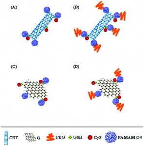
Specifically, the group investigated the protein binding capabilities, degree of cellular internalization, and cytotoxicity of carbon nanotubes (CNT) and graphene (G) sheets as well as CNT and G structures which had been conjugated with PAMAM dendrimers (G4) and polyethylene glycol (PEG).  These two polymers were chosen as conjugates as they are both known to improve the dispersibility and stability of various materials in solution.

Initial protein binding studies showed that the CNT and G conjugates exhibited weaker interactions with a model protein, bovine serum albumin (BSA), when compared to their non-conjugated counterparts.  These weaker interactions, which were thought to be primarily electrostatic in nature, could be extremely beneficial in limiting any possible functional interference which could arise from protein agglomeration on the nanostructure’s surface.  The group also studied the degree to which the different nanostructures were internalized within cells.  CNT conjugates were consistently found to be internalized in higher quantities than the G conjugates, presumably because their tubular shape promoted increased cell membrane penetration.  Finally, cytotoxicity studies illustrated that the conjugated nanostructures typically exhibited reduced toxicity levels compared to unmodified CNT or G, particularly at higher concentrations of the nanomaterial.

Overall, the study demonstrated that carbon nanostructures can have different biological activities depending on their shape and surface composition.  It also suggests that conjugation of carbon materials with polymers such as PEG and G4 might be an effective method for limiting protein binding and reducing cytotoxicity.


Structure effect of carbon nanovectors in regulation of cellular responses
Shashwat S. Banerjee, Archana Jalota-Badhwar, Prateek Wate, Sneha Asai, Khushbu R. Zope, Russel Mascarenhas, Dimple Bhatia and Jayant Khandare 
Biomater. Sci., 2013, Advance Article DOI: 10.1039/C3BM60082C

Ellen Tworkoski is a guest web-writer for Biomaterials Science.  She is currently a second year Ph.D. student in the biomedical engineering department at Northwestern University (Evanston, IL, USA).

Follow the latest journal news on Twitter @BioMaterSci or go to our Facebook page.

To keep up-to-date with all the latest research, sign-up to our RSS feed or Table of contents alert.

Digg This
Reddit This
Stumble Now!
Share on Facebook
Bookmark this on Delicious
Share on LinkedIn
Bookmark this on Technorati
Post on Twitter
Google Buzz (aka. Google Reader)

Treating cancer with nanoparticles

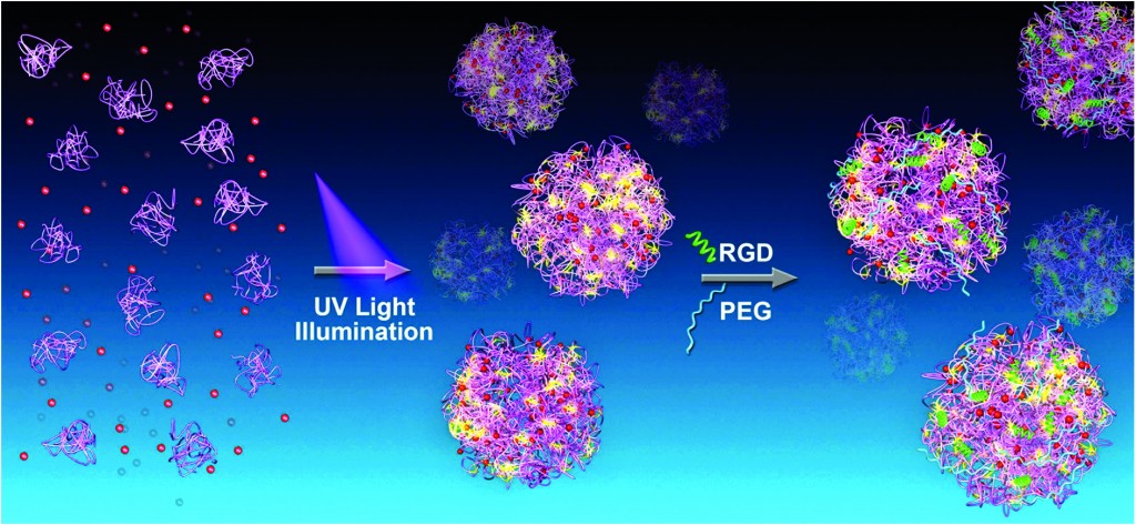
Cancer therapeutics is a burgeoning field of research, particularly because chemotherapy has serious side effects that include the devastation of healthy tissue and the possibility that some cancerous tissue survives the radiation. Research is therefore underway to specifically target cancerous tissue by using targeted drug delivery systems.   We know that nanoparticles can accumulate in cancer tissue, and Wei Wang’s group takes advantage of this fact to create a cancer-specific nanoparticle-based drug delivery system. 

The researchers produce nanoparticles that contain UV-sensitive proteins.  When exposed to UV light, the nanoparticle proteins assemble with a cancer drug and a cancer cell-targeting protein fragment.  The researchers aim to create a nanoparticle-drug complex that specifically attaches to cancerous cells.   When fluorescent nanoparticles were injected into cancerous mice, the researchers found that the particles specifically accumulated in the cancerous tissue.  Further, these nanoparticles were able to effectively kill cancer cells in a dish.  Finally, the researchers showed that these nanoparticles could decrease the size of a tumor in cancerous mice.  This research demonstrates an important breakthrough in cancer treatment. 

Photo synthesis of protein-based drug-delivery nanoparticles for active tumor targeting
Jinbing Xie, Ying Li, Yi Cao, Chun Xu, Mao Xia, Meng Qin, Jiwu Wei and Wei Wang
Biomater. Sci., 2013, Advance Article DOI: 10.1039/C3BM60174A

Debanti Sengupta recently completed her PhD in Chemistry from Stanford University.  She is currently a Siebel postdoctoral scholar at the University of California, Berkeley.  

Follow the latest journal news on Twitter @BioMaterSci or go to our Facebook page.

To keep up-to-date with all the latest research, sign-up to our RSS feed or Table of contents alert.
Digg This
Reddit This
Stumble Now!
Share on Facebook
Bookmark this on Delicious
Share on LinkedIn
Bookmark this on Technorati
Post on Twitter
Google Buzz (aka. Google Reader)

Cargo delivery to adhering myoblast cells from liposome-containing poly(dopamine) composite coatings

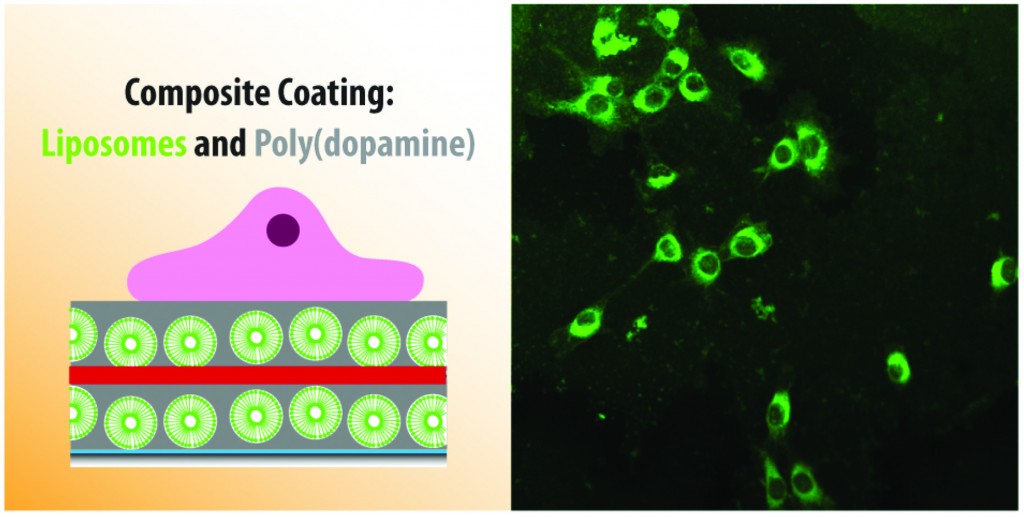
Most of us are accustomed to thinking of medicinal drugs as something we ingest or inject into our bodies.  However, if the drug can be introduced at the particular part of the body where it is needed the most, it will be more effective. Further, if we can slow down the release of the drug, we may be able to use less drugs for the same purpose.  With these long-term goals in mind, Brigitte Städler’s group has recently published a paper where they investigate the release of drugs from a biological coating.  This work can one day be used to modify the surface of implants in our body (commonly used examples include bone implants or pacemakers).  Modifying the surface of these implants would allow us to use them as drug depots in our body.  These implants can then release drugs more slowly into the bloodstream as opposed to all at once, thus making the drugs more effective.

The researchers used a structure known as a liposome – particles made out of fat that can potentially trap a drug in their centers – and surrounded them with a material that can be used to coat implant surfaces. They found that muscle cells could grow on these surfaces without dying, thus proving that the surfaces are not toxic. The liposomes were made out of a fat that fluoresces (emits light under certain conditions).  The muscle cells could absorb these fluorescent fats, which made the cells themselves fluorescent and allowed the cells to be tracked. Next, the researchers trapped a toxic drug in the liposomes, and found that the presence of the drug meant that they could control whether the cells survived or died.  While any kind of drug could in theory be trapped in the coating, a toxic drug could specifically be useful if the coating was near harmful or cancerous cells.

This work shows a great deal of promise in improving the way we deliver drugs to patients.

Cargo delivery to adhering myoblast cells from liposome-containing poly(dopamine) composite coatings
Martin E. Lynge, Boon M. Teo, Marie Baekgaard Laursen, Yan Zhang and Brigitte Städler 
Biomater. Sci., 2013, Advance Article DOI: 10.1039/C3BM60107B

Debanti Sengupta recently completed her PhD in Chemistry from Stanford University.  She is currently a Siebel postdoctoral scholar at the University of California, Berkeley.  

Follow the latest journal news on Twitter @BioMaterSci or go to our Facebook page.

To keep up-to-date with all the latest research, sign-up to our RSS feed or Table of contents alert.

Digg This
Reddit This
Stumble Now!
Share on Facebook
Bookmark this on Delicious
Share on LinkedIn
Bookmark this on Technorati
Post on Twitter
Google Buzz (aka. Google Reader)

Nanofibrous/microfibrous biodegradable scaffolds for the regeneration and segregation of multiple tissue types

The advent of regenerative medicine has begun to expand the treatment options available to patients who require tissue reconstruction due to birth defects or injuries.  However, it still offers limited solutions for those patients whose wounds span multiple layers and types of tissues.  This is in part due to the shortcomings of the barrier membranes that are typically implanted in order to maintain tissue layer separation.  In this article, researchers from the University of Sheffield propose novel biodegradable bilayer and trilayer fibrous scaffolds that can effectively segregate tissue layers and maintain proper tissue structure during healing.

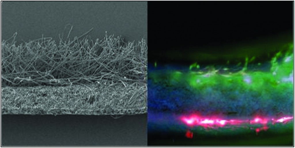
Electrospinning was used to create monolayers of microfibrous polylactic acid (PLA), microfibrous poly ε-caprolactone (PCL), and nanofibrous polyhydroxy-butyrate-co-hydroxyvalerate (PHBV).  Initially, fluorescently labeled fibroblasts and mesenchymal progenitor cells (hESMP) were co-cultured on separate sides of each of the monolayer scaffolds.  After seven days, cell mixing was observed on both the PLA and PCL scaffolds.  However, cell segregation was still clearly evident on the PHBV scaffold, indicating that its nanofibrous structure was acting as a barrier to cell penetration. 

The researchers then created bilayer membranes of PHBV-PLA and PHBV-PCL that exhibited similar barrier properties and maintained high levels of cell viability.   These bilayer scaffolds were designed such that the different degradation rates of the composite polymers were comparable to the different growth rates of hard and soft tissue, thus making them better candidates for the treatment of conditions, such as cleft palate, which require bone and soft tissue segregation.  Finally, a PLA-PHBV-PLA trilayer membrane was designed with the goal of supporting two different types of soft tissue growth.  Subsequent results confirmed that the nanofibrous PHBV layer effectively separated the fibroblasts and keratinocytes that were cultured in the two PLA layers.

Ultimately the researchers were able to demonstrate that nanofibrous scaffolds are capable of promoting cell viability while maintaining separation between different cell types.  The methodology used is simple and reproducible and the resulting scaffold is biocompatible and biodegradable.

Development of bilayer and trilayer nanofibrous/microfibrous scaffolds for regenerative medicine
Frazer J. Bye, Julio Bissoli, Leanne Black, Anthony J. Bullock, Sasima Puwanun, Keyvan Moharamzadeh, Gwendolen C. Reilly, Anthony J. Ryan and Sheila MacNeil 
Biomater. Sci., 2013, 1, 942-951 DOI: 10.1039/C3BM60074B

Ellen Tworkoski is a guest web-writer for Biomaterials Science.  She is currently a second year Ph.D. student in the biomedical engineering department at Northwestern University (Evanston, IL, USA).

Follow the latest journal news on Twitter @BioMaterSci or go to our Facebook page.

To keep up-to-date with all the latest research, sign-up to our RSS feed or Table of contents alert.

Digg This
Reddit This
Stumble Now!
Share on Facebook
Bookmark this on Delicious
Share on LinkedIn
Bookmark this on Technorati
Post on Twitter
Google Buzz (aka. Google Reader)