Archive for the ‘Hot Article’ Category

New insights in to PDT induced necrotic cell death in glioblastoma

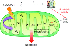
5-ALA-PDT induces RIP3-dependent necrosis in glioblastomaNew insights in to the mechanism of action of 5-aminolevulinic acid (5-ALA) based photodynamic therapy (PDT) against glioblastoma reveal the involvement of receptor-interacting protein 3 (RIP3) in necrosis induction.

Glioblastoma are one of the most frequent and deadly brain tumours in human adults; prognosis with this type of cancer is very poor but 5-ALA-PDT has been emerging as a promising treatment.  In this treatment 5-ALA is metabolized into the protoporphyrin IX (PPIX) photosensitizer inside mitochondria and PPIX preferentially accumulates into cancer cells due to their ferrochelatase deficiency.

This study from Jacques Piette and co-workers in Belgium is a result of the observation that 5-ALA-PDT treatment of glioblastoma leads to cell death by necrosis rather than apoptosis.  It has become clear that cell death from necrosis is actually a programmed event rather than the accidental cell death it was long thought to be; this has coined the term necroptosis.  Recent studies into the mechanism of necroptosis have revealed that a key effector is RIP3, a Ser/Thr kinase belonging to the RIP kinase family. 

Jacques Piette and co-workers show that 5-ALA-PDT-induced necrosis in glioblastoma cells was dependent on RIP3 and that PDT-mediated singlet oxygen production is the cause of the RIP3-dependent necrotic pathway activation. 

Further details are presented in their recently published Photochemical and Photobiological Sciences article which is free to access until 30th of November.

5-ALA-PDT induces RIP3-dependent necrosis in glioblastoma
Isabelle Coupienne, Grégory Fettweis, Noemí Rubio, Patrizia Agostinis and Jacques Piette
Photochem. Photobiol. Sci., 2011, DOI: 10.1039/C1PP05213F

Digg This
Reddit This
Stumble Now!
Share on Facebook
Bookmark this on Delicious
Share on LinkedIn
Bookmark this on Technorati
Post on Twitter
Google Buzz (aka. Google Reader)

Iron, oxidative stress and solar ultraviolet A radiation

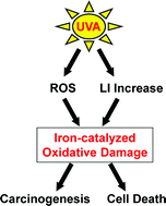
C1PP05204GA recently published perspective from Charareh Pourzand, Rex Tyrrell, co-editor-in-chief of PPS, and co-workers at the University of Bath gives an overview of recent findings on intracellular labile iron (LI) distribution and trafficking.  Using UVA radiation as an example, it also discusses mechanisms and consequences of oxidant-induced alterations in the intracellular pool of LI.  This perspective forms part of an upcoming themed issue on UVA.

Iron is a vital nutrient and is required for a wide variety of cellular processes due to its facile redox chemistry and affinity to oxygen.  However, when the redox-active chelatable ‘labile’ form exceeds the normal binding capacity of the cell this labile iron (LI) can become toxic.  In the presence of reactive oxygen species LI can catalyse the formation of oxygen-derived free radicals that overwhelm the cell’s antioxidant defence mechanism and result in cell damage.  Although intracellular iron homeostasis is tightly regulated acute exposure of skin cells to UVA induces an increase in intracellular LI and this appears to play a key role in the increased susceptibility of skin cells to UVA-mediated oxidative membrane damage and necrotic cell death.

To find out more follow the link below.  Full text is FREE until November 11th.

Iron, oxidative stress and the example of solar ultraviolet A radiation
Asma Aroun, Julia Li Zhong, Rex M. Tyrrell and Charareh Pourzand
Photochem. Photobiol. Sci., 2012, DOI: 10.1039/C1PP05204G

Digg This
Reddit This
Stumble Now!
Share on Facebook
Bookmark this on Delicious
Share on LinkedIn
Bookmark this on Technorati
Post on Twitter
Google Buzz (aka. Google Reader)

Investigations into the gender bias in skin cancer development reveal male unresponsiveness to UVA radiation

C1PP05224ANew insights into mechanisms behind gender differences in skin cancer development are revealed in this article from Vivienne Reeve and co-workers from Sydney, Australia.  A contributing factor for skin cancer development in mice and humans is immunosuppression as a result of UVB light; whilst the contribution of UVA is controversial it has been shown to antagonise immunosuppression by UVB.  One of the mechanisms for this UVA protection which has been demonstrated in mice is mediated via oestrogen receptor-β signalling, suggesting a gender bias in photoimmune response to UVA.

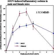
Gender bias is tested in this study by comparing the UV-induced inflammatory and immune responses in male and female hairless mice. The study uses solar simulated radiation (SSUV) to induce sunburn, the resulting inflammatory oedema is measured through the increase in irradiated skinfold thickness and levels of the pro-inflammatory cytokine interleukin (IL)-6 in the epidermis.  Immune function was assessed by the contact hypersensitivity (CHS) reaction and its suppression by SSUV was related to the level of expression of the immunosuppressive cytokine IL-10 in the epidermis.

Results showed that although male mice had less intense sunburn with lower expression of IL-6 the CHS reaction was more severely suppressed by SSUV than females and this also correlated with increased epidermal expression of immunosuppressive IL-10. Exposure to UVB alone had equal effects in male and female mice, however, UVA irradiation induced immunoprotection in females but this effect was reduced or not present in males.  The results indicate that males have a relative unresponsiveness to the photoimmune protective effects of UVA.  This could explain the known gender bias in skin cancer development in both mice and humans.

Interested in knowing more?  Read the full article here.  Free until 4th November.

Gender differences in UV-induced inflammation and immunosuppression in mice reveal male unresponsiveness to UVA radiation
Vivienne E. Reeve, Munif Allanson, Diane Domanski and Nicole Painter
Photochem. Photobiol. Sci., 2011, DOI: 10.1039/C1PP05224A

Digg This
Reddit This
Stumble Now!
Share on Facebook
Bookmark this on Delicious
Share on LinkedIn
Bookmark this on Technorati
Post on Twitter
Google Buzz (aka. Google Reader)

Photoexcitation of crystalline trityl-alkyl ketones

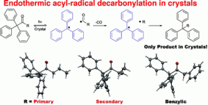
C1PP05240C In this recently published communication John R. Scheffer and Miguel A. Garcia-Garibay and their teams from the University of British Columbia and the University of California Los Angeles demonstrate that the solid state photoexcitation of several triphenylmethylalkyl ketones results in the loss of CO and the exclusive formation of radical–radical combination products. Furthermore, differences in reactivity suggest a stepwise mechanism with the unprecedented formation of primary and secondary radicals in some of the radical pair intermediates in the solid state.

Interested in knowing more? Read the full article here. Free until October 21st!

Stable radicals during photodecarbonylations of trityl-alkyl ketones enable solid state reactions through primary and secondary radical centers
Gregory Kuzmanich, Arunkumar Natarajan, Yanhui Shi, Brian O. Patrick, John R. Scheffer and Miguel A. Garcia-Garibay
Photochem. Photobiol. Sci., 2011, DOI: 10.1039/C1PP05240C

Digg This
Reddit This
Stumble Now!
Share on Facebook
Bookmark this on Delicious
Share on LinkedIn
Bookmark this on Technorati
Post on Twitter
Google Buzz (aka. Google Reader)

Tanning salons and skin cancer

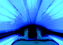
Tanning salons and skin cancerIn a recently published perspective on the use of sunbeds Jean-François Doré and Marie-Christine Chignol discuss prevalence of their use and examine the evidence suggesting that use of indoor tanning increases the risk of skin Cancer.  The authors conclude that the use of sunbeds should be strongly discouraged and banned under the age of 18. To find out more, read the full article for free until September 16th!

Tanning salons and skin cancer
Jean-François Doré and Marie-Christine Chignol
Photochem. Photobiol. Sci., 2011, Advance Article
DOI: 10.1039/C1PP05186E, Perspective

This article is part of an upcoming Themed Issue on UVA radiation with Guest Editors Rex Tyrell (Bath, UK), Vivienne Reeve (Sndney, Australia) and Evelyne Sage (Orsay, France).

Digg This
Reddit This
Stumble Now!
Share on Facebook
Bookmark this on Delicious
Share on LinkedIn
Bookmark this on Technorati
Post on Twitter
Google Buzz (aka. Google Reader)

UV wavelength-dependent DNA damage and skin cancer

In this perspective, Gerd P. Pfeifer and Ahmad Besaratinia discuss the influence of wavelength on the nature of the DNA damage and the mutagenic properties of solar UV radiation.  It forms part of an upcoming themed issue on the biology of UVA.

UV wavelength-dependent DNA damage and human non-melanoma and melanoma skin cancer

Skin Cancer is currently the most common tumour diagnosed in many countries and the numbers of cases have increased dramatically over the last few decades.  Skin cancers are one of the few examples of human cancer where the causative agent is known with high certainty as the epidemiological evidences clearly indicates that solar UV radiation is associated with skin cancer.  This means it can act as a useful system to dissect various steps of carcinogenesis, allowing initial events at the DNA level and specific genes that are frequently mutated to be identified.

Read the full article for free until 2nd September.

UV wavelength-dependent DNA damage and human non-melanoma and melanoma skin cancer
Gerd P. Pfeifer and Ahmad Besaratinia
Photochem. Photobiol. Sci., 2011, DOI: 10.1039/C1PP05144J

Digg This
Reddit This
Stumble Now!
Share on Facebook
Bookmark this on Delicious
Share on LinkedIn
Bookmark this on Technorati
Post on Twitter
Google Buzz (aka. Google Reader)

Two pronged attack on antibiotic resistant bacteria with photodynamic antimicrobial chemotherapy and antimicrobial peptides

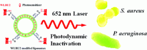
A novel bacteria-targeting liposomal formulation has been developed by a team in Germany which improves photodynamic antimicrobial chemotherapy (PACT) by using the bacteria-targeting ability of antimicrobial peptides (AMP).

In PACT laser light is used to trigger a photosensitizer (PS) to an activated state, this results in the transfer of energy to surrounding oxygen leading to generation of reactive oxygen species (ROS).  These ROSs oxidise biological molecules resulting in cytotoxicity.  PACT has attracted significant attention in the last decades as a potential strategy to treat bacterial infections, especially antibiotic resistant species due to a broad spectrum of action and a belief that bacteria will not easily develop resistance to ROSs, furthermore PACT is more phototoxic to bacteria than mammalian cells.

Antimicrobial peptide-modified liposomes for bacteria targeted delivery of temoporfin in photodynamic antimicrobial chemotherapy Tempoforin is one of the most potent PSs but it cannot be administered alone as it is highly hydrophobic.  A suitable delivery system is necessary and liposomes are an attractive choice and have been utilised before, however, when liposomes containing tempoforin are applied to an infection site they are also applied to cells nearby resulting in side effects.  To avoid this is it necessary to target the liposomes to bacteria.

A promising source of bacteria-targeting ligands may come from AMPs which are cationic amphipathic peptides found ubiquitously in most classes of life forms where they are part of the innate immune system.  They are able to interact with and penetrate negatively charged lipids in bacterial membranes forming membrane pores resulting in damage.  Their mode of action, like PACT, results in a broad range of action and low susceptibility to antibiotic resistance.

Alfred Fahr and co-workers used a synthetic AMP called WLBU2 which has broad antimicrobial activity and also shows specific and effective binding to various microbes.  By formulating tempoforin-loaded liposomes conjugated on their surface with WLBU2 they were able to improve PACT efficiency on both gram-positive and gram-negative bacteria.

To find out more read the full article.  Free to read until August 19th.

Antimicrobial peptide-modified liposomes for bacteria targeted delivery of temoporfin in photodynamic antimicrobial chemotherapy
Kewei Yang, Burkhard Gitter, Ronny Rüger, Gerhard D. Wieland, Ming Chen, Xiangli Liu, Volker Albrecht and Alfred Fahr
Photochem. Photobiol. Sci., 2011, DOI: 10.1039/C1PP05100H

Digg This
Reddit This
Stumble Now!
Share on Facebook
Bookmark this on Delicious
Share on LinkedIn
Bookmark this on Technorati
Post on Twitter
Google Buzz (aka. Google Reader)

How the sun can affect your immunity

Mary Norval discusses with Anna Simpson how ultraviolet (UV) radiation can suppress immune responses and the consequences of this for our health. Mary Norval and Gregory Woods have recently published a Perspective on ‘UV-induced immunosuppression and the efficacy of vaccination’

Mary Norval

Mary Norval is Professor Emeritus at the University of Edinburgh in Scotland. Her research interests are based on the effects of UV radiation on human health, especially immunological aspects.

How did you become interested in the effects of UV on immunosuppression? 

I trained as a virologist in the 1970s and became interested in skin infections, including cold sores, caused by the herpes simplex virus (HSV). HSV remains latent in the body following the first infection but can be reactivated, resulting in the re-emergence of skin lesions. Exposure to the sun is a common trigger for the reactivation. I was intrigued by why and how this should happen; the experiments took me into what was then a new area, subsequently called photoimmunology, and converted me from a virologist into an immunologist in the process. 

So how does UV radiation cause immunosuppression? 

It is difficult to provide a simple answer to this question as several multistep pathways are possible, depending on factors such as the antigen (a molecule that stimulates an immune response when introduced into the body) in question and the quantity and spectrum of the UV radiation. In essence, there are several UV absorbing compounds in the skin that initiate the process. In particular, DNA forms cyclobutane pyrimidine dimers and urocanic acid, a compound present in the skin, undergoes a trans to cis double bond isomerisation. As a result of these structural changes, a wide range of immune mediators (molecules released by immune cells in response to invasion by the antigen) is produced. These have effects on several cell populations, inducing some to migrate from the skin to the draining lymph nodes. There they undergo an abnormal interaction with immune effector cells (cells that cause antigen breakdown), leading to the generation of regulatory cells that mediate suppression of the immune response. The time between the UV exposure and antigen administration needs to be less than a few days for the immunosuppression to occur and, once generated, the regulatory cells are life-long. 

What are the implications of this for vaccinations and why is it such an important topic? 

In several mouse models, UV irradiation before or after vaccination was demonstrated to suppress the animals’ ability to respond immunologically. In humans, there is evidence that exposure to UV radiation or the sun can suppress certain immune responses. Thus UV exposure may have the potential to lower the efficacy of human vaccination. It is important to find out whether this is the case or not as vaccination represents a major public health strategy throughout the world to control an increasing range of common infections, particularly in childhood. 

It seems that this potentially serious issue has been overlooked. Why do you think this is? 

It is true that very few attempts have been made to investigate this area. This could be due to ignorance regarding the potential of sun exposure to suppress immunity or due to the multidisciplinary nature, protracted time and expense of such research. Additionally, it is obviously not ethical to artificially irradiate individuals, especially children, before vaccination or to infect them with live microorganisms to test the efficacy of vaccination. This means that an epidemiological approach may provide the most information, for example to assess the vaccine-specific immune responses in people vaccinated in the winter months compared with the summer months. 

Your research isn’t all doom and gloom for sun lovers as you have previously published on the benefits of UV radiation due to its role in vitamin D formation. What do you feel is the best way to strike a balance? 

Most of the vitamin D in the majority of people is produced by exposure to the UV radiation in sunlight. As vitamin D has many health benefits, it is important to ensure sufficient sun exposure but without increasing the risk of developing skin cancer. A simple message that fits everyone under all circumstances is not possible as there are many variables including skin colour, body fat and age as well as latitude of residence, weather conditions, season of the year and time of day. Various models have been developed in an attempt to provide some guidance and recently, measurements of vitamin D levels following real sun exposure have been published. These indicate that casual midday summer sun exposure should be enough to provide an adequate vitamin D level in most individuals who have their face and arms uncovered. Sunburn should be avoided as it is considered the major environmental risk factor for all forms of skin cancer. Protective measures should be taken when in areas of high sun intensity, such as by seeking shade, covering up and using sunscreens. 

What scientific achievement are you most proud of? 

I am most proud of the collaborations I started to investigate the role of urocanic acid in immunosuppression. In addition, I demonstrated that UV radiation could downregulate immune responses in HSV infections, the first time that UV had been shown to affect microbial immunity. 

If you weren’t a scientist what would you be? 

Possibly I might have become a professional golfer, although when I was a student, there was no professional circuit for women. 

Read Mary and Gregory’s Perspective by following the link below:

UV-induced immunosuppression and the efficacy of vaccination
Mary Norval and Gregory M. Woods
Photochem. Photobiol. Sci., 2011, DOI: 10.1039/C1PP05105A
Digg This
Reddit This
Stumble Now!
Share on Facebook
Bookmark this on Delicious
Share on LinkedIn
Bookmark this on Technorati
Post on Twitter
Google Buzz (aka. Google Reader)

Understanding heme biosynthesis regulation for 5-aminolevulinic acid-based photodynamic therapy

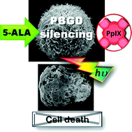
The centrality of PBGD expression levels on ALA-PDT efficacyA team from Bar-Ilan University, Israel, have revealed the importance of porphobilinogen deaminase (PBGD) expression levels on the efficacy of 5-aminolevulinic acid-based photodynamic therapy (ALA-PDT)

ALA-PDT is a method used worldwide for the treatment of various malignancies.  It utilises the heme synthesis pathway to accelerate the synthesis of heme precursors, porphyrins.  Light irradiation of the tumour excites the prophyrin molecules and induces the formation of reactive oxygen species resulting in cell death.  The mechanisms of porphyrin synthesis regulation are not fully understood, but this study provides a big advance.

The team investigated the effect of expression levels of two main enzymes in heme biosynthesis, ALA dehydratase (ALAD) and PBGD on the capacity of K562 cells to undergo cell death following ALA-PDT.  They discovered a novel regulation feedback of PBGD on ALAD activity which is fundamental for the regulation of the heme synthesis pathway.

Interested in knowing more?  Read the full article for free until 13th July.

The centrality of PBGD expression levels on ALA-PDT efficacy
Avital Schauder, Tamar Feuerstein and Zvi Malik
Photochem. Photobiol. Sci., 2011, Advance Article
DOI: 10.1039/C1PP05085K, Paper

Digg This
Reddit This
Stumble Now!
Share on Facebook
Bookmark this on Delicious
Share on LinkedIn
Bookmark this on Technorati
Post on Twitter
Google Buzz (aka. Google Reader)

Hunting elusive green fluorescent proteins

After a 40 year hunt, scientists have tracked down the genes responsible for fluorescent proteins in Obelia medusa – a type of jellyfish. Knowledge of these genes could lead to new fluorescent protein tags for use in cell biology. 

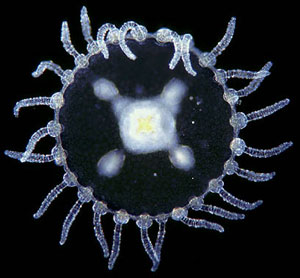
Obelia medusa jellyfish

Three different fluorescent protein colours - green, cyan and yellow - were discovered in the Obelia medusa jellyfish

The discovery of the gene encoding green fluorescent protein (GFP) in the jellyfish Aequorea victoria paved the way for GFP to be introduced as a fluorescent tag in cell biology. It is used to track the positions and interactions of proteins in cells, and led to the Nobel prize in chemistry in 2008. Fluorescent proteins similar to GFP were discovered in Obelia medusa during biochemical studies in the 1970s; however, despite repeated attempts, nobody was able to clone the gene responsible for making these proteins. 

Now, Mikhail Matz and his team at the University of Texas at Austin, US, have solved the problem by taking a different approach. Obelia has two main stages of life, a polyp attached to a surface and a free swimming jellyfish. Previous attempts to identify the genes responsible for the fluorescent proteins focused on the polyp. ‘The trick this time was to use the jellyfish as the starting point,’ explains Matz. To their surprise, the team discovered not one, but three different fluorescent protein colours: green, cyan and yellow. This is the first time that three different colours of fluorescent protein have been found in an organism outside of the Anthozoa (a class of organisms that includes sea anemones and corals). 

Matz and the team also found a variety of fluorescent colours and proteins in non-bioluminescent parts of the jellyfish. This is a point that Marc Zimmer, an expert in fluorescent proteins at Connecticut College, US, says is important. ‘I am always amazed that we still don’t know the function of GFP-like proteins. This work is particularly interesting since it reports on green, cyan and yellow FPs cloned from Obelia, where they are located in both bioluminescent and non-bioluminescent cells, confirming prior suggestions that their function may be biochemical and does not always involve fluorescence.’ 

Knowledge of the gene encoding GFP allowed it to be introduced as a fluorescent reporter in a wide variety of cell biology experiments. Now that the equivalent genes have been discovered in Obelia,  scientists will be able to discover whether these GFP homologs could make similarly useful tools. 

Taken from a Chemistry world story written by Russell Johnson on the 26th May 2011   

Read more about this research here:
Multi-colored homologs of the green fluorescent protein from hydromedusa Obelia sp.
Galina V. Aglyamova, Marguerite E. Hunt, Chintan K. Modi and Mikhail V. Matz, Photochem. Photobiol. Sci., 2011
DOI: 10.1039/c1pp05068k

Digg This
Reddit This
Stumble Now!
Share on Facebook
Bookmark this on Delicious
Share on LinkedIn
Bookmark this on Technorati
Post on Twitter
Google Buzz (aka. Google Reader)