Archive for the ‘Nanoscale’ Category

Siligraphene gets serious about solar cells: Nanoscale article featured in Chemistry World

Theoretical scientists have predicted SiC7 – a new phase of the graphene–silicene hybrid siligraphene, which if synthesisable, could revolutionise flexible optoelectronic devices such as solar cells.

A charge density map reveals delocalised π bonds in SiC7 siligraphene's irregular hexagonal rings

SiC7 siligraphene has an interesting structure with a graphene-like honeycomb lattice, but unlike graphene, its hexagonal rings are irregular. The scientists expect SiC7 siligraphene to be much better than SiC2 siligraphene and single-layer black phosphorus at absorbing sunlight. Additionally, broken symmetry caused by silicon doping would create band gaps that boost its optoelectronic properties.

Interested to know more? Read the full article by Suzanne Howson in Chemistry World.

The original article is free to access and can be read at:

SiC7 siligraphene: a novel donor material with extraordinary sunlight absorption
Huilong Dong, Liujiang Zhou, Thomas Frauenheim, Tingjun Hou, Shuit-Tong Lee and Youyong Li
Nanoscale, 2016,8, 6994-6999.

Digg This
Reddit This
Stumble Now!
Share on Facebook
Bookmark this on Delicious
Share on LinkedIn
Bookmark this on Technorati
Post on Twitter
Google Buzz (aka. Google Reader)

Go with the (lateral) flow

Lateral flow assays (LFA) have become a promising tool in the diagnostics market in recent years due to their low cost, ease of use and short assay times. The simplicity of their use extends to the inclusion of, typically, gold nanoparticle labels that provide a visual confirmation, due to their distinctive red colour, of a particular target analyte. However, limitations, such as quantitative analysis and assay sensitivity, can hinder their broader use in diagnostics.

To overcome these limitations, researchers from South Korea have utilised surface enhanced Raman scattering (SERS) to detect staphylococcal enterotoxin B (SEB) on a LFA strip. The authors have opted to use Raman reporter-labelled hollow gold nanospheres (HGNs) instead of the conventional gold nanoparticle labels. Using HGNs, the authors were able to detect as low as 0.001 ng/mL of SEB, which is about three orders of magnitude more sensitive than the conventional ELISA-based method for SEB.

These researchers demonstrates that SERS can be utilised to overcome the limitations of conventional LFAs in order to achieve greater assay sensitivity without deviating from the original LFA design.

Application of a SERS-based lateral flow immunoassay strip for the rapid and sensitive detection of staphylococcal enterotoxin B
Joonki Hwang, Sangyeop Lee and Jaebum Choo
Nanoscale, 2016, DOI: 10.1039/C5NR07243C

Dr Lee Barrett is a guest web writer for the Nanoscale blog. Lee is currently a postdoctoral researcher in the Centre for Molecular Nanometrology at the University of Strathclyde. His research is currently focused on the development of nanoparticle-based sensors and surface enhanced Raman scattering (SERS). Follow him on twitter @L_Bargie

Digg This
Reddit This
Stumble Now!
Share on Facebook
Bookmark this on Delicious
Share on LinkedIn
Bookmark this on Technorati
Post on Twitter
Google Buzz (aka. Google Reader)

Emerging topic: Two-dimensional materials

Two-dimensional materials research is an emerging topic area, in particular their application in electronic devices. To help our readers stay up to date we have collected papers published in Nanoscale into one online web collection entitled 2D materials for electronic devices.

Editor’s Choice selection from Associate Editor, Professor Andrea Ferarri

A highly conducting graphene film with dual-side molecular n-doping
Youngsoo Kim, Jaesung Park, Junmo Kang, Je Min Yoo, Kyoungjun Choi, Eun Sun Kim, Jae-Boong Choi, Chanyong Hwang, K. S. Novoselov and Byung Hee Hong
Nanoscale, 2014,6, 9545-9549

“Graphene has many record properties. It is transparent like (or better than) plastic, but conducts heat and electricity better than any metal, it is an elastic film, behaves as an impermeable membrane, and it is chemically inert and stable. Thus it seems ideal as the next generation transparent conductor. There is a need to find a substitute for indium tin oxide (ITO) in the manufacturing of various types of displays and touch screens, due to the brittleness of indium that makes it difficult to use them when flexibility is a requirement. Graphene is an ideal candidate for such a task. Thus, coupled with carbon’s abundance, this presents a more sustainable alternative to ITO. Prototypes of graphene-based displays have been produced and commercial products seem imminent.

One drawback is that, in order to beat ITO’s conductivity, graphene needs to be doped. Kim et al. report a dual-side molecular doping method, demonstrating effective work function modulation, high carrier density and significant reduction of sheet resistance in large area graphene samples grown by chemical vapour deposition. With further optimization, their approach may enable a variety of practical applications of graphene films requiring low sheet resistance comparable to indium tin oxide (ITO), as well as high transparency and flexibility.”

Here are some popular articles from the collection:

Graphene-analogue carbon nitride: novel exfoliation synthesis and its application in photocatalysis and photoelectrochemical selective detection of trace amount of Cu2+
Hui Xu, Jia Yan, Xiaojie She, Li Xu, Jiexiang Xia, Yuanguo Xu, Yanhua Song, Liying Huang and Huaming Li
Nanoscale, 2014, 6, 1406-1415

Electrochemical properties of CVD grown pristine graphene: monolayer- vs. quasi-graphene
Dale A. C. Brownson, Sarah A. Varey, Fiazal Hussain, Sarah J. Haigh and Craig E. Banks
Nanoscale, 2014,6, 1607-1621

Transparent conductors composed of nanomaterials
Michael Layani, Alexander Kamyshny and Shlomo Magdassi
Nanoscale, 2014, 6, 5581-5591

Access the full collection here!

Digg This
Reddit This
Stumble Now!
Share on Facebook
Bookmark this on Delicious
Share on LinkedIn
Bookmark this on Technorati
Post on Twitter
Google Buzz (aka. Google Reader)

HOT article: Ionizing radiation improves glioma-specific targeting of superparamagnetic iron oxide nanoparticles conjugated with cmHsp70.1 monoclonal antibodies (SPION–cmHsp70.1)

In this HOT article, tumour-specific targeting using superparamagnetic iron oxide nanoparticles (SPIONs) combined with ionising radiation is investigated. Heat shock protein Hsp70 is known to be expressed by cells exposed to stressful conditions or specifically on the membrane of highly aggressive tumour cells.  SPIONs, which are negative contrast agents for magnetic resonance imaging (MRI), were synthesised to contain the Hsp70 specific antibody (cmHsp70.1) to increase uptake into various tumour cells.

Conjugation of the cmHsp70.1 was accomplished by introducing amine groups to the dextran polymer coating of SPIONs, allowing covalent attachment to the carboxylic acid functional groups of the antibody through carbodiimide coupling. This was shown to dramatically increase loading of iron oxide into various tumour cells, compared to unconjugated SPIONs. By exposing cells to a non-lethal dose of ionising radiation, additional uptake could be achieved as cells express more of the heat shock mHsp70.

The differences in uptake can be seen in the figure below in both in vivo and in vitro environments using MRI and fluorescence microscopy, respectively. This approach has the potential to be clinically relevant for both diagnosis and therapy of tumours.

Figure 1 Targeting of the orthotopic C6 glioma by SPION–cmHsp70.1 conjugates. (A) Magnetic resonance images for the control, non-irradiated animals treated with SPIONs, SPION–cmHsp70.1 particles and irradiated rats (10 Gy) treated with SPION–cmHsp70.1. Images were obtained in RARE-T1, TurboRARE-T2 and FLASH regimens. Retention of the nanoparticles in the tumor presented as hypotensive zones on T2-weighted and gradient echo images (red arrows). (B) Immunofluorescent images of the brain tumor stained with anti-Hsp70 antibodies (green). Nuclei stained with DAPI (blue). Nanoparticles detected using reflective laser scanning at 488 nm (red). Scale bar, 40 μm.

Ionizing radiation improves glioma-specific targeting of superparamagnetic iron oxide nanoparticles conjugated with cmHsp70.1 monoclonal antibodies (SPION–cmHsp70.1)
Maxim A. Shevtsov, Boris P. Nikolaev, Vyacheslav A. Ryzhov, Ludmila Y. Yakovleva, Yaroslav Y. Marchenko, Marina A. Parr, Valerij I. Rolich, Anastasiya L. Mikhrina, Anatolii V. Dobrodumov, Emil Pitkin and Gabriele Multhoff
Nanoscale, 2015,7, 20652-20664, DOI: 10.1039/C5NR06521F, Paper

Dr Mike Barrow is a guest web writer for the Nanoscale blog, he currently works as a Postdoctoral Researcher at the University of Liverpool. Twitter: @mikesyb

Digg This
Reddit This
Stumble Now!
Share on Facebook
Bookmark this on Delicious
Share on LinkedIn
Bookmark this on Technorati
Post on Twitter
Google Buzz (aka. Google Reader)

HOT article: Responsive polymer brushes for controlled nanoparticle exposure

In this article, Scheutjens-Fleer self-consistent field (SF-SCF) theory simulations were used to accurately predict the position of a nanoparticle attached to the end-group of a responsive polymer chain surrounded by a majority of non-responsive polymer chains in a mixed brush system.

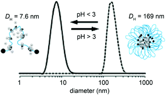
Certain stimuli such as pH or temperature can be used to create a defined ‘on-off’ switch between a protected ‘off’ state, where the responsive polymer chain is contracted thus burying the nanoparticles well within the non-responsive components, or an ‘on’ state where the nanoparticle is exposed to the medium through swelling of the responsive polymer chains.

The solvent quality was parameterised by the Flory-Huggins interaction parameter (χ) and for nearly all investigated systems there was a sharp transition at the so-called critical χ value between the on-off state.   Design variables for mixed polymer brushes such as grafting density, chain length and nanoparticle size were investigated and all had an effect on the critical χ value, with a larger particle size and grafting density leading to an increase in χ. Fixing the polymer chain length for both responsive and non-responsive polymer chains led to the most optimal switching.

The authors are planning to develop materials from these findings and use the mixed polymer brush-nanoparticle systems as rapidly responsive (bio)sensors with single molecule sensitivity.

Responsive polymer brushes for controlled nanoparticle exposure
Namik Akkilic, Frans A. M. Leermakers and Wiebe M. de Vos
Nanoscale, 2015,7, 17871-17878, DOI: 10.1039/C5NR05150A

Dr Mike Barrow is a guest web writer for the Nanoscale blog, he currently works as a Postdoctoral Researcher at the University of Liverpool. Twitter: @mikesyb

Digg This
Reddit This
Stumble Now!
Share on Facebook
Bookmark this on Delicious
Share on LinkedIn
Bookmark this on Technorati
Post on Twitter
Google Buzz (aka. Google Reader)

Highlights from ChinaNANO

The Nanoscale Editorial Office is delighted at the success of the Nanoscience Symposium which occurred at the ChinaNANO 2015 conference in Beijing from 3rd-5th September. This one-day symposium included talks from the Nanoscale Associate Editors about their latest research covering topics such as carbon nanomaterials, nanoelectronics, bionanomaterials, nanophotonics, nanoclusters and nanocrystals.

Some articles submitted to Nanoscale from delegates of the ChinaNANO 2015 conference can be found below.

Surface coating of siRNA-peptidomimetic nano-self-assemblies with anionic lipid bilayers: Enhanced gene silencing and reduced adverse effects in vitro
Xianghui Zeng, Anne Marit de Groot, Alice Sijts, Femke Broere, Erik Oude Blenke, Stefano Colombo, Willem van Eden, Henrik Franzyk, Hanne M Nielsen and Camilla Foged
Nanoscale, 2015, DOI: 10.1039/C5NR04807A

Strong metal-support interaction in novel core-shell Au-CeO2 nanostructures induced by different pretreatment atmospheres and its influence on CO oxidation
Zhihua Wang, Huifen Fu, Ziwei Tian, Dongmei Han and Fubo Gu
Nanoscale, 2015, DOI: 10.1039/C5NR06929G

Fabrication of ultra-thin silicon nanowire arrays using ion beam assisted chemical etching
Zhiyuan Tan, Wenjia Shi, Chungang Guo, Quan Zhang, Liang Yang, Xiaoling Wu, Guo-an Cheng and Ruiting Zheng
Nanoscale, 2015,7, 17268-17273, DOI: 10.1039/C5NR02876K

Ethanol-assisted gel chromatography for single-chirality separation of carbon nanotubes
Xiang Zeng, Jinwen Hu, Xiao Zhang, Naigen Zhou, Weiya Zhou, Huaping Liu and Sishen Xie
Nanoscale, 2015,7, 16273-16281, DOI: 10.1039/C5NR04116C

There was a great turn-out and we received a lot of useful feedback from the attendees.

A prominent highlight of the event was the reception organised by the Royal Society of Chemistry to celebrate the launch of Nanoscale Horizons as well as the Nanoscale and Nanoscale Horizons partnership with the National Center for Nanoscience and Technology (NCNST) in Beijing which included cutting an enormous cake!

Left to right: Xiaodong Chen, Fiona McKenzie, Chunli Bai and Xingyu Jiang

Digg This
Reddit This
Stumble Now!
Share on Facebook
Bookmark this on Delicious
Share on LinkedIn
Bookmark this on Technorati
Post on Twitter
Google Buzz (aka. Google Reader)

Micro meets nano: Multicolour gold nanoprobes detect micoRNA biomarkers in serum

Researchers from Chinese Academy of Sciences have investigated the use of molecular beacons (MBs) for the simultaneous detection of multiple microRNA (miRNA) biomarkers.

The MBs were immobilized onto gold nanoparticles (AuNPs) via poly-adenine (poly-A) spacer. In addition, the authors used short oligonucleotides (oligos) consisting of 5 As in order to fill the gaps between MBs on the AuNP surface to ensure stability in salt solutions and obtain greater fluorescence signals.

MBs with 3 different fluorophores were employed in order to enable multiplexed detection of miRNAs in simulated serum samples. The authors reported that each MB specifically bound to its corresponding miRNA target in the presence of 10% fetal bovine serum (FBS), thereby demonstrating the applicability of this method for real biological sample detection.

In recent years, miRNA has emerged as a potential biomarker for many types of diseases, such as cancer, neurological disorders and cardiovascular disease. The need for rapid and sensitive assays for miRNA detection is therefore of great interest. Wang et al. have demonstrated a step in this direction with the research presented here.

Elaborately designed diblock nanoprobes for simultaneous multicolor detection of microRNAs
Chenguang Wang, Huan Zhang, Dongdong Zeng, Wenliang Sun, Honglu Zhang, Ali Aldalbahi, Yunsheng Wang, Lili San, Chunhai Fan, Xiaolei Zuo and Xianqiang Mi
Nanoscale, 2015,7, 15822-15829, DOI: 10.1039/C5NR04618A

Dr Lee Barrett is a guest web writer for the Nanoscale blog. Lee is currently a postdoctoral researcher in the Centre for Molecular Nanometrology at the University of Strathclyde. His research is currently focused on the development of nanoparticle-based sensors and surface enhanced Raman scattering (SERS). Follow him on twitter @L_Bargie

Digg This
Reddit This
Stumble Now!
Share on Facebook
Bookmark this on Delicious
Share on LinkedIn
Bookmark this on Technorati
Post on Twitter
Google Buzz (aka. Google Reader)

HOT article: Microgel coating of magnetic nanoparticles via bienzyme mediated free-radical polymerization for colorimetric detection of glucose

Researchers from Qigang Wang’s group have developed a new strategy for the fabrication of core shell magnetic microgels for glucose detection, which is one of the most frequently used tests in clinical environments. Magnetic nanoparticles containing carboxylic acid groups on the surface were used as a starting material to covalently bind the enzyme glucose oxidase (GOx). A second enzyme, horseradish peroxidase, was also attached through a bifunctional polyethylene glycol polymer which ensured a working distance between the two immobilised enzymes. The microgels could be formed by adding glucose and acetylacetone (ACAC) with PEGMA and crosslinker PEGDA. One of the by-products of glucose oxidation, hydrogen peroxide, reacts with ACAC to form radicals that polymerise the monomers resulting in a gel-like coating formed around the enzyme containing particles.

The preparation of the magnetic core–shell microgels.

After polymerisation the enzymes retained their reactivity. Even after 7 days storage, 96% catalytic activity was observed with respect to a fresh sample. The high selectivity towards glucose was demonstrated with other sugars e.g. fructose, lactose and maltose. It is thought that this strategy could be extended to the detection of other biomolecules through new oxidase-HRP systems, as well as being easily translatable to clinical fields.

Microgel coating of magnetic nanoparticles via bienzyme-mediated free-radical polymerization for colorimetric detection of glucose
Qing Wu, Xia Wang, Chuanan Liao, Qingcong Wei and Qigang Wang
Nanoscale, 2015,7, 16578-16582. DOI: 10.1039/C5NR05716G

Dr Mike Barrow is a guest web writer for the Nanoscale blog, he currently works as a Postdoctoral Researcher at the University of Liverpool. Twitter: @mikesyb

Digg This
Reddit This
Stumble Now!
Share on Facebook
Bookmark this on Delicious
Share on LinkedIn
Bookmark this on Technorati
Post on Twitter
Google Buzz (aka. Google Reader)

Call for Nanotechnology Data Reporting Guidelines

Dr. Mervi Heiskanen

Dr. Stephanie Morris

The authors of this article are both Program Managers based at the US National Cancer Institute’s (NCI) Center for Biomedical Informatics and Information Technology and Office of Cancer Nanotechnology Research, respectively, working towards improving quality of and access to published experimental data.

In recent years, there has been increasing interest in improving how nanomaterials are defined and characterized due to a lack of specific nano-related metadata standards in the Nanotechnology field. The nano-community agrees data reporting guidelines would facilitate data reproducibility and reuse.

The recent collaboration between Elsevier Journals and the NCI cancer Nanotechnology Laboratory (caNanoLab) data portal is an important step towards providing researchers with easy access to high quality nanotechnology data for reproduction and re-use. However, access to data is only useful if information about experimental details is available. Is there something we can do to improve usability of nanotechnology data?

The lack of high quality nanotechnology research data is a known challenge, further complicated by the diversity and growing number of nanomaterials. The OMICs communities (e.g., genomics and proteomics) have pioneered the development of databases such as the Gene Expression Omnibus (GEO) Database and the Worldwide Protein Data Bank (wwPDB), as well as standard guidelines for recording data. These guidelines define the minimum information that must be reported and stored, and facilitate data reproducibility and integration across different datasets to enable further analysis by the research community. Many of these reporting guidelines can be accessed at Biosharing, and are required by journals for data deposition during the manuscript submission process.

Several STM journals already require authors to adhere to minimum characterization requirements, particularly when reporting new chemical compounds, which reviewers are asked to evaluate to ensure reproducibility and reliability of the research. Nature and its sister journals have further enforced this for their life science articles by implementing an initiative which includes the submission of a checklist by authors intended to remind them to provide sufficient experimental details to enable reproducibility. However, we need to agree on a nanotechnology-specific checklist, and extend the reproducibility initiative to include other relevant journals. This needs to be a collaborative effort driven by the research community, editors and publishers, regulatory agencies, and funding organizations in order for this to become common practice and lead to improvements in data reuse.

The nanotechnology community is in the early stages of defining metadata that should be included in data submissions, and recognizes the metadata will differ by research field (e.g., biomedicine, ecological studies, and health and safety). Examples of nanomaterial databases working towards this goal include caNanoLab, which uses MinChar, and the Nanomaterial Registry’s Minimal Information about Nanomaterials (MIAN). However, there is no common minimal information guideline agreed upon by the larger nanotechnology community. The need for the development of a common reporting recommendation has been recognized by the NCI Nanotechnology Working Group (NCI Nano WG). With active participants from federal institutions, academia, and industry, primarily from the US and the NanoSafety Cluster in Europe, the NCI Nano WG can serve as a coordinating body for collecting community input across nanotechnology research fields. A nanotechnology minimal information standard is also of great interest to the US National Nanotechnology Initiative, which has developed a signature initiative on nanoinformatics (Nanotechnology Knowledge Infrastructure) that works with the nanotechnology community to provide resources and tools.

We believe that data submission guidelines combined with better access to data will improve data quality and reproducibility and will ultimately translate to advances in areas such as biomedical research, environmental safety, and nanomanufacturing. Collaboration and coordination among all stakeholders is needed to ensure data submission guidelines benefit all parties.

If everyone is moving forward together, then success takes care of itself.” – Henry Ford

DisclosureViews expressed in this web article are those of the authors and do not necessarily reflect the views or polices of the Department of Health and Human Services, nor does mention of trade names, commercial products, or organizations imply endorsement by the U.S. Government.

Digg This
Reddit This
Stumble Now!
Share on Facebook
Bookmark this on Delicious
Share on LinkedIn
Bookmark this on Technorati
Post on Twitter
Google Buzz (aka. Google Reader)

Hybrid nanoparticles: The fight against breast cancer heats up

Gold nanorods with cisplatin-polypeptide wrapping were developed for combinational photothermal therapy and chemotherapy of triple negative breast cancer.

Researchers from China have advanced the fight against breast cancer (BC) by developing a method that targets triple negative breast cancer (TNBC) – a highly aggressive subtype of BC and a form that is challenging to completely eradicate.

Their method consisted of the formation of gold nanorods (GNRs) with a cisplatin-polypeptide wrapping and folic acid (FA) functionalization (FA-GNR@Pt) for the simultaneous targeted photothermal therapy and chemotherapy. These hybrid nanoparticles combine the photothermal conversion properties of GNRs, superior biocompatibility of polypeptide poly(L-glutamic acid) (PGA), chemotoxicity of cisplatin and the tumour targeting ability of FA.  FA-GNR@Pt nanoparticles exhibited temperature increases both in vitro and in vivo using 655 nm NIR laser irradiation and, in combination with systemic administration in mice, were able to inhibit the proliferation and lung metastisis of the 4T1 breast tumour.

The research presented here takes significant steps in furthering the understanding of breast cancer, particularly TNBC, which have increased risk of metastisis.

Near infrared light-actuated gold nanorods with cisplatin–polypeptide wrapping for targeted therapy of triple negative breast cancer
Bing Feng, Zhiai Xu, Fangyuan Zhou, Haijun Yu, Qianqian Sun, Dangge Wang, Zhaohui Tang, Haiyang Yu, Qi Yin, Zhiwen Zhang and Yaping Li
Nanoscale, 2015, 7, 14854-14864.  DOI: 10.1039/C5NR03693C

Dr Lee Barrett is a guest web writer for the Nanoscale blog. Lee is currently a postdoctoral researcher in the Centre for Molecular Nanometrology at the University of Strathclyde. His research is currently focused on the development of nanoparticle-based sensors and surface enhanced Raman scattering (SERS). Follow him on twitter @L_Bargie.

Digg This
Reddit This
Stumble Now!
Share on Facebook
Bookmark this on Delicious
Share on LinkedIn
Bookmark this on Technorati
Post on Twitter
Google Buzz (aka. Google Reader)