Archive for the ‘Chemistry World Articles’ Category

Oil loving membranes for oil spill clean-ups: Nanoscale article in Chemistry World

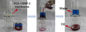
Researchers in China have made a new type of membrane that can separate oil from water and could potentially be used in oil spills, such as the one in the Gulf of Mexico. The membrane works by interacting differently with the substances as it is both superhydrophobic and superoleophilic, so that it repels water but attracts oil. This means that the oil is absorbed through the membrane, but the water can’t penetrate.

Oil and water separation using the membrane. The water and oil were dyed by methyl blue and oil red, respectively

The membrane is made from a polymerised fluorinated polybenzoxazine (F-PBZ) layer on top of cellulose acetate nanofibres. The scientists used an electrospinning technique (in which a viscous liquid is passed through a conducting needle to form a thread) to create a porous structure that makes the membrane even better at absorbing the oil.

Interested to know more? Read the full article in Chemistry World here…

Read the article from Nanoscale:

In situ polymerization approach to the synthesis of superhydrophobic and superoleophilic nanofibrous membranes for oil/water separation
Yanwei Shang ,  Yang Si ,  Aikifa Raza ,  Liping Yang ,  Xue Mao ,  Bin Ding and Jianyong Yu
Nanoscale, 2012, Accepted Manuscript
DOI: 10.1039/C2NR33063F

Digg This
Reddit This
Stumble Now!
Share on Facebook
Bookmark this on Delicious
Share on LinkedIn
Bookmark this on Technorati
Post on Twitter
Google Buzz (aka. Google Reader)

Colourful metal detection

The sensor changes from red to blue with an increasing mercury concentration

The sensor changes from red to blue with an increasing mercury concentration

Scientists in China have developed a sensor that can indicate the presence of heavy metal ions in a sample with a simple colour change.

Heavy metal ions such as mercury and lead are persistent pollutants in the environment and can cause severe health problems. Routine sampling of water sources is vital to evaluate the levels of toxic metals.

Current detection techniques involve the use of expensive equipment, require skilled technicians and are time consuming. A range of research has looked at developing detection techniques for in-field use and recently, interest has been focused on using aptamers. Aptamers are single-stranded DNA or RNA molecules that have been shown to reversibly bind with mercury and lead. Several sensors have been developed that incorporate aptamers but most have required complex labelling or detection techniques.

Read the full article in Chemistry World

Link to journal article
Colorimetric photonic hydrogel aptasensor for the screening of heavy metal ions
Bao-Fen Ye ,  Yuan-Jin Zhao ,  Yao Cheng ,  Ting-Ting Li ,  Zhuo-Ying Xie ,  Xiang-Wei Zhao and Zhong-Ze Gu
Nanoscale, 2012,4, 5998-6003, DOI: 10.1039/C2NR31601C

Digg This
Reddit This
Stumble Now!
Share on Facebook
Bookmark this on Delicious
Share on LinkedIn
Bookmark this on Technorati
Post on Twitter
Google Buzz (aka. Google Reader)

Nanoscale Editor features in Chemistry World

Nanoscale Associate Editor Professor Xiao Cheng Zeng‘s latest discovery has featured in Chemistry World this week. Zeng and colleagues have calculated the structure of a stable carbon dication with a coordination number of 7, higher than any yet seen experimentally. 

Read the Chemistry World article:  

Carbon clusters score lucky seven

14 August 2012 Andy Extance

  
The predicted cluster has a pentagon of titanium atoms around the central carbon, plus one above and one below. Credit: ACSUS and Chinese chemists say that they’ve calculated the structure of a stable carbon dication that would have a higher coordination number than any yet seen experimentally. Xiao Cheng Zeng from the University of Nebraska-Lincoln and colleagues have found that a carbon surrounded with seven titanium atoms can fulfil the necessary stability criteria. ‘We examined all first-row transition-metal elements and most main group elements,’ Zeng tells Chemistry World. ‘Only titanium fits thus far.’ 

 Surrounding carbon with more than four other atoms moves beyond conventional two-centre, two-electron bonds to arrangements sharing fractional numbers of valence electrons. The current record largest cluster seen experimentally was a hexacoordinate carbon structure synthesised by Japanese researchers in 2008. But theoretical physical chemists are curious to see how much further coordination numbers might be pushed.

Read full article 

Professor Zeng handles submissions to Nanoscale in the computational and theoretical fields, he also has a specialist interest in nanoclusters, computational nanocatalysis and computer-aided design and study of nanostructured materials.

Submit to Xiao Cheng Zeng’s Editorial Office today.

Digg This
Reddit This
Stumble Now!
Share on Facebook
Bookmark this on Delicious
Share on LinkedIn
Bookmark this on Technorati
Post on Twitter
Google Buzz (aka. Google Reader)

Pinning down cancer

US scientists have synthesised pin-shaped nanoparticles with magnetic and optical properties. The team, led by Xiaohua Huang at the University of Memphis, says that the nanoparticles ‘could be used for magnetic resonance imaging (MRI), early detection and photothermal therapy of cancer and other diseases’.

Transmission electron microscope image of the iron oxide–gold nanopins

Nanoparticles with both magnetic and optical properties are highly sought after in a number of fields, particularly medicine, where they could be efficient agents for imaging tumour cells using surface enhanced Raman scattering (SERS) and MRI.

To read the full article visit Chemistry World.

Synthesis and properties of near infrared-absorbing magnetic–optical nanopins
Saheel Bhana, Binod K. Rai, Sanjay R. Mishra, Yongmei Wang and Xiaohua Huang
Nanoscale, 2012, Advance Article
DOI: 10.1039/C2NR31291C

Digg This
Reddit This
Stumble Now!
Share on Facebook
Bookmark this on Delicious
Share on LinkedIn
Bookmark this on Technorati
Post on Twitter
Google Buzz (aka. Google Reader)

Back to carbon black

The electrochemical properties of amorphous carbon (right) and thermally reduced graphene oxide (left) are very similar

The electrochemical properties of amorphous carbon (right) and thermally reduced graphene oxide (left) are very similar

Scientists in Singapore have discovered the potential of a readily available material that could be used to replace expensive graphene analogues in a wide range of electrochemical processes.

Graphene consists of single layers of carbon atoms arranged in a honeycomb lattice structure. This structure provides unique properties, making graphene an important material for sensing and electrochemical applications.

Read the full article in Chemistry World

Thermally Reduced Graphenes Exhibiting Close Relationship to Amorphous Carbon
Colin Hong An Wong,  Adriano Ambrosi and Martin Pumera
Nanoscale, 2012, Accepted Manuscript
DOI: 10.1039/C2NR30989K

Digg This
Reddit This
Stumble Now!
Share on Facebook
Bookmark this on Delicious
Share on LinkedIn
Bookmark this on Technorati
Post on Twitter
Google Buzz (aka. Google Reader)

Two in one technique for biological imaging

A UK based team has combined two methods into a new technique to investigate cell-substrate interactions in biomedical research.

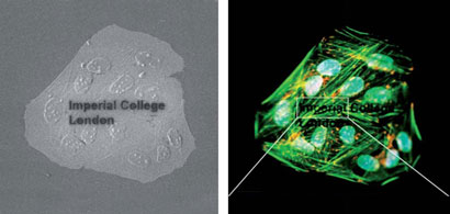
The new technique, correlative light-ion microscopy (CLIM), combines both ion and fluorescence microscopy to obtain topographical and biochemical information for the same area of a sample.

The idea for the technique came to Molly Stevens and her colleagues at Imperial College London, when they observed unknown structures while conducting characterisation tests on human tissue samples. ‘We realised that there was no simple and efficient method to correlate structural and biochemical information at the micro and nanoscale. Therefore, the only way forward was to ingeniously combine our available technology and expertise in order to develop a new imaging method,’ she says.

Scanning electron microscopy (SEM), which provides structural information, normally requires the sample to be dried and coated with a protective layer, while fluorescence microscopy – used to obtain biochemical information – usually needs the sample to be hydrated. The two techniques would appear to be mutually exclusive, or at the very least time consuming to correlate if run separately.

Both SIM (left) and fluorescence (right) microscopies can be performed on the same sample

Both SIM (left) and fluorescence (right) microscopies can be performed on the same sample

The new method works because instead of SEM, scanning ion microscopy (SIM) is used. This involves a beam of gallium ions instead of electrons, which does not interfere with the fluorescence signal, a problem with traditional SEM. Non-contradictory sample preparation also means that both tests can be run on the same sample.

‘I believe that the developed approach is a major breakthrough in the field as it generates new and useful information that is otherwise difficult to obtain,’ comments Ali Khademhosseini, from Harvard Medical School and Brigham and Women’s Hospital, US, who studies micro and nanoscale technologies to control cellular behaviour. ‘I anticipate its widespread use in many biological applications where both SEM and fluorescence are needed.’

Stevens believes the work can be helpful across many fields including biotechnology, biomaterials and cell biology, particularly when looking at in situ cell interactions. ‘Considering that fluorescence microscopes can be found in virtually any laboratory and scanning ion microscopes are more and more common, CLIM may well find very wide applicability in biomedical laboratories,’ she says.

Correlative light-ion microscopy for biological applications
Sergio Bertazzo, Thomas von Erlach, Silvia Goldoni, Pelin L Çandarlıoğlu and Molly M Stevens
DOI: 10.1039/C2NR30431G

Read the original article at Chemistry World

Digg This
Reddit This
Stumble Now!
Share on Facebook
Bookmark this on Delicious
Share on LinkedIn
Bookmark this on Technorati
Post on Twitter
Google Buzz (aka. Google Reader)

Speeding up wound healing

Scientists in China have developed a material that reduces the time required for a skin wound to heal.

A range of research has been conducted into the promising biomedical applications of chitosan, as it can clot blood effectively. Bingan Lu and his colleagues at Lanzhou University have developed a method to combine the benefits of chitosan with graphene, which has been shown to have antibacterial properties. 

Lu’s team mixed graphene with chitosan-polyvinyl alcohol (PVA) nanofibres using electrospinning (a process in which an electrical charge is applied to draw very fine fibres from the solution). The chitosan nanofibres combined with the graphene, forming thin membranes. 

The team applied the membranes to small skin wounds. Lu says that the membranes ‘covered the wounds like a band aid and, usually, one wound only needed one graphene-chitosan-PVA membrane’. They found that after 10 days, the wounds were significantly more healed than those without the membrane. 

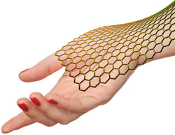
Graphene sheet on a hand

A membrane formed from chitosan, which clots blood effectively, and graphene, which is antibacterial, speeds up wound healing

Chunhai Fan, an expert in graphene materials at the Chinese Academy of Sciences, says that the work ‘shows a really interesting health application of graphene-based nanomaterials’ and adds that it ‘clearly shows that graphene-based antibacterial materials facilitate wound healing’.

To test the membrane further, Lu’s team used cell cultures to show that graphene is only detrimental to bacterial cells, and animal cells are unaffected. They suggest that this may be due to graphene transferring electrons through cell membranes. Bacterial cells are prevented from replicating by this process, as the electrons can reach the bacterial DNA, destroying it. Animal DNA is protected from the electrons by a second membrane. The team intends to investigate graphene’s antibacterial properties to confirm the mechanism.

Graphene-based composite materials beneficial to wound healing
Bingan Lu, Ting Li, Haitao Zhao, Xiaodong Li, Caitian Gao, Shengxiang Zhang and Erqing Xie
DOI: 10.1039/C2NR11958G

Read the original article at Chemistry World

Digg This
Reddit This
Stumble Now!
Share on Facebook
Bookmark this on Delicious
Share on LinkedIn
Bookmark this on Technorati
Post on Twitter
Google Buzz (aka. Google Reader)

Cleaning cadmium from blood: Nanoscale article featured in Chemistry World

With the development of modern industries, heavy metal pollution in humans is on the rise, say researchers in China, who have now designed a supermagnetic nanocomposite to effectively remove one of the pollutants – cadmium ions – from blood.

Removal of cadmium ions from a human blood sample with a magnet

The nanocomposite binds to cadmium ions in the blood and a magnet is used to attract the resulting complex for removal

Cadmium ions damage organs and are carcinogenic. Materials currently being studied to clear them from human blood don’t possess all the required properties for this purpose.

They either have good selectivity, high saturation magnetisation or good water dispersibility, but not all three. The new composite, made by Jun Jin and Jiantai Ma from Lanzhou University and colleagues, combines all of these properties.

The team built up the nanocomposite – PAD-PEG-Fe3O4@PEI – from four components. The first was magnetic iron oxide nanoparticles, chosen for their low toxicity. The team coated them with polyethylenimine (PEI), which binds to cadmium ions. The coating also reduces the chances of nanoparticle uptake by red blood cells, maximising their circulation time in the blood. Polyethylene glycol (PEG) was grafted onto this as an anchor for negatively charged 2,2′-(phenylazanediyl) diacetic acid (PAD), which counteracts interactions between the nanoparticles and plasma proteins or white blood cells.

Interested to know more? Read the full article in Chemistry World here…

 Read the paper from Nanoscale:

2, 2′-(phenylazanediyl) diacetic acid modified Fe3O4@PEI for selective removal of cadmium ions from blood
Jun Jin, Fang Yang, Fengwei Zhang, Wuquan Hu, Shao-bo Sun and Jiantai Ma
Nanoscale, 2012, Advance Article
DOI: 10.1039/C2NR11481J

Fancy submitting an article to Nanoscale? Then why not submit to us today!

Digg This
Reddit This
Stumble Now!
Share on Facebook
Bookmark this on Delicious
Share on LinkedIn
Bookmark this on Technorati
Post on Twitter
Google Buzz (aka. Google Reader)

Controlling termites with nanoparticles: Nanoscale article featured in Chemistry World

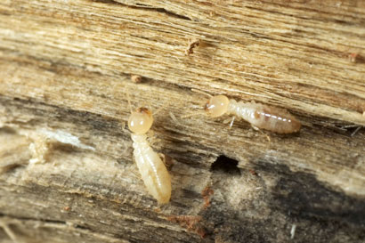
Scientists in Australia have found that mesoporous silica nanoparticles (MSNs) can store and deliver biocides in a controlled fashion over time, which could be beneficial to the timber industry with regards to termites.
C2NR11691J

Termites pose a significant threat to the industry throughout the tropics and subtropics. The conventional solution to this problem is to use agrochemical biocides such as dichloro-diphenyl-trichloroethane (DDT), aldrin, dieldrin, chlordane and heptachlor.

But these compounds cause environmental damage via bioaccumulation, threatening the existence of some species, particularly large predators at the top of the food chain. And attempts to destroy entire terminte colonies using them have been unsucessful. 

Now, Zhang Qiao and colleagues at the University of Queensland, have used the pore structure of mesoporous silica nanoparticles to adsorb biocides. They found that the nanoparticles released the biocide in a controlled manner. This slow release is important as the termintes will feed on and transfer the particles to other termites, eventually leading to colony destruction. 

The team chose four different types of MSN to test, using the agricultural biocide imidacloprid as a model. They found that MCM-48 particles had the highest adsorption capacity. ‘We can effectively load the biocide into MSNs and release it over 48 hours,’ says Qiao. ‘However, it is difficult to control the release because of the biocide’s water solubility and fast mass transport.’ 

Andrea O’Connor, an expert in nano and biomolecular engineering at the University of Melbourne, Australia, agrees that more control over release rates is needed. This would ‘minimise the early burst release and extend biocide delivery over biologically relevant time periods and dose rates’, she says. However, she adds that the system is simple and delivers the nanoparticles in a suspension into the site of an infestation ‘rather than relying on diffusion of released biocide through the environment, where it may be degraded or have undesirable adverse effects.’ 

Qiao adds that to effectively deliver the biocide over a period of about seven days, the MSNs need to be coated with other chemicals. The team is investigating a biodegradable polymer coating. 

Carl Saxton – Chemistry World 

Read the paper from Nanoscale: 

Adsorption and release of biocides with mesoporous silica nanoparticles
Amirali Popat, Jian Liu, Qiuhong Hu, Michael Kennedy, Brenton Peters, Gao Qing (Max) Lu and Shi Zhang Qiao
Nanoscale, 2012, Advance Article
DOI: 10.1039/C2NR11691J 

Fancy submitting an article to Nanoscale? Then why not submit to us today!

Digg This
Reddit This
Stumble Now!
Share on Facebook
Bookmark this on Delicious
Share on LinkedIn
Bookmark this on Technorati
Post on Twitter
Google Buzz (aka. Google Reader)

Chickpeas grow taller with carbon nanotubes

Carbon nanotubes can enhance plant growth without damaging plant cells, say scientists from India

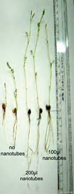
Chickpeas grow taller with carbon nanotubes

Sabyasachi Sarkar and colleagues from the Indian Institute of Technology in Kanpur treated chickpea plants with up to 6ug/ml of water soluble carbon nanotubes. They found that the nanotubes increased the growth rate in every part of the plant – in the roots, shoots and branches.

Sarkar thought that the channels could be replicated by carbon nanotubes. ‘We followed Thomas Edison’s recipe to make carbonised filament from bamboo or wood wool in his electric bulb to get the carbon nanotubes,‘ he adds. ‘Of course, we had to derivatise them to make them water soluble.’ The team achieved this by attaching carboxylic acid groups to the surface of the tubes.

The work seems to support the positive effect of carbon nanotubes,’ says Xiaohong Fang, an expert in the use of carbon nanotubes as molecular transporters in plants at the Chinese Academy of Sciences in China. However, she points out that the biological effects on plants may differ depending on the materials’ chemical and physical properties, plant type and cultivation conditions.

Read the rest of the Chemistry World story by Elinor Richards

View the Nanoscale article in full:

Growth stimulation of gram (Cicer arietinum) plant by water soluble carbon nanotubes
Shweta Tripathi, Sumit Kumar Sonkar and Sabyasachi Sarkar
Nanoscale,
2011, DOI: 10.1039/c0nr00722f

Digg This
Reddit This
Stumble Now!
Share on Facebook
Bookmark this on Delicious
Share on LinkedIn
Bookmark this on Technorati
Post on Twitter
Google Buzz (aka. Google Reader)