Author Archive

Nanoscale journals working together

Nanoscale & Nanoscale Horizons

The launch of Nanoscale Horizons seeks to build on and strengthen the nanoscience content already published in journals across the Royal Society of Chemistry portfolio. In particular, Nanoscale Horizons will complement and work alongside Nanoscale to provide a rounded view of innovation, and bridge the various disciplines in research across nanoscience and nanotechnology.

Nanoscale Horizons aims to publish first reports of exceptional significance in the field, therefore positioning itself as a premier journal in its field. The journal will publish leading research containing a clear conceptual advance and new insights into the topic presented. As such, there will be highly stringent criteria for publication, imposed by our Scientific Editors and Publishing Editors.

Nanoscale will continue to publish high impact research across the breadth of nanoscience and nanotechnology. The criteria for publication remain the same and will continue to be upheld by the journal’s Associate Editors to maintain Nanoscale’s reputation for publishing high quality, community-spanning research.

As with other Royal Society of Chemistry journals, the Nanoscale Horizons Editorial team will try to find the most suitable home for any manuscript that we receive. We endeavour to provide authors with the option to automatically transfer their manuscript to Nanoscale or another journal within our portfolio, where manuscripts are deemed more suitable for publication elsewhere.

To find out more about Nanoscale Horizons and keep up-to-date with all of the latest news, visit the webpage and sign up for the e-alerts.

Digg This
Reddit This
Stumble Now!
Share on Facebook
Bookmark this on Delicious
Share on LinkedIn
Bookmark this on Technorati
Post on Twitter
Google Buzz (aka. Google Reader)

Nanoscale’s new sister

Introducing Nanoscale Horizons – launching next year

The home for rapid reports of exceptional significance in nanoscience and nanotechnology is on its way.

Our newest journal will work alongside Nanoscale to provide a rounded view of innovation in nano research, and bridge the various disciplines involved with nanoscience and nanotechnology. We’ll be looking for high impact work in fields ranging from physics and chemistry to IT, healthcare and detection science.

A pioneering Editorial Board Chair

Our Editorial Board Chair is Professor Harold Craighead, Professor of Engineering at Cornell University, USA and a pioneer in nanofabrication methods. He will head up an expert editorial board, led by Executive Editor Dr Fiona McKenzie.

Rapid reports, cutting-edge research

The first issue in 2016 will lay the groundwork for what aims to be the journal of choice for outstanding research across a broad spectrum.

Articles published will benefit from wide exposure, and content published during 2016 and 2017 is free upon registration – giving maximum visibility to your research.

Nanoscale Horizons will be launching very soon. Sign up to our Email Alerts Service and make sure you’re among the first to hear the latest.

Digg This
Reddit This
Stumble Now!
Share on Facebook
Bookmark this on Delicious
Share on LinkedIn
Bookmark this on Technorati
Post on Twitter
Google Buzz (aka. Google Reader)

Hybridized doxorubicin-Au nanospheres exhibit enhanced Near-infrared surface plasmon absorption for photothermal therapy applications

Plasmon absorption stability of DAuNS and the triggered release of DOX

As cancer therapy evolves there is a desire to explore minimally invasive treatments which are applicable to all patients, regardless of concerns with drug resistance and tumour morphology. One such option being investigated is photothermal therapy (PTT), which seeks to achieve these aims through the selective uptake of photosensitizing agents by cancerous cells prior to their abalation using a near infa-red (NIR) light source.  However, currently the efficacy of PTT is reduced due to heterogeneous heat distribution, resulting in the accumulation of sub-lethal doses of the sensitizing agent within areas of the tumor.

Zhou and co-workers have sought to overcome this issue by creating a “double punch” strategy for tumour targeting using PTT. Through the implantation of hollow gold nanoshells with the chemical agent doxorubicin (DAuNS), a novel combination of a photosensitizing agent and a chemical targeting agent has been created. Furthermore, this unique, yet simple synthesis strategy is thought to be interconvertible with other drug and nanomaterial combinations, thus, widening the scope for potential PTT treatments.

Through comparisons with ‘single-strategy’ treatments of bare hollow gold nanoshells (HAuNS) or doxorubicin, the improved efficacy of the DAuNS is well established through both in-vitro and in vivo studies. This significant improvement can be attributed to the enhanced plasmon absorption in the NIR region of DAuNS in comparison to HAuNS (1.5 fold increase), with a more efficient photothermal conversion and a greater efficacy in tumor killing also established. These properties are only enhanced by the combined chemotherapeutic effect achieved through the deployment of the doxorubicin payload.

As this strategy obviates the concerns of genetic drug resistance and is a minimally invasive treatment, it could carry significant potential. This potential is only further enhanced by the ability to exchange different chemotherapeutic reagents, and as such this could be a significant breakthrough which aids future cancer therapies.

Hybridized Doxorubicin-Au Nanospheres Exhibit Enhanced Near-infrared Surface Plasmon Absorption for Photothermal Therapy Applications
Jialin Zhou, Zuhua Wang, Qingpo Li, Fei Liu, Yongzhong Du, Hong Yuan, Fu-Qiang Hu, Yinghui Wei and Jian You
Nanoscale, 2015, Advance Article. DOI: 10.1039/C4NR07279K.

Dr Derek Craig is a guest web writer for the Nanoscale blog. He is a Post Doctoral Research Fellow at the University of St. Andrews based in the fields of Biophotonics and Materials Science. With a background in chemistry, his work mainly focuses on the synthesis of nano to meso materials and the use of imaging techniques to study biological samples.
Digg This
Reddit This
Stumble Now!
Share on Facebook
Bookmark this on Delicious
Share on LinkedIn
Bookmark this on Technorati
Post on Twitter
Google Buzz (aka. Google Reader)

HOT article: Unfolding the secrets of DNA origami nanostructures

DNA origami machine components

DNA origami is an emerging field of nanotechnology that exploits the unique base-pairing capabilities of the DNA bases to produce complex and mechanically stiff self-assembled nanoscale geometries. Recently, DNA origami has found uses in applications such as single molecule sensing, drug delivery and templating molecular components.

This review, by researchers from Ohio State University, sets out to explore the mechanical nature of DNA nanostructures in order to understand how these structures will respond to physical interactions. The review covers three major areas of progress, including measuring and designing mechanical properties of DNA nanostructures, designing complex nanostructures based on imposed mechanical stresses and, finally, designing and controlling structurally dynamic nanostructures.

For researchers interested in the future possibilities of DNA origami, this review should provide an insightful first stop for discussing the mechanical design of DNA nanostructures.

Mechanical design of DNA nanostructures
Carlos E. Castro, Hai-Jun Su, Alexander E. Marras, Lifeng Zhou and Joshua Johnson
Nanoscale, 2015, Advance Article. DOI: 10.1039/C4NR07153K

Dr Lee Barrett is a guest web writer for the Nanoscale blog. Lee is currently a postdoctoral researcher in the Centre for Molecular Nanometrology at the University of Strathclyde. His research is currently focused on the development of nanoparticle-based sensors and surface enhanced Raman scattering (SERS). Follow him on twitter: @L_Bargie

Digg This
Reddit This
Stumble Now!
Share on Facebook
Bookmark this on Delicious
Share on LinkedIn
Bookmark this on Technorati
Post on Twitter
Google Buzz (aka. Google Reader)

Poster prize winners at Nano India 2015

Professor Ajay K. Sood awarding a Nanoscale poster prize at Nano India 2015

Many congratulations to G. Rajaraman and E. Manikandan for winning the Nanoscale poster prizes at Nano India 2015.

G. Rajaraman, from the Indian Institute of Technology, Bombay, won a prize for his poster entitled “New generation molecular nanomagnets”, and E. Manikandan’s poster was entitled “A fingerprint micro Raman spectroscopy study for few layered graphene formation on gold substrate by low energy ion implantation technique”. The prizes were awarded by Professor Ajay K. Sood from the Indian Institute of Science, Bangalore.

The conference was hosted by the Centre for Nanotechnology & Advanced Biomaterials (CeNTAB), SASTRA University, Thanjavur, on the 29-30th  January 2015. It is a national conference supported by the Department of Science & Technology (DST), and aims to promote the exchange of ideas and create a platform for collaborative research in interdisciplinary areas of nanoscience and nanotechnology. Further details are available on the conference website.

Digg This
Reddit This
Stumble Now!
Share on Facebook
Bookmark this on Delicious
Share on LinkedIn
Bookmark this on Technorati
Post on Twitter
Google Buzz (aka. Google Reader)

HOT article: Effects of morphology and chemical doping on electrochemical properties of metal hydroxides in pseudocapacitors

With a limited amount of fossil fuels in the ground, developing alterative strategies for energy production is of the utmost importance.  However, without a way to store that energy, these endeavors could be futile.

From the team of Gyeonghee Lee, Chakrapani V. Varansi, and Jie Liu (an Associate Editor here at Nanoscale), comes one of this month’s HOT papers, “Effects of morphology and chemical doping on electrochemical properties of metal hydroxides in pseudocapacitors.” In this paper, the team’s specific metal hydroxide of interest is Ni(OH)2.

image file: c4nr06997h-s1.tif

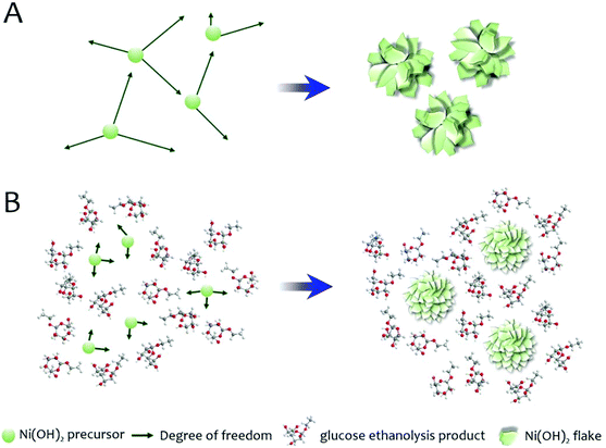
Schematic illustration of the proposed mechanism for the morphology modification of Ni(OH)2 by glucose in the solvothermal medium.

Lee et al. state “…that both morphology control and chemical doping positively affect the electrochemical performance of Ni(OH)2 when applied individually.”  In this paper, they set out to determine what happens to the electrochemical performance when morphology and doping are combined instead of applied individually.  In order to investigate the effects of both, they chose a solvothermal synthesis route for metal hydroxide flakes, creating both cobalt-doped (CoxNi1-x(OH)2) and undoped particles (Ni(OH)2) that have varying amounts of D-glucose added.  For both the doped and undoped particles, four sets of particles are created, where 0, 10, 20, and 50% of the urea normally used in the solvothermal process is replaced by the D-glucose.  All of the particles were characterized with x-ray diffraction (XRD) and scanning electron microscopy (SEM), with the SEM showing that increasing amounts of glucose decreased flake size.  XRD and thermogravimetric analysis also revealed that increasing D-glucose amounts created an increase in the amount of interlayer water, with XRD also indicating degree of crystallinity decreased with increasing D-glucose.

In order to understand how the glucose affects morphology, the authors ran the solvothermal synthesis without the metal precursor. XRD and nuclear magnetic resonanace (NMR) data indicates the presence of ethyl-substituted glucose (formed during the synthesis process) instead of D-glucose.   The authors conclude that “…ethyl glucoside molecules can hinder the precursor diffusion and thus inhibit the metal hydroxide growth during synthesis.”

The electrochemical performance of all of the types of particle is evaluated using cyclic voltammetry (CV), galvanostatic charge–discharge, and electrochemical impedance spectroscopy (EIS).  Increasing glucose levels on undoped particles increased specific capacitance and cobalt doping is also shown to improve specific capacitance.  Combining the two showed that specific capacitance increased up to 50% of Co doping, but was less effective above 50%.

Lee et al. find that “…the addition of glucose in the ethanol-mediated solvothermal synthesis effectively reduces the particle size of metal hydroxide flakes. The specific capacitance is improved as a result of the increased surface area and reduced particle size.”  In terms of specific capacitance, the higher interlayer water levels found for increasing glucose levels may make ion mobility easier in the materials, enhancing the specific capacitance of the doped-Ni(OH)2.  In the doped materials, the specific capacitance was increased with low doping and decreased with high doping.

In designing future iterations of Ni(OH)2 batteries, careful attention will need to be placed on both morphology and doping levels in order to find the best balance for electrochemical performance.

Effects of morphology and chemical doping on electrochemical properties of metal hydroxides in pseudocapacitors
Gyeonghee Lee, Chakrapani V. Varanasi and Jie Liu
Nanoscale, 2015, 7, 3181-3188. DOI: 10.1039/C4NR06997H

Stephanie E. Vasko is currently a Senior Research Assistant at The Rock Ethics Institute at the Pennsylvania State University in State College. Her research focuses on science communication, STEM education, and the intersections between art, craft, and science.  You can follow her on Twitter at @stephanievasko.

The opinions and views expressed in this piece are those of the author and do not represent or reflect the opinion, views, or policy of the Pennsylvania State University, the Rock Ethics Institute, or the National Science Foundation.

Digg This
Reddit This
Stumble Now!
Share on Facebook
Bookmark this on Delicious
Share on LinkedIn
Bookmark this on Technorati
Post on Twitter
Google Buzz (aka. Google Reader)

Shepherding cells – moving in the right direction?

image file: c4nr06594h-f1.tif

Schematic representation depicting the ability for an external magnetic field to attract magnetic nanoparticles which have been internalised within cells. For details please view the full article.

Superparamagnetic nanoparticles are widely used for non-invasive imaging techniques such as magnetic resonance imaging (MRI) due to their ability to only become magnetised under the influence of an external magnetic field. In this article, it is demonstrated that labelling cells with magnetite nanoparticles can allow for manipulation of both direction and speed of the migration of cells using an external magnet. Bespoke nanoparticles were synthesised with a positive charge to induce internalisation into two different cell lines that are important for wound repair. Placing labelled cells under the influence of an external magnetic field resulted in 2D migration of cells towards the magnet, whereas non-labelled cells (in a magnetic field) and labelled cells with no magnetic showed no directional movement.  The migration could be monitored by bright field and fluorescent microscopy as the nanoparticles contained a fluorescent tag.  The possibility of controlling cell mobility is suggested to have importance in not only cell therapies, but also tissue engineering and cell tracking. This tailored synthesis approach could also allow tracking of cells in vivo using a bi-modal imaging approach of dual MRI and whole animal fluorescence.

Manipulating Directional Cell Motility Using Intracellular Superparamagnetic Nanoparticles
Michael Bradshaw, Tristan Clemons, Diwei Ho, Lucia Gutierrez, Francisco Lazaro, Michael House, Timothy Guy St Pierre, Mark Fear, Fiona Wood and Swaminathan Iyer
Nanoscale, 2015, Advance Article. DOI: 10.1039/C4NR06594H

Dr Mike Barrow is a guest web writer for the Nanoscale blog. He currently works as a Postdoctoral Researcher at the University of Liverpool.

Digg This
Reddit This
Stumble Now!
Share on Facebook
Bookmark this on Delicious
Share on LinkedIn
Bookmark this on Technorati
Post on Twitter
Google Buzz (aka. Google Reader)

Introducing our new Associate Editors: Xiaogang Liu, Hongxing Xu and Yves Dufrêne

We are pleased to introduce three new Associate Editors for Nanoscale: Xiaogang Liu, Hongxing Xu and Yves Dufrêne.

Professor Xiaogang Liu

Xiaogang is Dean’s Chair Professor in the Department of Chemistry at the National University of Singapore. He obtained his B. Eng from the Beijing Technology and Business University in China, and MS degree in Chemistry from East Carolina University, US. After completing his PhD at Northwestern University in the US, Professor Liu worked as a Postdoctoral Associate at the Massachusetts Institute of Technology (MIT), before joining the National University of Singapore. His research interests encompass supramolecular chemistry, materials science, and bioinorganic chemistry, specifically controlling assemblies of dynamically interacting biological molecules and understanding the relationship between structure and physical properties.

Xiaogang says: “I’m thrilled to take on a new role as an Associate Editor for Nanoscale, a forum that enables researchers to share their exciting work in the diverse field of nanoscience and nanotechnology. I look forward to working with members of our community and do hope to continue to improve the quality of the journal.”

Professor Hongxing Xu

Hongxing is Professor, the director of the Center for Nanoscience and Nanotechnology, and the Vice Dean of the School of Physics and Technology and the Institute for Advanced Studies at Wuhan University. His research is focused on surface enhanced spectroscopy and nanoplasmonics, in particular, phenomena, mechanisms, devices and applications based on surface plasmon resonances in novel metal nanostructure systems.

Professor Yves Dufrêne

Yves is a Research Director of the National Fund for Scientific Research and a Professor at the Université Catholique de Louvain (UCL), Belgium. He obtained his Bioengineering degree and PhD at UCL, then worked as a postdoctoral researcher at the Naval Research Laboratory, USA, before returning to UCL. He is interested in nanobioscience and nanobiotechnology, specifically in the development and use of advanced nanoscale techniques for analyzing biological systems. His research focuses on studying the nanoscale surface architecture, biophysical properties and molecular interactions of living cells – particularly microbial pathogens – using atomic force microscopy (AFM). The goals are to further understand key cellular functions, like cell adhesion, and to contribute to the development of nanoscopy techniques for the life sciences.

We are delighted to welcome Yves to the Nanoscale Editorial Board. He comments: “I am very honored and excited to become Associate Editor of such a great journal, definitely one of the very best in nanoscience. My main mission will be to promote publication of top-quality research in the fast moving area of nanobioscience and nanomedicine.”

Digg This
Reddit This
Stumble Now!
Share on Facebook
Bookmark this on Delicious
Share on LinkedIn
Bookmark this on Technorati
Post on Twitter
Google Buzz (aka. Google Reader)

HOT article: A tunable submicro-optofluidic polymer filter based on guided-mode resonance

image file: c4nr07233b-f3.tif

SEM images of the polymer submicro-channels and photographs of the fabricated submicro-optofluidic PGMR filter with empty channels and with water filled channels, respectively.

Optical filters are routinely included in devices used for communications, displays and bio-sensing. One such class of optical filters, which have been frequently used, are guided-mode resonance (GMR) filters. Inherent errors with common GMR filters are assigned to fabrication difficulties and as a result, new classes of reconfigurable GMR filters have been created.

In this HOT article, Jin and co-workers have proposed and devised a novel polymer-based GMR (PGMR) which can be incorporated into lab on a chip devices and become tuned by the optical properties of the fluidic mixture present. The simple and low cost PGMR filter was fabricated in three stages, using two-beam interference lithography, floating nanofilm transfer, and finally, thermal bonding technology.

The tunability of this class of PGMR was tested by filling the fluidic channels with a range of liquid mixtures of differing refractive indices. The resulting PGMR exhibited high reflection efficiency in the visible wavelength region, a narrow band tuning range and a high tuning efficiency. The successful incorporation of the PGMR in an optofluidic device operating in the visible wavelength region opens up the possibility of the inclusion of such PGMR devices in future lab on a chip devices.

A tunable submicro-optofluidic polymer filter based on guided-mode resonance
Guohui Xiao, Qiangzhong Zhu, Yang Shen, Kezheng Li, Mingkai Liu, Qiandong Zhuang and Chongjun Jin
Nanoscale, 2015, 7, 3429-3434. DOI: 10.1039/C4NR07233B.

Dr Derek Craig is a guest web writer for the Nanoscale blog. He is a Post Doctoral Research Fellow at the University of St. Andrews based in the fields of Biophotonics and Materials Science. With a background in chemistry, his work mainly focuses on the synthesis of nano to meso materials and the use of imaging techniques to study biological samples.
Digg This
Reddit This
Stumble Now!
Share on Facebook
Bookmark this on Delicious
Share on LinkedIn
Bookmark this on Technorati
Post on Twitter
Google Buzz (aka. Google Reader)

HOT article: Glance into the nanoparticle-imprinted mirror antenna (NIMA)

An ultrahigh-sensitivity plasmonic antenna (NIMA)

Researchers using surface enhanced Raman scattering (SERS) are always on the look out for new substrates that take advantage of coupled metallic nanoparticles to improve sensitivity.  In this HOT article, researchers from Taiwan have introduced NIMAs (nanoparticle (NP)-imprinted mirror antennas) for exactly this purpose.

The researchers deposited Ag on a polycarbonate substrate and used Si molds to create 2D periodic nanostructures, which were then used to create NIMAs by self-assembling Ag nanoparticles onto the Ag mirrors.  The formation of 2D nanoclusters on the mirror results in more intense Raman signals as a result of electromagnetic coupling between the NPs in the clusters. NIMAs have several advantages over other SERS-active substrates. For example, NIMAs gain broadband enhancement from single structures, rather than from various substrates with different morphologies. Also, using a deeper, more consistent structure and tuning the surface plasmon resonance (SPR) modes can drastically improve the SERS enhancement observed from NIMAs.

The researchers have produced a SERS-active substrate that is compatible in the visible to near-infrared regime and is capable of detecting rhodamine 6G at a concentration as low as 10-15 M.  These attractive advantages should be enough for any SERS researcher to reflect on the possibility of adopting NIMAs as a sensing platform in the future.

Chen-Chieh Yu, Yi-Chuan Tseng, Pao-Yun Su, Keng-Te Lin, Chang-Ching Shao, Sin-Yi Chou, Yu-Ting Yen and Hsuen-Li Chen
Nanoscale, 2015, Advance Article. DOI: 10.1039/C4NR05902F.

Dr Lee Barrett is a guest web writer for the Nanoscale blog. Lee is currently a postdoctoral researcher in the Centre for Molecular Nanometrology at the University of Strathclyde. His research is currently focused on the development of nanoparticle-based sensors and surface enhanced Raman scattering (SERS).

Digg This
Reddit This
Stumble Now!
Share on Facebook
Bookmark this on Delicious
Share on LinkedIn
Bookmark this on Technorati
Post on Twitter
Google Buzz (aka. Google Reader)