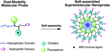
The synthesis of nanoprobes for application in more than one imaging technology is becoming more popular. So-called “bi-modal” probes can reduce limitations of single imaging modalities such as sensitivity or penetration depth. In this Hot article, the synthesis of two amphiphilic, dual-modality, optical imaging/MRI nanoprobes is reported. Each probe, containing both a fluorophore and a gadolinium complex, was specifically engineered using hydrophobic and hydrophilic components so they would self-assemble above the critical micelle concentration (CMC), which in turn would improve the MRI performance. The materials could offer potential advantages compared to conventional, unimolecular probes. Flow cytometry was used to confirm that both negatively charged assemblies were efficient at labelling KB-3-1 (human cervical cancer) cells at different labelling concentrations and incubation periods, through measurement of cell fluorescence. Cells viability was not compromised for each condition. The authors are now looking to further improve performance of self-assembled cell tracking agents through synthetic manipulation of construct size and surface charge.
Design and assembly of supramolecular dual-modality nanoprobes
Shuang Liu, Pengcheng Zhang, Sangeeta Ray Banerjee, Jiadi Xu, Martin G. Pomper and Honggang Cui
Nanoscale, 2015, 7, 9462-9466. DOI: 10.1039/C5NR01518A
Dr Mike Barrow is a guest web writer for the Nanoscale blog. He currently works as a Postdoctoral Researcher at the University of Liverpool.


