Archive for the ‘Miscellaneous’ Category

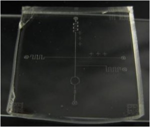
Simple visualization of a microfluidic acoustic pump’s sound path

R. Rambacha, C. Schecka, V. Skowroneka, L. Schmida and T. Frankea,b,*
aMathematisch-Naturwissenschaftliche Fakultät, Lehrstuhl für Experimentalphysik I, Soft matter and Biological physics, Universität Augsburg, Germany
b Department of Physics and School of Engineering and Applied Science, Harvard University, USA.
*E-mail: tfranke[at]seas.harvard.edu; Thomas.Franke[at]physik.uni-augsburg.de

Why is this useful?


Microfluidic PDMS-chips are widely used in many labs. Recently, the use of acoustics in combination with PDMS devices has attracted much attention in the field because it is simple to use and allows for unique control of minute amounts of fluid, cells and particles on a microfluidic chip. This trend is also reflected by a series of tutorial papers1 and front covers2,3 in Lab on a Chip that are dedicated to this topic.

The core component of these chips is a versatile interdigital transducer (IDT), consisting of a pair of interlocked electrodes on a piezoelectric substrate. It can be tailored to meet the demands of a specific application, e.g. on microfluidic chips the IDT is often used as an acoustic pump. The overall shape of the electrode arrangement determines the fluid actuating sound-path on the chip and makes the difference between specific IDTs. For better aligning the IDT with other fluidic components, such as a PDMS channel, testing the functionality of an IDT and probing completely new IDT-designs, it is often necessary to visualize IDT’s acoustic path or the focal point of a focused IDT. However this visualization still remains elusive and requires expensive equipment such an AFM, SEM or a vibrometer. These methods are not available in every lab and very time-consuming. We show a simple and quick way to visualize the IDT’s sound path. The only components needed are isopropanol and a microscope, which are both available in almost every microfluidic lab.

What do I need?


  • Sample microfluidic chip with IDT and frequency generator
  • Isopropanol
  • Microscope

What do I do?


1. Place the chip with the IDT on the microscope.

2. Put a few drops of isopropanol onto the chip to wet its surface.

3. Turn on the IDT and observe with microscope.

4. Then, knowing the position of the acoustic wave path, place the PDMS onto the chip.

5. Because there is still alcohol on the chip, firm bonding of the plasma treated PDMS to the chip is delayed and there is still some time (10min or so) to align the PDMS precisely under the microscope. When the alcohol has evaporated the PDMS eventually bonds firmly to the chip and is ready to use.

Covering the chip with an isopropanol film before turning on the IDT.

Covering the chip with an isopropanol film before turning on the IDT.

Optical micrograph of the chip with a straight IDT. An interference pattern of the SAW can be seen.

Optical micrograph of the chip with a straight IDT. An interference pattern of the SAW can be seen.

Focused tapered IDT excited with two different frequencies (left: 120MHz, right: 240MHz). The acoustic path shifts with higher frequency towards the center of the IDT.

Focused tapered IDT excited with two different frequencies (left: 120MHz, right: 240MHz). The acoustic path shifts with higher frequency towards the center of the IDT.

Two opposed tapered IDTs at (from top to down) 161MHz, 163MHz and 165MHz.

Two opposed tapered IDTs at (from top to down) 161MHz, 163MHz and 165MHz.

Visualization of a focused tapered IDT’s acoustic paths at 90MHz.

Visualization of a focused tapered IDT’s acoustic paths at 90MHz.

Demonstrating the visualization of the focused IDT’s acoustic path. Depending on the material’s anisotropy the focal point is shifted towards or away from the IDT.

Demonstrating the visualization of the focused IDT’s acoustic path. Depending on the material’s anisotropy the focal point is shifted towards or away from the IDT.

What else should I know?


  • With other fluids such as water, glycerin and ethanol, the visualization effect was less pronounced. The effect was most evident with isopropanol.
  • By knowing the applied frequency and the distance between the electrodes of the tapered IDT at the excited spot (it is equal to the wavelength), this is also a very simple and quick method to calculate the material’s surface speed of sound with an error below 5%.
  • One can determine the material’s anisotropy by measuring the difference between experimental and geometrical distance of the focal points of a focused IDT4.

References


[1] H. Bruus, J. Dual, J. Hawkes, M. Hill, T. Laurell, J. Nilsson, S. Radel, S. Sadhal and M. Wiklund. Lab on a Chip, 2011, 11, 3579
[2] J. Shi, S. Yazdi, S. Lin, X. Ding, I. Chiang, K. Sharp and T. Huang. Lab on a Chip, 2011, 11, 2319
[3] L. Schmid and T. Franke. Lab on a Chip, 2013, 13, 1691
[4] J.B. Gree, G.S. Kino and B.T. Khuri-Yakub. IEEE 1980 Ultrasonnics Symposium, 69

Digg This
Reddit This
Stumble Now!
Share on Facebook
Bookmark this on Delicious
Share on LinkedIn
Bookmark this on Technorati
Post on Twitter
Google Buzz (aka. Google Reader)

A simple trick to open up clogged microfluidic chip

Stefania Mazzitelli and Claudio Nastruzzi,
Department of Life Sciences and Biotechnology, University of Ferrara, Ferrara, Italy
Email: mzzsfn[at]unife.it

Why is this useful?


An event any microfluidic researcher never wants to occur is channel clogging. Unfortunately this drawback is not unusual, especially when working with narrow channel microchips and polymeric solutions or cell suspensions. On many occasions, microchips perfectly fitting the experiment appear to be irremediably lost and on the way to the rubbish bin.

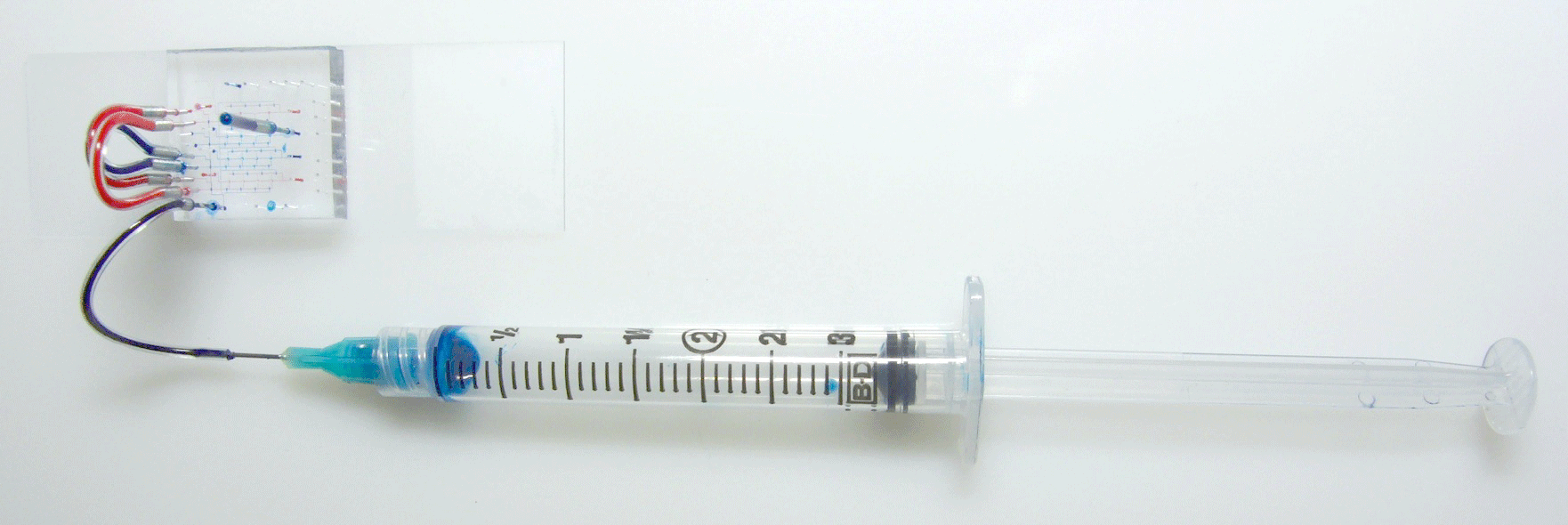
Presented in this tip is a very simple and inexpensive solution that can revitalize a clogged microfluidic chip. We have tested this simple protocol both on glass or PDMS microchips with positive results, moreover we solved severe clogging (as evidenced by optical microscopy) caused by cell clustering as well as by the frequent occurrence of polymer precipitation within the microchannels. Our method solves the issues reported above using an extremely simple approach and a microwave oven.

What do I need?


  • 21 gauge hypodermic needle [1]
  • Plastic tube (ETEF, FEP or PTFE) 1/16” OD, 0.75mm ID [2]
  • A 50 mL syringe [3]
  • A microwave oven

What do I do?


1–5 Using the 21 gauge hypodermic needle and the FEP (fluorinated ethylene-propylene) tube, build up the plastic port for microfluidic interfacing (the needle should fit perfectly in the FEP tube assuring a tight, fluid proof inlet, see panel 4).

6–9 Insert the FEP tube into a microfluidic port. Depending on the position of the clog (determined by microscopic observation) insert the tube in the port as far away from the clog as possible.
10–12 Pump, by hand held syringe, filtered distilled water into the chip, applying as much pressure as possible. In the clog is constituted of lipophilic materials or hydrophobic polymers, replace the water with ethanol, isopropanol, acetone or their mixtures with water.

13 Put the microfluidic chip into a standard kitchen microwave oven for 5 min at 500-700 watts.

NOTE: before treating the chip in microwave, REMOVE the metallic needle or the entire port.

14–15 Remove the microfluidic chip from the microwave oven, reinstall the port and flush, as soon as possible, water (or solvent) into the channels. In case one treatment in the microwave does not open up the channels, repeat the entire procedure.

16 Your beloved chip is now open and ready for a new set of experiments.

Pictures 1-8

Images 9-16

References


[1] http://www.picsolution.com/
[2] http://www.upchurch.com
[3] http://www.artsana.com

Digg This
Reddit This
Stumble Now!
Share on Facebook
Bookmark this on Delicious
Share on LinkedIn
Bookmark this on Technorati
Post on Twitter
Google Buzz (aka. Google Reader)

A simple, effective and cheap means to reduce bubbles in microchannels

Brian Miller*, Dr. Helen Bridle, Dr Stewart Smith
School of Engineering, The University of Edinburgh, G1 John Muir Building,, Kings Buildings ,Edinburgh, EH9 3JL
Email: B.miller[at]ed.ac.uk
Tel: +44 131 650 7860

Why is this useful?


A common issue that arises when attempting to fill microfluidic channels is that of air bubbles becoming trapped against surfaces such as walls and features within the channel. Many different techniques have been used to minimise/eliminate these including pre-filling with surfactant dosed water combined with soaking in an ultra-sonicated bath1, attempting to fill very shortly after O2 plasma treatment while surfaces are hydrophilic2 and bubble traps integrated into the design3-5.

Presented in this tip is a cheap post-manufacture solution for the reduction/elimination of bubbles when filling devices. This method takes advantage of the 10-fold increase in solubility of CO2 gas when compared to O2 and N2. By pre-filling your device with pure CO2 the trapped gas is dissolved away rapidly in comparison to air.

What do I need?


• CO2 “ Cornelius keg charger” and CO2 canister (Amazon B000NV9CE6)
• DI/RO water
• 0.01” ID, 1/16” OD PEEK tubing (Sigma-Aldritch Z226661)
• PEEK Tubing to Luer Lock Adaptor + Ferrule (LS-T116-100 and LS-T116-300 Mengel Engineering)
• AN-4 to 1/8” Female NPT adaptor (Aeroquip#023-FCM2721)
• Male Luer Lock x 1/8″ NPT male adaptor (Cole Parmer PN: OU-31507-84)

What do I do?


Assemble the metal adaptors onto the keg charger as illustrated in Fig.1. Cut a ~3ft to 3 ½ ft length of PEEK tubing and seal into the luer-lock adaptor. This acts as a flow resistor that will reduce the output pressure from ~60 bars to ~ 1-2 bars. Connect the PEEK adaptor to the Luer lock on the charger (Fig.2).

Place PEEK tubing into a device input (Fig.3). Ours mount directly into the device having used a 1.2mm hole punch to create input ports on the PDMS devices. Depending on your device ports you may need to develop a unique solution suitable for your preferred porting method.

Use your fingers to block off any other inputs and use the trigger of the keg charger to release CO2 into your device. Use your fingers to block off the output ports and depress keg charger trigger to fill the other input channels.

Immediately connect DI/RO water filled tubing to your device and begin filling. Remember to pre-fill the tubing with water so that air trapped in the tubing is eliminated.

 Please see the time-lapsed videos showing a bubble completely dissolved (SI.1) in ~10 mins (flow rate ~50ul/min in a 50um high device). Also note that for very large bubbles this technique slowly decreases in efficacy until it appears a saturation point is reached (SI.2) after roughly 30 mins. It is recommended to “massage” any very large bubbles towards the outputs and ideally break them up into smaller pockets of trapped gas. Compare these to a video of a medium sized (1mm radius) bubble of normal air to see little to no reduction at a similar flow rate over 10 mins SI.3)

Figure 1

Fig.1: Charger Adaptor Assembly

Figure 2

Fig.2: Assembled CO2 filling instrument with PEEK tubing flow resistor

Figure 3

Fig.3: Filling a device with pure CO2

References


[1] D. W. Inglis, N. Herman and G. Vesey, Biomicrofluidics, 2010, 4.
[2] I. Wong and C.-M. Ho, Microfluidics and Nanofluidics, 2009, 7, 291-306.
[3] A. M. Skelley and J. Voldman, Lab on a Chip, 2008, 8, 1733-1737.
[4] W. Zheng, Z. Wang, W. Zhang and X. Jiang, Lab on a Chip, 10, 2906-2910.
[5] C. Lochovsky, S. Yasotharan and A. Gunther, Lab on a Chip, 12, 595-601.

Digg This
Reddit This
Stumble Now!
Share on Facebook
Bookmark this on Delicious
Share on LinkedIn
Bookmark this on Technorati
Post on Twitter
Google Buzz (aka. Google Reader)

Identifying multi layers or direction of flow with colored dots using Elmer’s Glue (polyvinyl acetate) and food coloring

Penny Burke and Teresa Porri
Cornell Nanobiotechnology Center, Cornell University, Ithaca, NY, USA

Why is this useful?


When making devices that are direction-specific and very small, you need to check them in the microscope each time to see which side to start your flow.  With this technique you can mark the PDMS with a color marker that does not interfere with the device.  When working with a multilayer device that has multiple valves and channels it is convenient to have identification markers.

What do I need?


  • PDMS
  • Elmer’s Glue (polyvinyl acetate)
  • Food coloring
  • Applicator stick
  • 1ml syringe
  • 27ga blunt tip needle

What do I do?


  1. Put 2-3 ml of Elmer’s Glue in a small container and mix 1-3 drops of food coloring, depending on how bright you want the color to be. Mix a large enough amount of colored glue so that you can draw it up into the syringe without adding bubbles.  Larger volumes are easier to draw into the syringe.
  2. Express some of the glue out of the syringe so that you do not introduce any bubbles into the PDMS.
  3. Mix PDMS in the usual 10:1 ratio and pour over your wafer, checking for bubbles.
  4. Gently insert the syringe needle into the PDMS and inject a small amount of glue. Injected glue tends to stay where it is injected (Fig. 1).
  5. Carefully remove the syringe from the PDMS.
  6. Cure the PDMS as normal (Fig. 2).
Image of colored glue injected into PDMS

Fig.1: Injected glue tends to stay where it is injected

Cured PDMS

Fig. 2: Cure the PDMS as normal

What else should I know?


An applicator stick may be used instead of a syringe. Also, this procedure will work on top of the PDMS, but it will change its surface.

Digg This
Reddit This
Stumble Now!
Share on Facebook
Bookmark this on Delicious
Share on LinkedIn
Bookmark this on Technorati
Post on Twitter
Google Buzz (aka. Google Reader)

Adding colour to PMDS chips for enhanced contrast

Marco A. Cartas-Ayala and Suman Bose
Department of Mechanical Engineering, Massachusetts Institute of Technology, USA.

Why is this useful?


Most materials used to fabricate microfluidic devices are transparent to facilitate sample visualization (e.g. PDMS), but this property has several drawbacks too. Alignment and visualization of the channels is difficult when the channels are completely transparent, making bonding of polymer devices difficult. Additionally, when multilayer polymer devices are manufactured, sometimes it is necessary to distinguish between different layers to easily evaluate functionality. Finally, having a way to add permanently colour to any kind of transparent channel can become really handy when creating permanent exhibitions displaying the devices created in the lab.

What do I need?


  1. PDMS (Sylgard 184)
  2. SILC PIG. blue silicone pigment, from Smooth-On, Inc
  3. 3 mL Syringe
  4. Blunt pieces of stainless tube (1/2 inch long, diameter smaller than PDMS holes, from New England Small Tube)
  5. Tygon tubing that fits the blunt needles and the stainless pieces of tubing
  6. Blunt needles for 1 mL syringe (diameter selected accordingly to tygon tubing diameter)

What do I do?


  1. Mix PDMS (Sylgard 184) in the recommended 10:1 ratio
  2. Add to the mix 5% w/w of the rubber paint and mix completely. If the mixture is not mixed thoroughly, pockets of paint can be formed in the final mixture, if you have problems with the mix, reduce the paint ratio
  3. Degas the mixture for 30 minutes
  4. Load 0.1 mL of the sample into the syringe with the blunt needle and tubing
  5. Inject into the channels to visualize. Be careful to not introduce bubbles, while air in PDMS leaks out when enough pressure is applied, air has to be flown out from glass devices
  6. Cure PDMS at 70 C for 1 hour
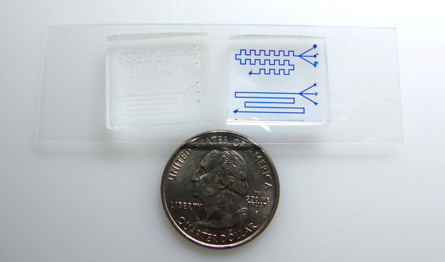
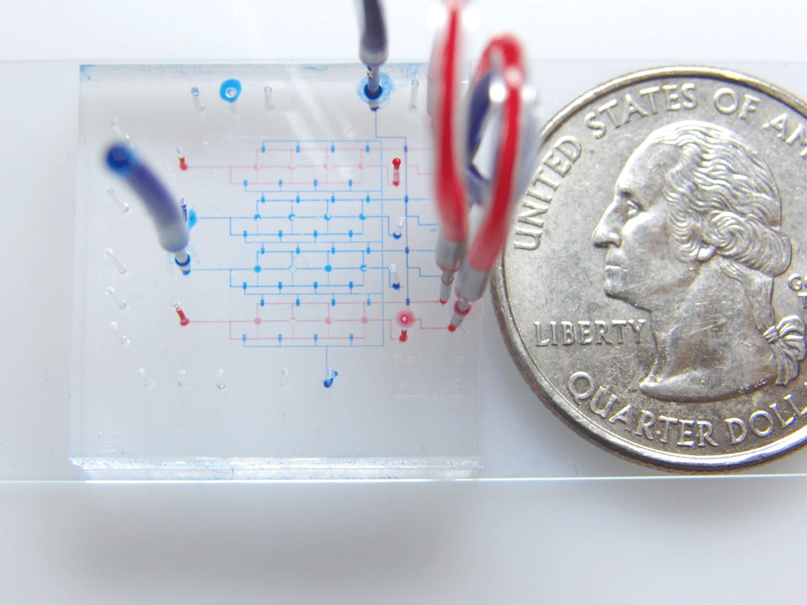
  7. Devices are ready for display. Notice the enhanced contrast of the colour filled channels vs the empty channels for the same device in Figure 2. While channels are visible only from some directions when they reflect light, colour-PDMS devices can be observed from every direction. Additionally, different device layers or areas can be specified by colour. In the figure control layers are blue and flow layers are red

Fig. 1 Injection of PDMS through the channels. Air trapped inside the syringe provides a way to regulate the pressure applied to the device to minimize de-bonding. Compressing the air to 1/3 of original volume should provide enough pressure to drive the PDMS through.


Fig. 2 Enhanced channel contrast after injection, devices on the left side have empty channels and devices on the right have color PDMS inside.

Fig. 3 Different device zones can be identified by color. Here control layer is blue and flow layer is red. Secondary regulation channels are practically invisible when not filled.

References


[1] http://www.upchurch.com
[2] http://www.smooth-on.com

Digg This
Reddit This
Stumble Now!
Share on Facebook
Bookmark this on Delicious
Share on LinkedIn
Bookmark this on Technorati
Post on Twitter
Google Buzz (aka. Google Reader)

Degassing a PDMS mixture without a vacuum desiccator or a laboratory centrifuge and curing the PDMS chip in an ordinary kitchen oven

Aung K. Soe and Professor Saeid Nahavandi
Centre for Intelligent Systems Research, Deakin University, Australia

Why is this useful?


Whitesides (2001) advocated that soft lithography can facilitate researchers to fabricate PDMS lab-on-a-chip devices with accessible and affordable resources [1]. A typical soft lithography fabrication laboratory requires master reverse molds, a PDMS pre-polymer mixer, scales for weighing, a vacuum desiccator, an oven and oxygen plasma treatment of PDMS surfaces to become hydrophilic [2].

In soft lithography, the PDMS elastomer is first mixed with the cross-linker (curing agent) in a weight ratio of 10:1. Stirring the mixture forms air bubbles. Traditional soft lithography protocols recommend using a vacuum desiccator before pouring the mixture onto the mold master. The PDMS pre-polymer is cured fully or partially inside the oven at 80 to 95 °C for 15 to 20 minutes, transforming the resin into solid silicone rubber. However, not all researchers who need to fabricate PDMS chips have all the resources stated.

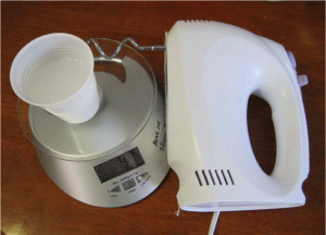
LaFratta (2010) demonstrated that a laboratory centrifuge can be used to degas PDMS [3], but a desktop laboratory centrifuge (to degas) and an oven (to cure) may not be accessible or affordable to all researchers. The technique described here uses a handheld electric mixer to mix and degas the PDMS pre-polymer. An ordinary kitchen oven is used to cure the PDMS chip.

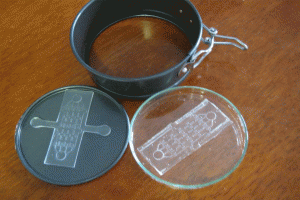
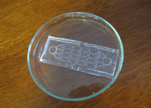
Since the food processor cannot be used for more than 3 minutes, the procedure is done in 2 sections, each lasting 2.5 minutes with 1 minute period between them. A Pyrex® petri dish is used to hold the reverse mold (master), so the dish will not deform inside the oven. The oven must be switched on and off so that the Pyrex does not crack.

What do I need?


  • SYLGARD 184 silicone elastomer base and curing agent (Dow Corning)
  • a low-cost hand-held electric mixer
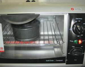
  • a kitchen grill oven with sustained heating capacity at 80 °C
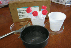
  • a baking cup (or a Pyrex petri dish) and polystyrene petri dishes for PDMS chip storage
  • a polished micro-machined or patterned reverse master mold that will not float on the PDMS pre-polymer
  • a scale that can work in units of grams
  • consumables: gloves, disposable cups, 2 or 4 plastic centrifuge tubes (15 ml capacity)

How do I do it?


1. Weigh the PDMS base and curing agent in the desired ratio in a disposable cup. 10:1 is the most common ratio, but any ratio can be used depending on the desired stiffness of the cured polymer.
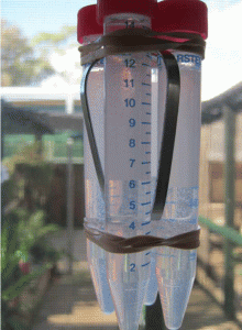
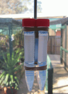
2. Mix base and curing agent together with the stirrer attachment of the hand-held electric mixer. Pour the pre-polymer evenly into 2 or 4 centrifuge tubes. If there is not enough pre-polymer for 2 or 4 tubes, fill 1 or 3 tubes and one more tube with water for balancing. It is important to balance the attachment during spinning. Tightly seal the centrifuge tube caps.
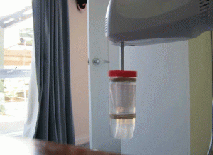
3. Tie the tubes to the stirrer attachments of the handheld mixer. Hold the mixer in a vertical position, switch on and spin for 2.5 minutes, then stop to give the spinner a rest for 1 minute. Turn the bottles 180 degrees and spin for another 2.5 minutes until all the bubbles have escaped from the pre-polymer mixture.
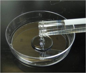
4. Pour the centrifuged PDMS onto the patterned reverse master mold in the baking cup or Pyrex petri dish.
5. Put the master mold container in the oven and cure at 80°C for 10 minutes if the baking cup is used. If the Pyrex petri dishes are used, the heat must be switched on for 5 minutes then off for 5 minutes since Pyrex dishes can crack under continuous heat. Switch the heat on and off for 30 minutes.
6. Peel the PDMS slab off the master using a sharp-tipped knife and store the PDMS chip in a polystyrene dish before autoclaving or treating with oxygen plasma.

Figure 1. Weighing, mixing and stirring

Figure 2. Before spinning

Figure 3. Spinning the tubes attached to the processor

Figure 4. After spinning

Figure 5. Acrylic reverse mold in the baking cup

Figure 6. Baking cup in ordinary kitchen oven at 80 °C

Figure 7. Peeling off PDMS slab from the baking cup

Figure 8. Storing the PDMS chip inside a petri dish


What else should I know?


The reverse master can be fabricated in microfluidics fabrication foundries at Stanford University or California Institute of Technology. Reverse masters can also be made by machining pcrylic (also known as PMMA and plexiglass) with subtractive CNC machines such as the Roland DG MDX 40. Three-dimensional additive printers can also be used. Third-party service companies can also do micro-machining and surface polishing if one wishes to outsource the master fabrication task.

References


[1] G. Whitesides et al., Soft lithography in biology and biochemistry, Annu. Rev. Biomed. Eng., 2001, 3(1), 335-373.
[2] A. Harsch et al., Pulsed plasma deposition of allylamine on polysiloxane: a stable surface for neuronal cell adhesion, J. Neurosci. Methods, 2000, 98(2), 135-144.
[3] C. N. LaFratta, Degas PDMS in two minutes, Chips & Tips (Lab on a Chip), 17 August 2010.

Digg This
Reddit This
Stumble Now!
Share on Facebook
Bookmark this on Delicious
Share on LinkedIn
Bookmark this on Technorati
Post on Twitter
Google Buzz (aka. Google Reader)

Degas PDMS in two minutes

Christopher N. LaFratta
Department of Chemistry, Tufts University, Medford MA, USA

Why is this useful?


Mixing the base and curing agent of PDMS inevitably incorporates air bubbles into the prepolymer.  These bubbles are usually removed after casting by degassing the sample under vacuum.  Vacuum degassing works well but requires about 30 minutes or more depending on the vacuum and amount of gas stirred in.  The technique described here uses a typical laboratory centrifuge to degas PDMS in two minutes.  The centrifuged PDMS will yield a bubble-free solid silicone rubber when cast on a smooth surface.

What do I need?


  • SYLGARD 184 silicone elastomer base & curing agent (Dow Corning)
  • 2 Centrifuge tubes (15 mL)
  • Clay Adams II compact centrifuge (3200 rpm or 1315 × g relative centrifugal force)

How do I do it?


  1. Weigh the PDMS base and curing agent (10:1) in a disposable centrifuge tube.
  2. Mix base and curing agent together with a wooden stick.
  3. Balance centrifuge with 2nd tube containing comparable amount of PDMS or water.  Place in centrifuge for 2 min.
  4. Cast centrifuged PDMS onto patterned wafer.
  5. Oven cure at 75°C for 1 h.

What else should I know?


If the PDMS is cast over high aspect ratio features, such as tall SU-8 photoresist lines, air bubbles may get folded in as the liquid PDMS flows over crevices.  These small isolated bubbles can usually be degassed under vacuum in about one minute.

A) Mixed PDMS in a centrifuge tube, note the air bubbles that have been mixed in. B) PDMS being degassed by centrifugation. C) Degassed PDMS after 2 minutes of centrifuging without any air bubbles.

Casting PDMS on a patterned silicon wafer

Cured PDMS cut from silicon wafer mold without air bubbles.

References


[1] C. N. LaFratta, T. Baldacchini, R. A. Farrer, J.T. Fourkas, M. C. Teich, B. E. A. Saleh, M. J. Naughton J. Phys. Chem. B, 2004, 108, 11256-11258.

Digg This
Reddit This
Stumble Now!
Share on Facebook
Bookmark this on Delicious
Share on LinkedIn
Bookmark this on Technorati
Post on Twitter
Google Buzz (aka. Google Reader)

An easy temperature control system for syringe pumps

Lorenzo Capretto, Stefania Mazzitelli and Claudio Nastruzzi
Department of Chemistry and Technology of Drugs, University of Perugia, Perugia, Italy

Why is this useful?


The most common way to pump liquid into microfluidic devices is by syringe or peristaltic pumps. Syringe pumps are usually preferred for their ease of use and for the most accurate and stable control of the flow rate. In addition, syringe pumps allow the use of disposable and sterile syringes very much facilitating all the protocols involving cells that require sterile conditions.

On the other hand, syringe-pumps have the disadvantage that regulating the temperature of the pumped liquid is difficult. This feature is particularly relevant for a number of microfluidic applications when the temperature of one or more liquid phases, pumped through the chip, should be strictly controlled. For instance, manipulation of animal cells (movement, sorting or encapsulation) usually requires a maintained temperature of 37°C. Moreover many protocols involving the use of polymers or labile compounds need controlled temperatures either below 0°C or above 40-50°C.

We propose a tip that, in an easy and cheap way, solves the problem of temperature control when using syringe-pumps, no matter which type of microfluidic devices are used.

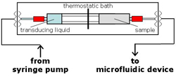
The general idea depends on the use of two coupled syringes (see Fig. 3B) acting as a pressure transducing system. The two coupled syringes, assembled as reported in the general scheme (Fig. 5), can be easily maintained at a fixed temperature by a cheap and flexible thermostatic bath, allowing liquid to be pumped into the chip at a constant, controlled temperature.

What do I need?


  • 1-30 mL polypropylene syringes (Artsana, Italy)[1]
  • Clips for conical joints KECKT (for 5 mL syringes use KECKT KC 14, Schott Duran, Germany, No.: 29 031 00)[2]
  • Teflon® FEP Tubing, 1/8″ OD (Upchurch Scientific, UK; No.: 1523)[3]
  • Quick Connect Luer Adapters, Female Luer to 1/4-28 Female (Upchurch Scientific, UK; No.: P-658)[3]
  • Nuts, for 1/8″ OD tubing (Upchurch Scientific, UK; No.: P-345)[3]
  • Flangeless Ferrules, for 1/8″ OD tubing (Upchurch Scientific, UK; No.: P-300)[3]
  • Standard Polymer Tubing Cutter for 1/16″ and 1/8″ OD tubing (Upchurch Scientific, UK; No.: A-327)[3]
  • Coping saw (X-ACTO, USA)[4]
  • Thermostatic bath equipped with a DC10 Immersion Circulator (Thermo Haake, Germany, No.:426-1001)[5] and with a screw clamp for a plexiglass bath (15x40x15 cm)

What do I do?


The procedure below refers to the assembly of a transducing system based on the use of 5 mL syringes.

1. Saw or cut the plungers of the two syringes, one at 1.5 cm from the plug (sample syringe) and the other at 6.5 cm from the plug (transducing syringe).

Figure 1.

2. Insert the longer plunger of the transducing syringe into the barrel of the sample syringe.

Figure 2.

3. Fix the barrels of the transducing and sample syringes with a KECK clip.

Figure 3.

4. Insert the Luer lock port for both transducing and sample syringes.

Figure 4.

5. Place the two syringe assembly into the thermostatic bath.

6. Operate the system following the general assembly scheme reported in Fig. 5A.

Figure 5A

Figure 5B

References


[1] www.artsana.com
[2] www.duran-group.com
[3] www.upchurch.com
[4] www.xacto.com
[5] www.thermo.com

Digg This
Reddit This
Stumble Now!
Share on Facebook
Bookmark this on Delicious
Share on LinkedIn
Bookmark this on Technorati
Post on Twitter
Google Buzz (aka. Google Reader)

Preventing suspension settling during injection

Ryan Cooper and Luke Lee
Department of Bioengineering, University of California-Berkeley, Berkeley, California

Why is this useful?


This technique was developed to solve the problem of cell suspensions settling to the bottom of the syringe during the time it took to load them into a device. It is a gentle method to keep mixtures of cells, beads and other particles in suspension.

What do I need?


  • 1 Loading Syringe
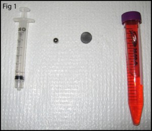
  • 1 Stainless Steel Ball Bearing (recommended diameter 1/3 to 2/3 the inside diameter of the syringe)
  • 1 Magnet

What do I do?


1. Clean the ball bearing with acetone followed by alcohol to remove any grease and sterilize its surface.

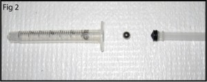
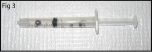
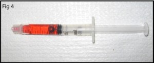
2. Pull the plunger out of the syringe (in a clean hood if sterile conditions are required) and drop the ball bearing into the tube, then reinsert the plunger (Fig. 2, Fig. 3).

3. Fill the syringe part way with the desired solution (Fig. 4)

4. Hold the syringe upright and tap its side to make the air bubbles inside float to the top of the syringe, then force them out with the plunger.

5. Once the air bubbles are out, finish filling the syringe (Fig. 5).

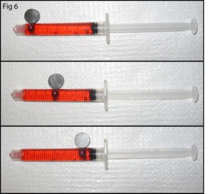
6. To prevent the solution inside the syringe from settling, simply move the magnet back and forth across the surface of the syringe, dragging the ball bearing back and forth inside the syringe and agitating the solution (Fig. 6).

7. Now you can take as long as you need to load the solution since you can prevent settling. If you do not have a magnet, the solution can be agitated by tipping the syringe and using gravity to move the bearing. Placing the entire syringe pump on a rocker plate could also be employed to move the bearing.

Digg This
Reddit This
Stumble Now!
Share on Facebook
Bookmark this on Delicious
Share on LinkedIn
Bookmark this on Technorati
Post on Twitter
Google Buzz (aka. Google Reader)

Avoiding bubble injection by droplet merging

Edmond W.K. Young, Aaron R. Wheeler and Craig A. Simmons.
University of Toronto, Canada.

Why is this useful?


In many microfluidics applications, the presence of bubbles can be undesirable because of their tendency to disturb fluid flow in microchannels.  For cell culture studies in particular, cells that are not strongly adhered to the underlying substrate can be sheared, detached, and entrained by passing bubbles, leaving behind regions depleted of cells.  Many microchannel designs therefore incorporate either bubble traps [1] or complex valve configurations [2] to eliminate bubbles from being introduced into the channels.  However, these elements add complexity, and thus are not ideal for use with rapid prototyping experiments, in which fluid is typically delivered by means of a syringe. We present a tip that avoids the common phenomenon of bubble injection by careful attention to reservoir fluid volumes.

What do I need?


  • PDMS-glass microchannel, irreversibly sealed, with polyethylene tubing (Intramedic) inserted into cored-out holes in the PDMS and sealed with epoxy (LePage)
  • Syringe, 1 mL (BD)
  • Syringe needle, 18 gauge (BD)
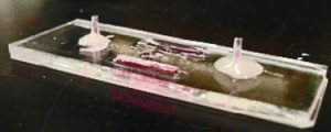
  • Fluid mediumTypical Microfluidic Chip

Figure 1. A Typical Microfluidic Chip

What do I do?


Prior to sample injection, the microfluidic device is primed with fluid and the inlet and outlet ports are not completely filled (Figure 1).  If a syringe needle is inserted into the inlet port (Figure 2a) at this stage, air will be trapped between the syringe needle and the liquid-air interface, causing bubbles to be injected into the microchannel. To avoid this phenomenon:

1. Insert syringe needle into outlet port (Figure 2b).  Note that this traps a bubble on the outlet side.

2. Slowly depress syringe to push fluid toward the inlet side.  Do so until fluid reaches the top of the inlet port and forms a small bead.
3. Remove syringe needle from outlet port.  Note that the liquid interface on the outlet side should now be lower than before Step 2.  However, the trapped bubble from Step 1 should now be eliminated.

4. Depress syringe to generate a small droplet at the tip of the needle (Figure 2c).

5. Touch the droplet at the needle tip to the bead of fluid at the inlet (Figure 2d).  Merging the droplet and the bead prevents formation of an air bubble.
6. Inject sample from syringe into channel as desired; no bubbles will be injected.

Figure 2a

Figure 2b

Figure 2c

Figure 2d

References


[1] E. Leclerc, Y. Sakai, and T. Fujii, Cell culture in 3-dimensional microfluidic structure of PDMS (polydimethylsiloxane). Biomed. Microdevices, 2003, 5(2), 109-114.
[2] L. Kim, M.D. Vahey, H.-Y. Lee, and J. Voldman, Microfluidic arrays for logarithmically perfused embryonic stem cell culture. Lab Chip, 2006, 6, 394-406

Digg This
Reddit This
Stumble Now!
Share on Facebook
Bookmark this on Delicious
Share on LinkedIn
Bookmark this on Technorati
Post on Twitter
Google Buzz (aka. Google Reader)