Archive for the ‘Emerging Investigators’ Category

Emerging Investigator Series – Aaron Streets

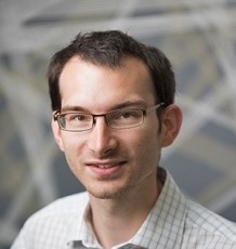
Lab on a Chip is very excited to introduce our most recent Emerging Investigator, Dr Aaron Streets! 

Aaron received a Bachelor of Science in Physics and a Bachelor of Arts in Art at the University of California, Los Angeles. He completed his PhD in Applied Physics at Stanford University with Dr. Stephen Quake. Aaron then went to Beijing, China as a Whitaker International Postdoctoral Fellow and a Ford postdoctoral fellow and worked with Dr. Yanyi Huang in the Peking University BIOPIC institute. Aaron joined the faculty of the University of California, Berkeley as an Assistant Professor in Bioengineering in 2016. He is currently a core member of the Biophysics Program and the Center for Computational Biology and he is a Chan Zuckerberg Biohub investigator. Aaron has received the NSF Early Career award and was recently named a Pew Biomedical Scholar.

You can read Aaron’s article, μCB-seq: microfluidic cell barcoding and sequencing for high-resolution imaging and sequencing of single cells, here.

If you’d like to know more about Aaron and his research, read the short interview with him below!


Your recent Emerging Investigator Series paper focuses on microfluidic cell barcoding and sequencing. How has your research evolved from your first article to this most recent article?

My first publication used a microfluidic-based dynamic light scattering apparatus to investigate protein crystallization. While this study relied on the integration of microfluidic control and optical analysis, it was really focused on the physics of biological macromolecule phase transitions. No cells, no sequencing, not even any microscopy.  The continuous thread that has persisted through my research has been the integration of multilayer microfluidic circuits with advanced optical analysis techniques, however we now take advantage of this technology to study single cells. This means that we rely on a lot of additional experimental and computational tools upstream and downstream of the microfluidics, including interfacing directly with biological samples that are input into our devices, as well as the library preparation, high-throughput sequencing, and ultimately genomic data analysis that are essentially the device outputs. So, while “lab-on-chip” technology is still at the core of our research, we rely heavily on a “chip-in-a-lab” paradigm.

What aspect of your work are you most excited about at the moment?

After a decade-and-a-half of working with microfluidic technology it is extremely energizing to see microfluidics percolate into all corners of biology and the biomedical sciences. It is particularly exciting to see microfluidic platforms become the primary driving technology that has advanced the field of single-cell biology. From the integrated microfluidic circuits that provided the first commercial single-cell RNA-seq platforms, to the droplet microfluidics and microwell technology that has brought ultra-high-throughput single-cell genomics to the masses and has fuelled the Human Cell Atlas Project. In our lab, we are excited about taking a deeper dive into single-cell measurements, and instead of optimizing throughput, really pushing the information content that can be extracted from a single cell. One of the beautiful aspects of integrated microfluidic circuits is that they pair incredibly well with advanced microscopy techniques while facilitating single-cell manipulation and sequential molecular biology protocols. We are excited about using these capabilities to make multimodal measurements on single cells, connecting genomic information to the spatial organization of molecules and other morphological phenotypes that are probed more efficiently with light.

In your opinion, what is the biggest advantage to using the μCB-seq platform compared to other methods?

It is easy to take a high-resolution image of a single cell, and it is easy (nowadays) to amplify and sequence the RNA or DNA from a single cell. It is still challenging, however, to take a high-resolution image of a single cell, and then pluck that same cell from the microscope slide and sequence it.  Microfluidic cell barcoding and sequencing (μCB-seq) provides a way to do just that for many cells in parallel. With this platform, we are really just using microfluidic chambers to help keep track of which cell was imaged before sequencing. To make this work, we use DNA barcodes that are pre-loaded into the microfluidic device so that each single-cell imaging chamber gets a unique cell barcode. After imaging each cell, that unique barcode gets imprinted into the cDNA from each respective cell so that we can pool all the cDNA from the lanes of our device and make a single sequencing library. After sequencing, the barcode will tell us which transcripts came from which cell, linking gene expression to image data.

What do you find most challenging about your research?

The most challenging aspects of our work tend to be figured out by the PhD students. These are the details you can’t find in a textbook like how to make a device robust to thermal cycling while under a microscope, or where did the cDNA go, or why isn’t this thing working? The truth is that there is a significant up-front investment in resources and time to get these platforms functional. Furthermore, depending on the type of microscopy, many of these tools cannot be ported into other labs. Once we get something like μCB-seq to the proof-of-principle stage there is another set of obstacles to make it a robust technology that we can train collaborators to use efficiently, so that the technology can have impact. Getting these tools into the hands of people who want to use them is still a non-trivial challenge, especially as the front-end and back-end of these experiments require more and more equipment and expertise. 

At which upcoming conferences or events may our readers meet you?

I would love to see everyone at the Physics and Chemistry of Microfluidics Gordon Research Conference in 2021. But if we can’t see each other in Tuscany, I look forward to seeing you on Zoom!

How do you spend your spare time? 

These days? Good question. I enjoy reading science fiction. I am an avid basketball fan. I love listening to live music. So I guess the answer is… reading science fiction.

Which profession would you choose if you were not a scientist?

I would be a world-famous artist. (Are we allowed to choose how successful we would be? Or just the profession?) Ok, I would be an artist… and a bartender. I studied art in undergrad at UCLA and I always imagined an alternate career making paintings and sculptures and art installations. 


If you’re interested in other articles in our Emerging Investigator Series, the whole collection can be found here

Digg This
Reddit This
Stumble Now!
Share on Facebook
Bookmark this on Delicious
Share on LinkedIn
Bookmark this on Technorati
Post on Twitter
Google Buzz (aka. Google Reader)

Emerging Investigator Series – Fabrice Gielen

Lab on a Chip is delighted to introduce our most recent Emerging Investigator, Fabrice Gielen! 

Dr. Fabrice Gielen is an Independent Research Fellow at the Living Systems Institute, University of Exeter. He obtained his PhD from Imperial College London during which he studied cellular membrane dynamics using a combination of single cell microfluidic trapping techniques and single molecule spectroscopy with Profs. Joshua Edel and Andrew deMello. He subsequently moved to Cambridge as a Post-Doctoral Research Associate with Prof. Florian Hollfelder to contribute to the field of ultra-high-throughput biocatalyst evolution. He is a Founder and Scientific Director of Drop-Tech Ltd, which commercializes droplet-on-demand platforms. His research interests include developing novel tools and methods to study and harness single cells with applications in bacteria-phage interactions, protein evolution and regenerative medicine.

Read Dr Gielen’s Emerging Investigator paper* “Deep learning guided image-based droplet sorting for on-demand selection and analysis of single cells and 3D cell cultures” and find out more about him and his research in the interview below. 

Picture of Emerging Investigator, Fabrice Gielen


Your recent Emerging Investigator Series paper focuses on sorting of single droplets for selection and analysis of single cells and 3D cell cultures. How has your research evolved from your first article to this most recent article?

 My first article (long ago already!) presented a microfluidic device for trapping and focussing microparticles within microchannels. The common theme with my present work is how precise control at the microscale enables us to perform novel types of biological experiments across populations. We can now truly benefit from the progress made in engineering, computing and optical setups to study biology, not just faster but in fundamentally different ways than was possible when I started my career.


What aspect of your work are you most excited about at the moment?

Studying single cells holds the promise to understand biology down to its fundamental unit. I joined the Living Systems Institute at the University of Exeter to exploit the advantages of droplet microfluidics in terms of single cell control and interrogation for the study of complex biological systems. Since then, I have developed multiple interests ranging from the behaviour of unicellular organisms, cellular differentiation or the discovery of novel antimicrobials. I believe there is also a large scope for other, yet unforeseen applications.


In your opinion, what is the biggest advantage to using your platform for classifying single droplet images compared to other methods?

 Images hold a lot of information and machines can uncover patterns where humans cannot. Recent technological progress allows us to combine image acquisition and analysis in real-time. Problems such as image classification used in medical diagnostics or selection to isolate rare cell types benefit enormously from machine learning tools that can identify objects only using human-labelled training data. Our paper shows one way of doing this is by using convolutional neural networks which can be trained to recognize several types of micro-objects simultaneously.


What do you find most challenging about your research?

The ability to combine in a single experiment microfluidic workflows, biological entities, and biochemical reactions has the flipside that successful and conclusive experiments happen only when all these variables coincide (which is sometimes rare!). On the bright side, microfluidics has gone a long way in terms of reproducibility and long-term functionality and the onus is now more on the biology.


At which upcoming conferences or events may our readers meet you?

I am planning to attend the MicroTAS 2020 (Palm Springs) and EMBL microfluidics (Heidelberg).  


How do you spend your spare time?

I like to take the family out to visit Devon and the Jurassic Coast, which I highly recommend to anyone visiting the region.


Which profession would you choose if you were not a scientist?

I would go for musician/composer, maybe pianist.


*Dr Gielen’s paper is free to access with an RSC account for the next month.

Digg This
Reddit This
Stumble Now!
Share on Facebook
Bookmark this on Delicious
Share on LinkedIn
Bookmark this on Technorati
Post on Twitter
Google Buzz (aka. Google Reader)

Emerging Investigators Series – Jacqueline Linnes

Dr. Jacqueline Linnes is an Assistant Professor in the Weldon School of Biomedical Engineering at Purdue University. She received her B.S. in Engineering from the Purdue University and Ph.D. in Biongineering from the University of Washington. She was a Fogarty Postdoctoral Fellow at the Division of Global Health Equity within Brigham and Women’s Hospital/Harvard Medical School and continued her postdoctoral training at Boston University in Biomedical Engineering. She has received numerous awards including the Mandela Fellows Global Innovation Challenge Award (2017), Fast Company’s World Changing Ideas Finalist (2018), and Marta E. Gross Assistant Professorship of Biomedical Engineering (2018).

Dr. Linnes’s research lab develops real-time detection technologies to prevent, diagnose, and better understand the pathogenesis of diseases. This work emphasizes the translation of fundamental microfluidics and biological assays into point-of-care diagnostics using human-centered design principles. Her extensive experience in translational research includes co-founding and managing early-stage field-testing and user feedback for two startup companies. She has co-developed point-of-care health diagnostics, wearable devices, and water purification technologies for use in the US, Bolivia, Nicaragua, Kenya, Zambia, and Haiti.

Read Dr Linnes’s Emerging Investigator article “Microfluidic Rapid and Autonomous Analytical Device (microRAAD) to Detect HIV from Whole Blood Samples” and find out more about her in the interview below.


Your recent Emerging Investigator Series paper focuses on detecting HIV using a microRAAD. How has your research evolved from your first article to this most recent article?

As a PhD student, I published on the causes of bacterial adhesion to proteins adsorbed to medical devices (in 2012). These infections are incredibly difficult to detect and I found that I didn’t want to just study the causes of infections but to develop the diagnostic tools themselves. I now use the molecular biology and surface analysis skills that I developed in my PhD to create point-of-care diagnostic devices in my own lab. A critical shift in my thinking came when I realized that both the technical skills and the problem solving mindset that I learned in my PhD research were transferable to entirely different fields. In my two postdocs, I worked on Global Health projects ranging from infection control to point-of-care diagnostic devices.  Now in my own research lab, we focus on developing, integrating and automating real-time detection technologies including point-of-care diagnostics and wearable devices, to meet the needs of underserved populations. This article is an example of a sample-to-answer test we developed to automate molecular detection of a pathogen (HIV) from whole blood sample at the point-of-care.


What aspect of your work are you most excited about at the moment?

I started my lab in 2015 with the vision that we could automate molecular detection in point-of-care diagnostics, so that the dvices could be used by anyone, anywhere in the world. I love that we have pulled together individuals with expertise in so many different fields from materials science, to electrical engineering, to molecular biology in order to make this technology work. A huge contingent of my lab and Dr. Stanciu’s lab, and at all levels, from undergraduate researchers to PhD’s have contributed to this project. Now we are bringing in more expertise in translational clinical research. I am currently in Kenya and just handed over a batch of these microRAADs to my colleague, Dr. Eddy Odari at Jomo Kenyatta University of Agriculture and Technology. Dr. Odari will be testing the MicroRAADs using real patient samples and I can’t wait to find out the results.


In your opinion, what is the biggest advantage to using your microRAAD compared to other methods of detecting HIV?

I know there’s still a ways to go, but I believe that the microRAAD platform will ultimately bridge the gap between laboratory-based molecular detection instruments and truly point-of-care diagnosis of HIV in the field.


What do you find most challenging about your research?

Designing technologies sample-to-answer molecular diagnostics that are both highly sensitive and remain robust and accessible to the clinicians, technicians, and patients who need them is incredibly challenging. In my lab, we find it critical to test out our ideas and prototypes via formal and informal usability studies to understand what can be done practically in the field settings that they are designed for. We redesign anything that isn’t actually usable in the real world.


At which upcoming conferences or events may our readers meet you?

I am at the 4th Africa International Biotechnology and Biomedical Conference in Nairobi and Mombasa, Kenya, and this October I will be attending the 2019 Biomedical Engineering Society Annual Meeting in Philadelphia, USA, and the 2019 MicroTAS conference in Basel, Switzerland.


How do you spend your spare time?

I have a 5 year old and a 3 year old so “spare time” is perhaps an overstatement, but we spend a lot of time outdoors at parks and playgrounds and my husband and I built a tree house in our backyard this summer.


Which profession would you choose if you were not a scientist?

That’s a tough one. I love my job as a biomedical engineering faculty member. I do think it would be fantastic to work at a science museum developing and building exhibits and outreach activities.


Can you share one piece of career-related advice or wisdom with other early career scientists?

Don’t underestimate the power that people play in your research. Play well with others, find a place that supports you in your efforts, seek out excellent employees and mentees, and make sure to invest in their development and in your own. Whenever possible, work directly with the people that you ultimately want to use your technology. It is both incredibly motivating and absolutely critical to making an impact that reaches beyond the confines of your own lab.

Dr Jacqueline Linnes

Dr Jacqueline Linnes (Picture credit: Purdue University photo/Rebecca Wilcox)

Digg This
Reddit This
Stumble Now!
Share on Facebook
Bookmark this on Delicious
Share on LinkedIn
Bookmark this on Technorati
Post on Twitter
Google Buzz (aka. Google Reader)

Emerging Investigator Series – Joo H. Kang

Dr. Joo H. Kang is currently an Assistant Professor of the Department of Biomedical Engineering, School of Life Sciences at Ulsan National Institute of Science and Technology (UNIST), Korea. He received double Bachelor’s degrees in Chemical Engineering and Life Science from Sogang University in 2002 and his M.S. and Ph.D. in Bioengineering from Korea Advanced Institute of Science and Technology (KAIST) in 2004 and 2008, respectively. He joined Children’s Hospital Boston, Harvard Medical School as a research fellow in 2009, and he continued his work at the Wyss Institute, Harvard University as a Wyss Technology Development Fellow from 2012-2016. He received several awards in his early career, including Postdoctoral Award for Professional Development from Harvard University, Wyss Technology Development Fellowship from Harvard University, Baxter Young Investigator Award from Baxter Inc., and Young Frontiers in Bio and Braining Engineering from KAIST. His research interests include multiscale biofluidic approaches for tackling infectious diseases and cancer, and miniaturized organ-mimicking microsystems.

Read Joo H. Kang’s Emerging Investigator article “Measurement of the magnetic susceptibility of subtle paramagnetic solutions using the diamagnetic repulsion of polymer microparticles” and find out more about him in the interview below:


Your recent Emerging Investigator Series paper focuses on measuring magnetic susceptibility of subtle paramagnetic solution using diamagnetic repulsion of polymer microparticles. How has your research evolved from your first article to this most recent article?

One of the research topics that interested me was to discriminate the subtle differences in the magnetic susceptibility of materials in a microfluidic regime. The first paper I published in regards to this (Kang,JH, et al., JACS, 2009) demonstrated the capability of discriminating the magnetic susceptibility of “solid microparticles” where they are diamagnetically forced to be located at a quasi-isomagnetic position in a microfluidic channel (a position where the differences of the magnetic susceptibility between the solid particles and surrounding media become nearly zero). When I was invited to make contribution to the Emerging Investigator Series of Lab on a Chip last year, I wanted to revisit this, and this time I aimed to assess the subtle magnetic susceptibility of “surrounding paramagnetic solutions”.

What aspect of your work are you most excited about at the moment?

As for the paper, I was surprised of the sensitivity of the device that can discriminate the magnetic susceptibility. We compared our results with those assessed by a conventional superconducting quantum interference device (SQUID), and found that our approach is even more sensitive than the conventional one. Likewise, we can unveil various scientific approaches when exploring fluidic regimes at the micro and nanoscale, and this is the most exciting aspect as being a part of the research community in this field.

In your opinion, what applications can your current approach be used for?

Because this is a platform technology, various applications are possible where we need to measure the magnetic susceptibility of paramagnetic solutions. Assessment of residual magnetic nanoparticles in biological samples, for examples, would be one of the potential uses. We could also use this platform to evaluate metal contamination of drinking water, such as chromium or iron oxide, which alters the magnetic susceptibility of water.

What do you find most challenging about your research?

Taking research from the “bench to products”. Since I started my independent research career, I realized that I have to make considerable efforts to get this happen while playing multiple roles at the same time. But I am enjoying it.

In which upcoming conferences or events may our readers meet you?

I am planning to attend microTAS 2019 that will be held in Basel, Switzerland this year.

How do you spend your spare time?

I am spending my time with my family, hiking, swimming, and playing soccer or games with my little son and daughter.

Which profession would you choose if you were not a scientist?

Probably an architect. This was one of the paths I was thinking of when I was a high school student.

Can you share one piece of career-related advice or wisdom with other early career scientists?

A clear vision on your own research and collaborators who you can share your vision with.

Digg This
Reddit This
Stumble Now!
Share on Facebook
Bookmark this on Delicious
Share on LinkedIn
Bookmark this on Technorati
Post on Twitter
Google Buzz (aka. Google Reader)

Emerging Investigator Series – Mei He

Dr. He is a tenure-track assistant professor at the University of Kansas. She received her PhD degree from the University of Alberta with professor Jed Harrison, and postdoctoral training from the University of California, Berkeley with professor Amy Herr. She is the vice chair of the ASABE Biosensor program and the Councilor of the American Electrophoresis Society. Dr. He is also the founder of Clara Biotech Inc. and the founder committee for the MidWest 3D technology society. Dr. He Received NIH Maximizing Investgator’s Research Award for Early Stage Investigators in 2019. She also received the Lab on Chip Outstanding Reviewer for the year of 2018. One of her publications also received the 2018 SLAS Technology Readers Choice Award. Her research interests include biomedical microfluidic devices and sensing approaches, 3D biomaterials, and nanodelivery, employed in programming and monitoring biomimetic immunity associated with extracellular vesicles.

Read Mei He’s Emerging Investigator article “3D-printing enabled micro-assembly of a microfluidic electroporation system for 3D tissue engineering” and find out more about her in the interview below:


Your recent Emerging Investigator Series paper focuses on “3D-printing Enabled Micro-assembly of Microfluidic Electroporation System for 3D Tissue Engineering”. How has your research evolved from your first article to this most recent article?

My first article observed the microscale evolution of porous polymer materials in the microfluidic channel when I was a PhD student. I found very interesting phenomena in the microfluidic device which actually inspired me to explore more surrounding dimensions, surface chemistry, and scales. Till to my recent article focusing on 3D geometric influence on cellular behavior and their extracellular vesicles secretion dynamics, the 3D dimension in microscale is intriguing in the biological system.

What aspect of your work are you most excited about at the moment?

I am very excited to take the microfluidic technology and phenomena into the biology world, as it will bring new investigation and discovery. Biology is still in its infancy stage, I am very excited to see how microfluidic technology could advance this growth.

In your opinion, what is the biggest impact your microfluidic electroporation system will have in tissue engineering?

Intracellular delivery of regulatory or therapeutic targets into the cell is very crucial in the field of tissue engineering and regenerative medicine. Current existing electro-transfection systems, including microfluidic platforms and commercial benchtop systems, are only able to study monolayer cell suspensions in vitro, which is incapable of clinical translation within in vivo tissue microenvironment. So developing a 3D, in vivo like tissue microenvironment with effective electro-transfection is very important to move to the clinical study in the future. We actually are more interested in downstream, precise control and manipulation of cellular machinery for secreting exosomes and extracellular vesicles under the transfection-induced stimulus, such technology is not existing yet but very important for understanding the interconnection of cargo internalization with cellular level responses elicited by exosomes delivery pathway.

What do you find most challenging about your research?

Building up an in vivo like tissue system with precise control is not straightforward. The environment in a controlled lab setting is totally different than in an in vivo biological system. So the analyzed information actually is not representative of the real situation in the human in vivo system. There are huge heterogeneities present in the cell population as well as human individuals, which poses the challenges for correctly understanding cellular system regulation, such as immunity, in our human body. Mimicking in vivo living system is very challenging, but crucial for understanding quite a few of mechanism and disease pathogenesis. Our research introduces new microfluidic technology and material solutions to solve such challenges.

In which upcoming conferences or events may our readers meet you?

I will attend next year Gorden Research Conference in Bioanalytical Sensors as well as the MicroTAS annual meeting.

How do you spend your spare time?

I have a 7-year-old boy and expecting a new baby girl this year. My spare time definitely is occupied by kids and watching them growing.

Which profession would you choose if you were not a scientist?

I always like to discover new things since I was a child. If I am not a scientist, I would like to be a greeting card designer or paleontologist.

Can you share one piece of career-related advice or wisdom with other early career scientists?

Being a life-long learner and always keeping strong scientific curiosity will definitely help with your research development. Get good mentors around you and you will appreciate their advice.

Digg This
Reddit This
Stumble Now!
Share on Facebook
Bookmark this on Delicious
Share on LinkedIn
Bookmark this on Technorati
Post on Twitter
Google Buzz (aka. Google Reader)

Emerging Investigator Series – Mathieu Odijk

Mathieu Odijk is an associate Professor at the University of Twente running his own research theme on Micro- and Nanodevices for Chemical Analysis. He received his PhD in Electrical Engineering from the University of Twente under the guidance of Albert van den Berg for his work on electrochemical microreactors for drug screening and proteomics applications in 2011. He has broaden his scope by various research visits at EPFL Lausanne (2012), the Wyss Institute at Harvard (2013), and MIT (2014).

The common aim of his research is to design novel devices to measure chemical quantities, pushing boundaries in applications to explore unknown territory. Often, this relates to faster, or better spatially resolved measurements at lower concentrations in small volumes. Micro- and nanofabrication techniques are used to enhance electrochemical, optical or mass spectrometric readout. The ultimate goal is to create new, yet robust tools for routine use in the lab or point-of-care applications.

Read Mathieu Odijk ’s Emerging Investigator article “A miniaturized push–pull-perfusion probe for few-second sampling of neurotransmitters in the mouse brain” and find out more about him in the interview below:

How has your research evolved from your first article to your most recent Emerging Investigator article?

I have nice memories about my first paper, as it was also submitted to Lab on Chip and immediately accepted without revisions (only one small question from 1 of the referee’s). The topic of that first paper was about the design of an electrochemical microreactor to study oxidative conversions in drug metabolism studies. We have been quite successful with that topic, now extending it also in the direction of studying electrochemical oxidative protein cleavage, and disulphide bond reduction using e.g. spectroelectrochemical means.

Many “ingredients” included in that first paper also are present in current projects such as microfluidics, advanced cleanroom fabrication, and analytical chemistry. These ingredients also form a key component of the focus area of my own research group (Micro- and nanodevices for Chemical Analysis).

 What aspect of your work are you most excited about at the moment?

It is my aim to push the boundaries of existing analytical tools with respect to limit detection, spatial or temporal resolution, or enhancing the number of repeats using high-throughput technology. I’m really excited about this latest paper demonstrating a miniaturized push-pull perfusion probe, as it is indeed improving both the spatial and temporal resolution by at least 1 order of magnitude compared to commercially available probes. As such, it is a nice showcase of what can be achieved by microfluidics.

In your opinion, what are the most important questions to be asked/answered in this field of research? 

I think it is really important to focus on the final application, and find good collaboration partners. If I take this push-pull perfusion probe as example, this research originated from discussions with neuroscientist who complained about a lack of temporal information from their existing microdialysis probes. However, quite a number of papers that describe probes with microfluidic channels only demonstrate in-vitro results. As we also found out in our project, bridging the gap towards in-vivo is certainly not trivial. It requires compromises in the technological area which you would not address if you stick to in-vitro experiments.

More generally I believe that the field has matured; lab on chip technology has become a means to achieve a higher goal. In this case this higher goal is to study neurochemical processes in the brain in more detail. However, I think this project also clearly demonstrates that there can be a lot of science in engineering. In this case we had to overcome challenges in microfabrication, fluid dynamics, mass-transport, protein chemistry, and adsorption kinetics.

What do you find most challenging about your research?

What I find really interesting is that my research is very multi-disciplinary in nature, crossing traditional boundaries such as “chemistry”, “physics”, or “biology”. However, that also poses a challenge as it is easy to develop a blind spot if you are exploring a new field of research. Again I would like to stress the importance of a good collaboration with experts in these fields to prevent failures at an early stage.

In which upcoming conferences or events may our readers meet you?

That is easy: I always try to attend MicroTAS.

How do you spend your spare time?

I’m a father of two small children, aged 1 and 4. If they leave me some spare time (and energy), I like to do woodworking, cycling, and indoor climbing. I also really like outdoors ice skating, but global warming is unfortunately interfering with the number of days ice skating is possible in the Netherlands.

Which profession would you choose if you were not a scientist?

I always wanted to be an inventor, with teacher as a close runner-up. I guess becoming a scientist is actually pretty close to that childhood dream. Any alternative profession should allow me to be able to either create new things, or educate other people (or both).

Can you share one piece of career-related advice or wisdom with other early career scientists?

At various points in my tenure track, I felt pressure to perform. This can be stressful and is most definitely counter-productive. Try to keep seeing/finding the fun in science, e.g. by asking your PhD students to share their Eureka moments in the lab with you. All the rest is of lesser importance.

Digg This
Reddit This
Stumble Now!
Share on Facebook
Bookmark this on Delicious
Share on LinkedIn
Bookmark this on Technorati
Post on Twitter
Google Buzz (aka. Google Reader)

Emerging Investigator Series – Han Wei Hou

Dr. Han Wei Hou is currently an Assistant Professor at the School of Mechanical and Aerospace Engineering and the Lee Kong Chian School of Medicine (LKCMedicine), Nanyang Technological University (NTU), Singapore. He received his BEng (First Class Hons) and PhD degree in Biomedical Engineering from the National University of Singapore in 2008 and 2012, respectively. Upon graduation, he did his postdoctoral training at Massachusetts Institute of Technology (MIT), and subsequently joined LKCMedicine at NTU as the inaugural LKCMedicine Postdoctoral Fellow in 2014.

Dr. Hou has over 30 peer-reviewed scientific publications, and his work has been featured in online science (ScienceDaily, TheScientist, Cancerforall and Genomeweb), healthcare (News Medical), as well as technology magazines (Gizmag, Nanowerk). He has received several scientific awards including the Singapore-MIT Alliance for Research and Technology (SMART) Graduate Fellowship (2009), Young Investigator Award at the 6th World Congress of Biomechanics (2010), and LKCMedicine Postdoctoral Fellowship (2014).


His current research focus on developing novel microfluidics point-of-care testing, and biomimetic organ-on-chip technologies for translational diabetes and cardiovascular diseases research. (Research group website: www.hwhoulab.com)

Read Han Wei Hou’s Emerging Investigator article “Integrated inertial-impedance cytometry for rapid label-free leukocyte isolation and profiling of neutrophil extracellular traps (NETs)” and find out more about him in the interview below:

Your recent Emerging Investigator Series paper focuses on Integrated inertial-impedance cytometry for rapid label-free leukocyte isolation and profiling of neutrophil extracellular traps (NETs). How has your research evolved from your first article to your most recent Emerging Investigator article?

My first article when I was an undergraduate student was on the study of cancer biomechanics using microfluidics. Since then, I worked on other blood-related diseases such as malaria, sepsis and diabetes, and gradually became more interested towards microfluidics-enabled studies of host inflammation and immune responses in metabolic diseases. Regardless of disease type, our key idea is to develop integrated label-free cell sorting and biosensing approaches so that it can be cheap, fast and readily translated to clinical use. In my opinion, this work is a nice combination of all aspects.

What aspect of your work are you most excited about at the moment?

With this paper, we can now use a drop of blood to assess immune heath within minutes in a single-step user operation. We believe this work has great translational potential, and we are actively seeking new collaborators to test other diseases with immune dysfunctions.

In your opinion, what is the next step from creating your device to it being used for point-of-care testing in diabetes? and what are the most important questions to be asked/answered in this field of research?

Through this work and other recent work by our group, we have showed that diabetic leukocytes have distinct dielectric differences which can be used for immune health risk stratification. The next few important questions to ask is why are they different, and how we can further develop our technologies/assays to improve prognostic capabilities.

What do you find most challenging about your research?

As our work is highly interdisciplinary, the most challenging aspects are about finding the right people (collaborators, students etc.) and asking the right scientific questions (not too basic science, not too clinical and not too engineering)!

In which upcoming conferences or events may our readers meet you?

MicroTAS 2019 (Basel) and Microfluidics & Organ-on-a-Chip Asia Conference 2019 (Tokyo)

How do you spend your spare time?

Family time! Nowadays I enjoy spending time with my 18-month-old daughter Hannah, who never fails to amuse me or tire me out. If time permits, I will try to catch some US late-night talk shows too!

Which profession would you choose if you were not a scientist?

Tough choice! I’m torn between being a Lego/toy designer and a pilot.

Can you share one piece of career-related advice or wisdom with other early career scientists?

Talk to people outside your research disciplines. Learn to unlearn things if necessary because science and technology is advancing so fast.


Digg This
Reddit This
Stumble Now!
Share on Facebook
Bookmark this on Delicious
Share on LinkedIn
Bookmark this on Technorati
Post on Twitter
Google Buzz (aka. Google Reader)

Emerging Investigator Series – Jessie S. Jeon

Dr. Jessie S. Jeon received her SB, SM, and PhD in Mechanical Engineering from Massachusetts Institute of Technology (2008, 2010, 2014), and worked as a research fellow at Beth Israel Deaconess Medical Center, (2014-2015). She has joined the KAIST faculty in the fall of 2015 as an assistant professor in the Department of Mechanical Engineering. Her research focuses on the development of microfluidic platform with applications in investigating biological systems. She plans to further develop the microfluidic system with the emphasis in fluidic aspects and also to extend its applications in mimicking various organ disease systems as well as other biological microenvironments. By doing so, she hopes to bridge the needs of biomedical research with the knowledge of mechanical engineering principles.

Read Jessie S. Jeon’s Emerging Investigator article “On-chip phenotypic investigation of combinatory antibiotic effects by generating orthogonal concentration gradients and find out more about her in the interview below: 

Your recent Emerging Investigator Series paper focuses on on-chip phenotypic investigation of combinatory antibiotic effects. How has your research evolved from your first article to this most recent article?

My group first worked on microfluidic-based single antibiotic testing platform where we could reduce the time it takes for antibiotic susceptibility testing (AST). As we learn more about AST, we realized that recently most studies on antibiotics focus on investigation of combinatory antibiotic effects. Since microfluidic platform enables combination of multiple channels, it was quite natural to try a combination of antibiotics in one chip.

What aspect of your work are you most excited about at the moment?

Broadly speaking, I am excited that we could potentially utilize our platform to screen for personalized medicine. That is to screen for patient specific therapy using microfluidic platform. The thought that our technology would contribute to enhance our lives definitely motivates me working on this topic.

In your opinion, what is the future of chip-based screening for clinical therapies?

I believe that with the development of lab-on-chips, we would be able to screen for the most optimal therapeutic strategy using a patient’s own cells, and this technology would bring the biggest impact to the society. This includes selection of strategy in terms of therapeutic methods as well as possibility in combinatory therapy either for antibiotics or anti-cancer drugs. That is also in line with my answer for the question above that I am very excited for the opportunities in personalized medicine with lab-on-a-chip technology.

What do you find most challenging about your research?

As a researcher in an interdisciplinary field, it is always challenging for me to identify meaningful biological and biomedical questions that I can address with my expertise. I realize that it is very important to keep keen relationships with clinicians and biologists.

In which upcoming conferences or events may our readers meet you?

I plan to attend the 2019 Annual Meeting of the Biomedical Engineering Society in coming October.

How do you spend your spare time?

I enjoy playing a variety of sports, mostly tennis these days, and I also try to spend more time with family on short trips whenever possible.

Which profession would you choose if you were not a scientist?

Perhaps I would be serving in military as I briefly took a part in the ROTC program when I was in college.

Can you share one piece of career-related advice or wisdom with other early career scientists?

While I’m still in a position needing much advice from others, I would like to share my thought that if you don’t give up, there will be opportunities to come.

Digg This
Reddit This
Stumble Now!
Share on Facebook
Bookmark this on Delicious
Share on LinkedIn
Bookmark this on Technorati
Post on Twitter
Google Buzz (aka. Google Reader)

RSC Analytical Chemistry journals Emerging Investigator Series

Our Analytical Chemistry journals Analyst, Analytical Methods and Lab on a Chip are committed to early career researchers in the analytical chemistry and engineering fields. Our Emerging Investigator Series provide a platform for early career researchers to showcase their best work to a broad audience.

If you have an independent career and are within 10 years of obtaining your PhD or within 5 years of your first independent position you may be eligible for our Analyst, Analytical Methods or Lab on a Chip Emerging Investigator Series.

Analyst Emerging Investigator Series

Series Editors: Ryan Bailey, Laura Lechuga and Jaebum Choo Find out more

Analytical Methods Emerging Investigator Series

Series Editors: Fiona Regan and Juan Garcia-Reyes Find out more

Lab on a Chip Emerging Investigator Series

Series Editors: Dino Di Carlo, Yoon-Kyoung Cho and Piotr Garstecki Find out more 

Appropriate consideration will be given to career breaks and alternative career paths.


Digg This
Reddit This
Stumble Now!
Share on Facebook
Bookmark this on Delicious
Share on LinkedIn
Bookmark this on Technorati
Post on Twitter
Google Buzz (aka. Google Reader)

Emerging Investigator Series – Jerome Charmet

Jérôme Charmet received the Diplôme d’Ingénieur in Microtechnology Engineering from HES-SO Arc in Switzerland in 1998, the M.Sc. degree in Biomedical Engineering from the University of Bern, Switzerland, in 2010, and the PhD degree from the University of Cambridge in 2015. Overall, he worked for more than 10 years in both industrial and academic positions, including Intel Corporation, the National Centre for Sensor Research of Dublin City University in Ireland, the Microtechnology Institute of HES-SO Arc in Switzerland and the Centre for Misfolding Diseases of the University of Cambridge, UK. He joined the University of Warwick as an Assistant Professor in 2016 where he is developing integrated microfluidic platforms to study complex fluids and biological environments with applications in diagnosis, monitoring and drug screening/discovery. Read more about his group research here.

Read his Emerging Investigator article “Resolving protein mixtures using microfluidic diffusional sizing combined with synchrotron radiation circular dichroism” and read about him in the interview below:

Your recent Emerging Investigator Series paper focuses on protein mixtures using microfluidic diffusional sizing combined with synchrotron radiation circular dichroism. How has your research evolved from your first article to this most recent article?

It has evolved quite a lot, in fact it was not even directly related to microfluidics!

What aspect of your work are you most excited about at the moment?

I have started to explore organs-on-a-chip platforms with some colleagues biologists and I find it quite fascinating. But to be honest, every aspects of my work is exciting. I have a great team and collaborators I really enjoy to work with on a daily basis!

In your opinion, what applications can your current approach be used for?

In the manuscript we have used diffusional sizing to resolve the secondary structure of a complex mixture of proteins using synchrotron radiation circular dichroism, but the approach can be applied to other biomolecules with other bulk measurement techniques. We are taking advantage of laminar flow to separate the mixture into “controllable” fractions. By measuring the mixture and the different fractions, we can retrieve information about each component in the mixture.

What do you find most challenging about your research?

It’s multidisciplinary nature.. but it is also one of the most rewarding (when it works).

In which upcoming conferences or events may our readers meet you?

I’m just back from MicroTAS 2018 in Kaohsiung (Taiwan). Next, I will be attending the 8th Annual UK and Ireland Early Career Blood Brain Barrier Symposium 2018 in Oxford.

How do you spend your spare time?

Hiking, running … and these days spending as much time as possible with my 1 year old son.

Which profession would you choose if you were not a scientist?

I got to work with art conservator-restorers (…for some reason) and it is something I would definitely enjoy.

Can you share one piece of career-related advice or wisdom with other early career scientists?

It will sound a bit cheesy, but I will say “believe in your own ideas and importantly, find the right environment to develop them”.

Digg This
Reddit This
Stumble Now!
Share on Facebook
Bookmark this on Delicious
Share on LinkedIn
Bookmark this on Technorati
Post on Twitter
Google Buzz (aka. Google Reader)