A vertical microfluidic probe developed by researchers in Switzerland can create a range of immunohistochemistry staining conditions on a single tissue sample.
Immunohistochemistry is a process of detecting antibody biomarkers and is regularly used to reveal abnormal cells in tissue sections. Pathologists conducting immunohistochemistry tests often work with very limited samples for which they don’t know the optimal staining conditions. Under-staining can give a false negative, but over-staining causes loss of contrast and can generate false positives.
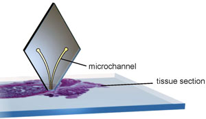
The diamond-shaped probe is positioned above the tissue sample and scans horizontally across it. The microchannels inject and aspirate liquids at a distance of 1-30µm from the tissue section
Now, a microfluidic device developed by Emmanuel Delamarche and colleagues at the IBM Zurich Research Laboratory, Rüschlikon, offers local staining of tissue sections, which allows a range of staining conditions to be used on one section. The microfluidic probe is positioned vertically above the tissue section and scans horizontally across it. The head of the probe has two apertures at its apex. The liquid is injected onto the tissue section from one aperture and aspirated at the second. ‘Instead of incubating the entire tissue section, the probe can scan it with a variable speed so as to vary locally the incubation time. This is a simple trick, but it should give an optimal staining contrast at least on one spot of the sample,’ explains Delamarche. Tissue sections are prepared for staining in the conventional fashion and post-staining processing is also unchanged.
Delamarche and the team have shown proof of concept with their current research, but in the future, they hope it will receive clinical validation. In addition, the probe could be used for fundamental research. ‘Developing a novel tissue staining method to detect various biomarkers is critical to obtain a more accurate and sophisticated understanding of drug discovery and clinical pathology,’ explains Je-Kyun Park, an expert in bioengineering at the Korea Advanced Institute of Science and Technology, South Korea. Using the microfluidic probe to scan multiple locations with different conditions could help analyse tissue microarrays.
Micro-immunohistochemistry using a microfluidic probe
Robert D. Lovchik, Govind V. Kaigala, Marios Georgiadis and Emmanuel Delamarche
Lab Chip, 2012, Advance Article
DOI: 10.1039/C2LC21016A
Original article published at Chemistry World


")