Archive for February, 2017

A simple, bubble-free cell loading technique for culturing mammalian cells on lab-on-a-chip devices

Sahl Sadeghi1­* and Meltem Elitas1

1 Faculty of Engineering and Natural Sciences, Sabanci University, 34956, Istanbul, Turkey

* Sahl Sadeghi wrote the paper.


Purpose

Lab-on-a-chip (LOC) devices significantly contribute different disciplines of science. Polydimethylsiloxane (PDMS) is one of the main materials, which is widely used for the fabrication of biological LOCs, due to its biocompatibility and ease of use. However, PDMS and some other polymeric materials are intrinsically water repellant (or hydrophobic), which results in difficulties in loading and operating LOCs. The eminent consequence of hydrophobicity in LOCs for biological systems is the entrapment of air bubbles in microfluidic channels. Although the oxygen plasma treatment of PDMS reduces the surface hydrophobicity for a certain period of time, the hydrophilic property of PDMS vanishes over time1. The persistent problem of bubbles in the microfluidics led to several studies conducted to overcome it. Some of these solutions suggested implementing bubble traps2,3, surface treatment of LOCs through hydrophilic coatings4, and using actively controlled bubble removal systems 5,6.

Although the aforementioned design complexities are introduced to LOCs in order to reduce the clogging problem caused by the bubbles, these modifications also result in higher production cost, complex operation, and long device preparation time. In many single-cell experiments without losing or damaging the rare cells, these cells needs to be introduce into the LOCs. Here, we present a simple method that enables loading a small number of cells without introducing bubbles in the microfluidics channels.


Materials

·         PDMS (Dow Corning Sylgard 184 Silicon Elastomer Kit)
·         Pipette tips (20-200 ul, Eppendorf, # 3120000917)
·         Pipetman (Gilson, P200, #69989-5)
·         Aqueous ethanol 70% (ZAG Chemistry)
·         Cell culture medium (DMEM, PAN Biotech, #P04-01548)
·         Mammalian cells (MCF7, ATCC-HTB-22)
·         Sterile syringe (BD 10 ml Syringe, Luer-Lok Tip, #300912)
·         Sterile Hamilton syringe (Hamilton, 100 ul SYR, #84884)


Procedure

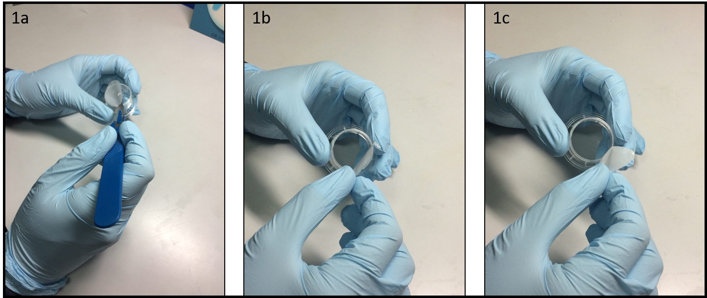
Step 1: Insert two 200-ul pipet tips at the inlet and outlet ports of the PDMS device as illustrated in Figure 1.

Step 2: Introduce a 70% aqueous ethanol into the inlet-pipet tip using a pipetman. Thus, the inner surface of microfluidic channels will be disinfected and the fluid flow will be tested within the micro channels as it is applied in many other protocols for LOCs7,8.

Step 3: Gently apply pressure pressing the pipetman to force ethanol solution flow through the micro channels and cavities of the PDMS device. Take care to avoid applying negative pressure from the outlet-pipet tip, which might create air leakage through the pipet connections. Besides, applying a negative pressure will directly affect the amount of a gas dissolved in the liquid according to Henry’s law9 that might contribute formation of more bubbles inside the micro-channels. The positive pressure will facilitate removal of the air bubbles via dissolving them.

Step 4: After flushing the chip with ethanol solution, inspect the chip to ensure bubble removal. In case of air bubbles, repeat the steps 2 and 3.

Step 5: Fill the syringe (10 ml) with medium or phosphate buffered saline (PBS). Take care to remove the air bubbles inside the syringe, mount and lock the needle on the syringe. Then, flow medium through the needle too make sure the needle is full of medium without any bubble. Insert the needle in the inlet-pipet tip; gently apply positive pressure to replace the ethanol with medium. Next, collect the excess medium from the outlet-pipet tip.

Step 6: Fill the inlet pipet with fresh medium in such a way that due to certain height (h) between the levels of the medium in the inlet and outlet pipet tips, very slow medium flow will be established inside the micro-channels.

Step 7: Load your cells into the Hamilton syringe and take care to ensure that there is no air bubble inside its needle and syringe. Insert the needle of the Hamilton syringe into the inlet-pipet tip as explained in Step 5, Figure 1. Introduce the cells via applying gentle positive pressure to the syringe. Established flow streams in the PDMS chip will deliver the released cells to the desired positions in the chip. Flow rate can be arranged adjusting the applied positive pressure and amount of medium collected in the inlet and outlet pipet tips. The excess supernatant from the outlet-pipet tip can be collected, and fresh medium can be supplied through the inlet-pipet tip during the experiment.


References

  1. Tan, S.H., N.T. Nguyen, Y.C. Chua, and T.G. Kang,. Biomicrofluidics, 2010. 4(3).
  2. Zheng, W.F., Z. Wang, W. Zhang, and X.Y. Jiang,. Lab on a Chip, 2010. 10(21): p. 2906-2910.
  3. Wang, Y., D. Lee, L.S. Zhang, H. Jeon, J.E. Mendoza-Elias, T.A. Harvat, S.Z. Hassan, A. Zhou, D.T. Eddington, and J. Oberholzer, . Biomedical Microdevices, 2012. 14(2): p. 419-426.
  4. Wang, Y.L., C.E. Sims, and N.L. Allbritton,. Lab on a Chip, 2012. 12(17): p. 3036-3039.
  5. Karlsson, J.M., M. Gazin, S. Laakso, T. Haraldsson, S. Malhotra-Kumar, M. Maki, H. Goossens, and W. van der Wijngaart,. Lab on a Chip, 2013. 13(22): p. 4366-4373.
  6. Cortes, D.F., T.-X. Tang, D.G.S. Capelluto, and I.M. Lazar,. Sensors and Actuators B: Chemical, 2017. 243: p. 650-657.
  7. Benavente-Babace, A., D. Gallego-Perez, D.J. Hansford, S. Arana, E. Perez-Lorenzo, and M. Mujika,. Biosensors & Bioelectronics, 2014. 61: p. 298-305.
  8. Yesilkoy, F., R. Ueno, B.X.E. Desbiolles, M. Grisi, Y. Sakai, B.J. Kim, and J. Brugger,. Biomicrofluidics, 2016. 10(1).
  9. Henry, W., Phil. Trans. R. Soc. Lond., 1803. 93: 29–274.

Figure 1 – Schematic of cell loading procedure in a microfluidic PDMS device.


Digg This
Reddit This
Stumble Now!
Share on Facebook
Bookmark this on Delicious
Share on LinkedIn
Bookmark this on Technorati
Post on Twitter
Google Buzz (aka. Google Reader)

A novel low cost method to prepare a cross-linked gelatin membrane for potential biological applications

Gabriele Pitingolo1 and Valerie Taly1

 1 INSERM UMRS1147, CNRS SNC 5014, Université Paris Descartes, Equipe labellisée Ligue Nationale contre le cancer 2016. Paris, France.

email: gabriele.pitingolo@parisdescartes.fr


Why is this useful?

In recent years, several gelatin types (i.e. GelMA, A or B) have been used in pharmaceutical formulation and tissue engineering due to their excellent biocompatibility, propensity to cell differentiation and availability at low cost.1 The use of gelatin as biomaterial is also advantageous for the possibility to tune the mechanical properties of the substrate, changing the concentration in water and the degree of cross-linking. However, Transwell® is the most used permeable support with microporous membranes and is a standard method for culturing cells.2 This commercial type of support has been widely used to study the molecular secretion by different cell types and also to reproduce several in vitro physiological barriers (i.e. blood brain barrier).3 Transwell® cell culture inserts are convenient, because they are sterile and easy-to-use, but they are very expensive (~ 300 $ for a 12 pack) and possess a limited range of biomaterial properties because they are made from polyester or polycarbonate. Furthermore, as shown by Falanga and colleagues, these porous membranes are often integrated onto microfluidic chip for permeability studies.4

Recently, Yong X. Chen et al. proposed an alternative method to prepare a suspended hydrogel membrane platform for cell culture, designing a complicated protocol to synthesize the GelMA and to fabricate an open-grid structure made of polylactic acid (PLA) polymer using a commercial printer.5 Our tip shows a novel low-cost method for preparing a cross-liked gelatin membrane as a permeable support useful for potential biological applications. In addition, the proposed protocol doesn’t require the use of sophisticated fabrication technologies or expensive materials. It uses gelatin from porcine skin and the formaldehyde vapor technique to cross-link the gelatin membrane. As proof of concept, we integrate the gelatin membrane into a microfluidic chip, to show the possibility to develop a platform for comparable studies in static and dynamic conditions.

Furthermore, to demonstrate the resistance to culture temperature (around 37° C) of the prepared gelatin membrane, we tested the mechanical properties (Young’s modulus) before and after the incubation time (2 days). Finally, we observed the preservation of the mechanical properties and structural integrity that makes the membrane usable for studies with cell culture.


What do I need?

  • Transwell insert
  • Porcine gelatin type A
  • Formaldehyde solution
  • Scalpel
  • PMMA milled chamber or similar


What do I do?

  1. Remove the porous membrane from the Transwell® insert (Fig. 1a-1c) or alternatively use a similar homemade support. To facilitate this step is convenient to use a scalpel to incise the membrane along the entire diameter.


  1. After the removal of the membrane, the Transwell® support is ready to use. Position the structure at the center of the PMMA chamber (depth 2 mm at least) (Fig 2a) and pour liquid 10% w/v gelatin, without bubbles, onto the PMMA chamber and inside the Transwell® support (Fig 2b). After 2 min of stabilization, put the system in the fridge, for at least 10 minutes.


  1. After the gelation time (10 minutes at 4°C) it is possible to remove the formed gelatin membrane from the PMMA chamber, with the aid of a scalpel to facilitate the detachment (Fig 3a-3b). As shown in Figure 3c the gelatin membrane it appears very flat, an ideal characteristic for cell cultivation (Fig 3c). To guarantee the preservation of the mechanical properties during the cell culture step, cross-link the gelatin membrane using the classical protocol “cells-biocompatible”, such as glyceraldehyde6, formaldehyde7 and glutaraldehyde8 methods or natural products such as genipin.9


  1. In this case, we used the vapor formaldehyde method to cross-link the prepared gelatin membrane and to obtain a system with lower aqueous solubility, higher mechanical strength and stability against enzymatic degradation. We exposed the gelatin membrane to formaldehyde vapors for 1 day. In Figure 4a we show the difference, after 48 h of culture conditions, between a sample cross-linked (left) and not (right). The final depth of the cross-linked gelatin membrane is around 1 mm, however, it is possible to change the depth tuning the liquid gelatin amount. Finally, we calculated, before and after the incubation time, the young’s modulus of the cross-linked gelatin membrane, observing a similar value of 40 kPa (compression test by using hydraulic testing system Instron DX).


  1. Integration of the gelatin membrane into a microchip. In this section, we detail the integration of the gelatin membrane into a microfluidic chip. As example, we used the same geometry proposed in our previous work4, to make a device for a permeability experiment (Figure 5a). As shown in Figure 5b, just pour the liquid gelatin into the smaller drilled microchannel, using a pipette to form a thin uniform layer of gelatin (Figure 5c). After gelation at 4° C, cross-link the formed gelatin membrane using the previously described method, the result is shown in Figure 5d. To bond the different PMMA-PDMS substrates we propose here a magnetic approach recently developed by our group10, to preserve the gelatin membrane by physical-chemical stress as in the case of solvent evaporation and plasma bonding. Figure 5e shows the final chip.


Conclusions: In this tip, a novel biocompatible gelatin permeable support was obtained by using a simple and low cost fabrication method. Vapor formaldehyde method or other chemical crosslinking approach can be applied to crosslink the integrated gelatin membrane for the use as potential scaffolds for cell culture. Furthermore, the Young’s modulus and the thickness of the permeable membrane can be adjusted by changing the initial concentration of the gelatin, the degree of cross linking and the amount of liquid gelatin. Finally, we showed the integration of the gelatin membrane into a modular microchip. Therefore, we propose an easy and low cost method to prepare a permeable gelatin membrane for cell biology and for other applications.


Acknowledgements

This work was carried out with the support of the Pierre-Gilles de Gennes Institute equipment (“Investissements d’Avenir” program, reference: ANR 10-NANO 0207).


References

  1. Geckil, Hikmet, et al. “Engineering hydrogels as extracellular matrix mimics.” Nanomedicine 5.3 (2010): 469-484.
  2. https://www.corning.com/worldwide/en/products/life-sciences/products/permeable-supports/transwell-guidelines.html
  3. Guarnieri, Daniela, et al. “Shuttle‐Mediated Nanoparticle Delivery to the Blood–Brain Barrier.” Small 9.6 (2013): 853-862.
  4. Falanga, A. P., Pitingolo, G., Celentano, M., Cosentino, A., Melone, P., Vecchione, R. & Netti, P. A. (2016). Shuttle‐mediated nanoparticle transport across an in vitro brain endothelium under flow conditions. Biotechnology and Bioengineering.
  5. Chen, Yong X., et al. “A Novel Suspended Hydrogel Membrane Platform for Cell Culture.” Journal of Nanotechnology in Engineering and Medicine 6.2 (2015): 021002.
  6. Sisson, Kristin, et al. “Evaluation of cross-linking methods for electrospun gelatin on cell growth and viability.” Biomacromolecules 10.7 (2009): 1675-1680.
  7. Usta, M., et al. “Behavior and properties of neat and filled gelatins.” Biomaterials 24.1 (2003): 165-172.
  8. Talebian, A., et al. “The effect of glutaraldehyde on the properties of gelatin films.” Kemija u industriji 56.11 (2007): 537-541.
  9. Bigi, A., et al. “Stabilization of gelatin films by crosslinking with genipin.” Biomaterials 23.24 (2002): 4827-4832.
  10. Pitingolo Gabriele, et al. “Fabrication of a modular hybrid chip to mimic endothelial-lined microvessels in flow conditions.” Journal of Micromechanics and Microengineering” (Accepted manuscript)
Digg This
Reddit This
Stumble Now!
Share on Facebook
Bookmark this on Delicious
Share on LinkedIn
Bookmark this on Technorati
Post on Twitter
Google Buzz (aka. Google Reader)