Researchers in Canada have shrunk bubbles to single-micrometre diameters, suitable for use in ultrasound.
Source: Royal Society of Chemistry
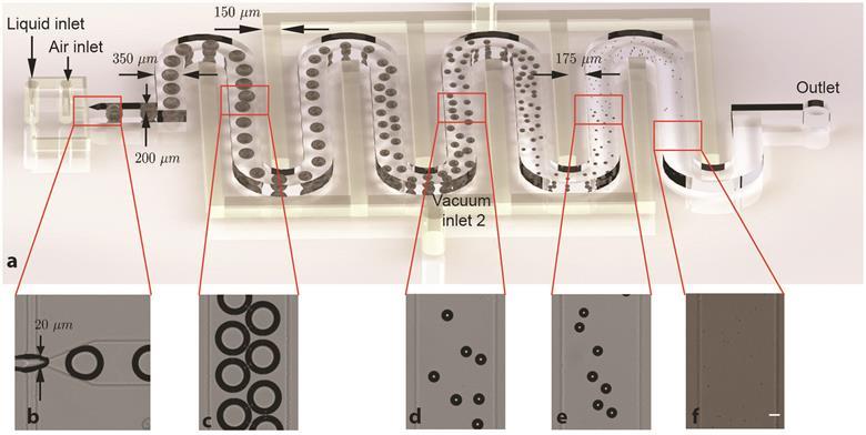
Bubbles shrink down to 1–7µm in diameter as they flow through the device’s serpentine microchannel
Microbubbles are commonly used in ultrasound imaging as they improve the visual distinction between blood and surrounding tissues. Bubbles are injected intravenously, and under ultrasound they are excited at their resonant frequency. This resonance means they scatter a much higher proportion of the ultrasound than the surrounding tissues, allowing clear imaging of blood vessels.
The bubbles needed for ultrasound are around 2µm in diameter. Current microfluidic techniques cannot produce bubbles this small, and the techniques used to generate these microbubbles generally use physical agitation or shearing. The bubbles produced often have a large size distribution, and filtration is needed to separate out those suitable for use.
Read the full story by Laura Fisher in Chemistry World.
This article is free to access until 24 May 2017
V Gnyawali et al, Soft Matter, 2017, DOI: 10.1039/C7SM00128B


")