Author Archive

Overwhelmed to death: an anti-cancer gene therapy approach paired with an immune-activating distress signal

Frontline therapies for treating colorectal cancer have shortcomings. These include their inability to impede local tumor recurrence and metastatic spread to distant sites such as the abdomen.  

Researchers have now utilized a gene therapy approach that simultaneously compromises cancer cell survival while activating immune system cells with cancer-killing abilities.

Gene therapy – an advanced technique developed to insert or inject therapeutic genes into human cells – has shown some success in treating the disease. In a previous study, Xiao and co-investigators at State Key Laboratory of Biotherapy, and the Department of Thoracic Oncology Cancer Center, West China Hospital, Sichuan University, had used a gene therapy approach to induce cancer cell death. Their study found that Vesicular Stomatitis Virus Matrix Protein (VSVMP), when inserted into a cancer cell,  compromises the cellular skeletal framework, which is made up of structural proteins. Cell death ensued as a consequence.

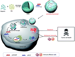
In the current study, the research team further armed with VSVMP gene delivery vessel with Interleukin-12 (IL-12) – a protein known to recruit and switch on the cancer-killing functions of immune cells.

The novel drug particles are based on Heparin-polyethyleneimine (HPEI) nanoparticles. To overcome the high toxicity and non-biocompatible nature of PEI, the team used a method to covalently conjugate this substance with heparin.

Their results, based on lab-grown cancer cells and animal studies, suggest that this novel complexed drug molecule (particle size: 53nm) increases tumor cell death, reduces division frequency, and stimulates the recruitment and activation of two types of cancer-killing cells: T cells and NK cells.

Specifically, the drug inhibited the growth of C-26 colon cancer cells. Animal studies showed that the drug reduced tumor weight. Metastatic spread of tumor cells to the abdomen was also reduced. The team proposes that the drug-derived IL-12 induces a secondary cascade of chemical mediators, which in turn recruit and activate cancer-killing immune cells. Their data supports this proposal. Interestingly, their study also found that the complexed drug molecule did not show adverse side effects within the major organs.

Read the full article here:

Nanoparticles co-delivering pVSVMP and pIL12 for synergistic gene therapy of colon cancer

Yuanyuan Xiao, Yuping Yang, Yujiao Wu, Chunmei Wang, Hao Cheng, Wei Zhao, Yang Li, Beibei Liu, Jianlin Long, Wenhao Guo, Guangping Gaoa and Maling Gou

Digg This
Reddit This
Stumble Now!
Share on Facebook
Bookmark this on Delicious
Share on LinkedIn
Bookmark this on Technorati
Post on Twitter
Google Buzz (aka. Google Reader)

A closer look at clean dishes: detection of domestic detergent residues with LIBS technology

Dish detergents help keep our dishes clean; however, the long term health effects of detergent residues on tableware and cookware is yet to be discussed publicly. Studies suggest that certain household detergents may be linked with disturbances in hormone regulation in humans.

As a first step in uncovering the role of domestic dish detergent in affecting health, a research team comprising scientists from China Agricultural University, China Research Center of Intelligent Equipment for Agriculture, and Beijing Academy  of Agriculture and Forestry Sciences, have developed a method to detect detergent residue rapidly and in real-time, i.e., the process does not involve dissolving, preparing, or conditioning the residue prior to detection.

This method is based on a process called Laser Induced Breakdown Spectroscopy (LIBS). Detergent residues are first vaporized by a high-power laser, leading to the generation of vaporized plasma. At the end of the laser pulse, these atoms and ions spontaneously return from a higher energy state to a lower energy state. This energy decay is associated with the emission of optical radiation of specific wavelengths. The emitted radiation is collected and channeled toward a spectrometer, which converts wavelength information into readable numbers that scientists can record and analyze.

Although the LIBS technology has existed for several years, and is used routinely by researchers in the field, this study let by Zhao an colleagues is the first to use this method to measure household detergents.

Graphical abstract for C7RA04304J

Analogous to how a sensor at a grocery store is programmed to recognize barcodes printed on different items, the scientists used the numbers generated by the spectrometer to generate ‘signatures’ to help them recognize the different detergents used in the study. Using this method, the team found that detergent detection in real-time can be more flexible, used with tableware of different shapes, used to measure trace amounts of detergent, compatible with dry and wet dishes, and safe on tableware.

To demonstrate the utility of the method to real world applications, the team conducted a series of timed dish washes and residue analyses. Their results suggest that a 16-minute rinse removes detergent residues. They also suggest that this information will be useful in designing and programming commercial dishwashers.

This study may someday inspire public health advocates to take a closer look at the prevalence of dish residues in public and household settings. When this day arrives, LIBS technology for residue detection may be pivotal in conducting  studies to better understand the relationship between dish residues and overall wellbeing.

Read the full article here:

Detection of domestic detergent residues on porcelain tableware using laser induced breakdown spectroscopy
Xiande Zhao, Daming Dong, Yang Lic and Chunjiang Zhao
RSC Adv., 2017, 7, 28689-28695 (Open Access)

Digg This
Reddit This
Stumble Now!
Share on Facebook
Bookmark this on Delicious
Share on LinkedIn
Bookmark this on Technorati
Post on Twitter
Google Buzz (aka. Google Reader)

Can this quantum sized double-edged sword help diagnose and treat breast cancer?

In a study led by Ko and colleagues at the Department of Dental Materials, School of Dentistry, Kyung Hee University, Korea, researchers armed graphene QDs with two therapeutic moieties: a HER-targeting antibody meant to help the therapeutic QDs find HER2-expressing breast cancer cells; and doxorubicin (DOX) – a chemotherapeutic drug used widely in treating breast cancer.

Consistent with previously established criteria (size, shape etc.) for drug carriers, the current study found that the estimated size of 222 nm makes the nanocarriers good candidates for further development toward diagnostic and therapeutic applications. Further, the nanocarriers had excitation and emission wavelengths of 370 nm and 450 nm respectively, making them glow in the ultraviolet range and as a result, optimal for medical imaging applications. The research team showed through chemical binding analysis that anti-HER antibodies were firmly bound to the QDs, and that the QDs were hydrophilic. The team conducted thermal stability studies and showed that the nanocarriers were stable at temperature ranges  much greater than the physiological body temperature range.


The study also analyzed whether the therapeutic nanocarriers were able to specifically target and enter breast cancer cells, release the DOX payload under specific pH and temperature conditions, and subsequently induce breast cancer cell death. Using a HER2-expressing breast cancer cell line, the team showed that the nanocarriers could kill cells in a dose dependent manner. A temperature of 37oC and pH of 5.5 were optimal for DOX release. Results in fluorescent microscopy studies suggested that DOX was released immediately after the nanocarriers entered HER2-expressing cells.


This study proposes that graphene-based QDs, when armed with anti-HER antibodies and DOX, have great potential for translation. In addition, with biomarker-based treatment decisions entering clinical practice in oncology settings, QD-based therapeutic nanocarriers are likely to have a notable impact on cancer therapy.
Read the full article here:

Graphene quantum dot-based theranostic agents for active targeting of breast cancer
N. R. Ko, M. Nafiujjaman, J. S. Lee, H.-N. Lim,a Y.-k. Lee and I. K. Kwon

Digg This
Reddit This
Stumble Now!
Share on Facebook
Bookmark this on Delicious
Share on LinkedIn
Bookmark this on Technorati
Post on Twitter
Google Buzz (aka. Google Reader)

Killing cancer cells with a DNA-based molecular bridge

Monoclonal antibodies (mAbs) are antibodies made by clones of immune cells derived from a common parent cell. These synthesized molecules have achieved widespread clinical utility in the treatment of cancer owing to their high degree of specificity to proteins present on the surface of cancer cells, lower toxicity compared to other classes of targeted therapies, and improved treatment outcomes among patients with advanced stage cancer.

Non-Hodgkin Lymphoma (NHL) is a type of cancer where a subtype of immune cells called B-cells exhibit unrestrained cell division. The abnormal B-cell, now called a malignant B-cell, produces more abnormal cells like it. CD20 is a protein present on the surface of malignant B-cells. Rituximab (RTX) is used to treat patients with NHL because it can bind CD20 and consequently trigger cell death.

To address the growing need for CD20 targeted therapeutics, Cong and colleagues at the Department of Laboratory Diagnosis/Thoracic Surgery, Changhai Hospital Affiliated to The Second Military Medical University, Shanghai, China, developed molecules called aptamers that can bind to CD20 with greater specificity and strength compared to RTX.

Graphical Abstract

Graphical Abstract

Aptamers are molecules made up of  single stranded DNA that form complex 3D structures and can bind to target proteins, analogous to mAbs. The team used a method called cell-SELEX to retrieve an enriched pool of highly specific CD20-binding aptamers starting with their initial aptamer library. The aptamers used in the study were obtained after 15 rounds of selective refinement.


The study finds that Anti-CD20 DNA Aptamer (ACDA) can bind surface CD20 in NHL cells with greater strength compared to RTX. In the past, experiments have shown that cross-linking surface CD20 with mAbs (i.e. extracellular cross-linking) is a potent method of inducing cell death. A major limitation is that extracellular cross-linking cannot be realized in vivo. Cong et al. develop a method to link two ACDA molecules with polyethyleneimine (PEI) linker’, forming a molecular bridge  – the P-ACDA – capable of spanning the distance between and cross-linking two CD20 molecules. The study finds that P-ACDA led to substantially more cell death compared to ACDA.

Aptamers as a novel class of targeted therapies are expected to outperform mAbs because they do not evoke the body’s endogenous immune response (i.e. less immunogenic) and therefore in good compliance with current FDA recommendations. They are also easier to store since they are stable across a broad temperature range ,less expensive to manufacture, show consistency between production batches and can bind to both protein as well as non-protein targets. For these reasons, the clinical relevance of aptamers in treating HNL and potentially other cancers must be watched closely in the years to come.

Read the full article here:

Cong Wu, Wei Wan, Ji Zhua, Hai Jina, Tiejun Zhao and Huafei Li

Digg This
Reddit This
Stumble Now!
Share on Facebook
Bookmark this on Delicious
Share on LinkedIn
Bookmark this on Technorati
Post on Twitter
Google Buzz (aka. Google Reader)

Within The Heart: Regenerating Diseased Heart Valve Leaflets

Hair and nails grow back, but heart valves cannot regenerate naturally. This fact is being challenged by a team of scientists who have developed a method that could someday regrow defective heart valves.

Valvular interstitial cells (VIC), the most prevalent cells in the heart valve, are developmentally locked in a state of quiescence, preventing their division within the body. In people suffering from inflammation-induced or inborn heart valve defects, damaged valves have to be replaced surgically with either mechanical (made with artificial polymers) or bioprosthetic (made with heart tissue) valves.

People with artificial valves are required to take blood thinners for extended periods and make significant changes to their lifestyle. In addition, artificial valves in younger patients do not grow and remodel with time, causing additional complications during adulthood. These issues are further compounded by an estimate suggesting that over 850,000 patients will require heart valve transplants by the year 2050.

Soumen Jana and colleagues at the Division of Cardiovascular Diseases, Mayo Clinic, USA approached this problem from a different angle. Relying on studies showing that VICs can be isolated from heart valves and grown in a laboratory setting, the team developed a nanofibrous membrane-based scaffolding structure to support the growth of VICs. Similar to the cross-cross patterns formed by ropes in a hammock, their polycaprolactone polymer scaffold comprises randomly oriented nanofibers (~457 nm in diameter) to form a cradle within which VICs from defective valves can be grown.

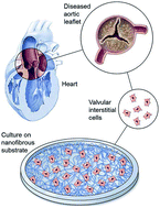
Within the body, VICs are arranged as monolayer sheets, forming a veneer on the outer surfaces of heart valve leaflets. They form this complex structure by depositing collagen – a cellular cementing protein that allows cells to interlock like a cobblestone path to form a sheet-like structure. The researchers reasoned that by artificially simulating conditions ideal for VIC growth within the body, they could create VIC sheets in the laboratory and examine their similarity to naturally occurring VICs in heart valve leaflets.

The study found that VICs from healthy valves showed greater levels of cell division on the scaffold compared to VICs from defective valves. Interestingly, the scaffold induced collagen deposition from VICs obtained from both healthy and defective valves. The study also looked at a series of genes and proteins important in VIC growth. Patient derived VICs grown on nanofibrous scaffolds deposited appropriate amounts of cementing proteins necessary for leaflet regeneration.

Clinical trials aimed at regenerating heart valves with chemical drugs and DNA modifying methods are under way. In their study, Jana and colleagues suggest VIC regeneration as a novel idea. They also engineer a scaffold that supports VIC growth, demonstrate its practicality and highlight its ability to be translated into a clinically impactful technology.

Read the full article here:

Regeneration ability of valvular interstitial cells from diseased heart valve leaflets
Soumen Jana, Rebecca Hennessy, Federico Franchi, Melissa Young, Ryan Hennessya and Amir Lerman

Digg This
Reddit This
Stumble Now!
Share on Facebook
Bookmark this on Delicious
Share on LinkedIn
Bookmark this on Technorati
Post on Twitter
Google Buzz (aka. Google Reader)

Serving up anti-cancer cocktails: design, synthesis and evaluation of hybrid inhibitors

Scientists in the area of cancer drug development constantly find ways to target cancer’s Achilles heel. Bcr-Abl is a protein expressed in 95% of all Chronic Myelogenous Leukemia (a type of blood cancer) cases. It remains activated (i.e. switched ON) and instructs cancer cells to divide indefinitely. Bcr-Abl has remained an attractive target for therapy. Yet another attractive therapeutic target is Histone Deacetylase 1 (HDAC1) – a protein known to control cell survival via its ability to influence the turning on and turning off of certain genes.

Previous studies show that the drugs Dasatinib and MS-275 efficiently inhibit the cancer promoting activities of Bcr-Abl and HDAC1 respectively.  Clinical trials also suggest that these drugs, when used independently in separate studies, can be used to treat a variety of solid as well as blood-borne cancers. Bcr-Abl and HDAC1 are components of distinct cellular wiring systems, referred to as signalling pathways, which sustain cell survival and division. Single agent drugs, or drugs that stifle a single cancer-promoting pathway, weed out most cancer cells but also set the stage for drug-resistant cells. Reports suggest that both Dasatinib and MS-275 are associated with cancer drug resistance.

Multi-target inhibitors are a new and evolving class of cancer drugs that can simultaneously inhibit at least two signalling pathways. These compounds  have emerged as a potential solution in circumventing cancer drug resistance. Chen and colleagues at the Department of Organic Chemistry, School of Science, China Pharmaceutical University, China, designed and produced a series of hybrid drug molecules which combine the attributes of the HDAC inhibitor MS-275 with the Bcr-Abl inhibitor Dasatinib.

To determine the effect of the hybrid drugs on cancer cell survival, the research team tested the drug’s ability to halt the growth of three cell types exhibiting features of leukemia, kidney cancer and prostate cancer respectively. They found that all drugs in the series were toxic to cancer cells, with leukemia and kidney cancer cells showing the greatest degree of sensitivity to the hybrid drugs.

To better understand how the hybrid drugs interacted with the Bcr-Abl and HDAC1 active sites (i.e. the ON switch), the team relied on computer-generated three-dimensional models of the hybrid drugs, Bcr-Abl and HDAC1 proteins. Using a method similar to finding the right key for a lock, a computer program found that a hybrid compound termed 6a, which happened to be the most potent compound in the series, fit most snugly into both Bcr-Abl  and HDAC1 active sites. In theory, 6a would prevent both Bcr-Abl and HDAC1 from becoming activated (i.e they remain switched OFF).

On the basis of these observations, this study strengthens the paradigm that chemically melding two cancer drugs to form a novel single molecule may prove to be an effective clinical strategy for anticancer treatment. On a broader scale, this is one among many studies advocating for the use of multi-target agents in cancer treatment, highlighting an imminent upsurge in single molecule combination therapies.

Read the full article here:

Digg This
Reddit This
Stumble Now!
Share on Facebook
Bookmark this on Delicious
Share on LinkedIn
Bookmark this on Technorati
Post on Twitter
Google Buzz (aka. Google Reader)

A hydrogel-based trojan horse for antitumor therapy

Paclitaxel (PTX) is among the most widely used chemotherapeutic agents in clinical settings. The drug imposes its anticancer effect by preventing cell division. Cancer cells learn to resist PTX over time by various mechanisms including creating alterations in the protein targeted by PTX and rewiring of cell survival pathways to evade cell death.

Clinicians combine PTX with suberoylanilide hydroxamic acid (SAHA) to suppress cancer drug resistance and improve treatment outcome. The benefits of combination therapy include improved accumulation of the drug at cancer sites, the ability to trigger cell death by complementary or synergistic mechanisms and longer retention of the drug within patients. Given the strong rationale for combination therapies, Shu and colleagues at the Department of Pharmaceutical Analysis, Key Laboratory on Protein Chemistry and Structural Biology, China developed a novel peptide hydrogel which encapsulates PTX and SAHA within a single co-delivery nano-carrier.

Graphical Abstract for C6RA19917H The researchers loaded PTX and SAHA onto the same nano-carrier in the following sequence: (1) an amino acid-based self assembling hydrogel precursor (Nap) was prepared, (2) PTX was conjugated to the self assembling hydrogel to form a pro-drug and (3) the pro-drug was allowed to encapsulate SAHA, forming the final drug (Nap-PTX-SAHA). The researchers subsequently characterized the mechanical features of their novel drug delivery system and tested it using a mouse model of liver cancer.

The study found that the Nap-PTX-SAHA hydrogels could be injected at room temperature into test mice, suggesting that no specialized equipment or storage conditions were necessary to administer the drug. The study also found that SAHA is released more readily than PTX from Nap-PTX-SAHA hydrogels. This could mean that cancer cells will be exposed to the two chemotherapy agents at different times, allowing for a one-two punch based tumor killing strategy.

When administered to tumor-bearing mice, the Nap-PTX-SAHA regimen was found to decrease tumor volume up to 2-fold compared to mice treated traditionally with PTX or SAHA alone. Interestingly, the researchers also noted that Nap-PTX-SAHA was associated with fewer side effects, as evidenced by normal eating behavior and weight in test mice. Interestingly, Nap-PTX-SAHA was absorbed lesser in non-target organs such as the heart, spleen and kidneys.

On the basis of these promising preclinical studies, the authors propose that Nap-PTX-SAHA represents a promising candidate for clinical trials in the years to come.

Read the full article here:

Digg This
Reddit This
Stumble Now!
Share on Facebook
Bookmark this on Delicious
Share on LinkedIn
Bookmark this on Technorati
Post on Twitter
Google Buzz (aka. Google Reader)

Micelles meet transplantation medicine: How a novel nanoparticle based immune cell blocker might benefit human organ transplantation

Organ transplantation saves lives. According to the Organ Procurement and Transplantation Network, U.S Department of Health and Human Services, over 22,000 organs transplantation surgeries have been conducted between January and September 2016.

Ischemia Reperfusion Injury (IRI) is a well characterized cardiac transplantation-related complication wherein the host tissue (graft), deprived of blood supply for prolonged periods, undergoes damage when blood supply is restored post-implantation. Immune cells at the interface of the graft and recipient tissue mediate damage by releasing inflammation-promoting chemicals and free radicals.

In a study led by Nadig and colleagues at the Department of Surgery, Division of Transplant, Medical University of South Carolina, USA, researchers first acknowledge the central role played by endothelial cells (EC) in promoting IRI-associated tissue damage and subsequently developed a novel pH-sensitive, immunosuppressive drug-loaded, targeted micelle nanoparticle to curb the damaging effects of ECs. The team choose rapamycin as their immunosuppressive drug of choice given its dual roles in limiting cytotoxic immune cell functions and in protecting tissues that make up blood vessels.

While treating patients with immunosuppressive drugs prior to surgery is currently a standard practice, a major drawback of this approach is that these drugs prevent immune system activity throughout the body, placing patients at risk for diseases including diabetes and cancer. As an initial step in addressing this limitation, Nadig et al. coated the micelles with cyclic arginine-glycine-aspartate moieties, which specifically bind to and integrin protein (the alpha v beta 3 receptor protein) present almost exclusively on ECs. As a finishing touch, the team attached fluorescent chemical compounds to allow for tracking and visualization in their studies.

Graphical abstract of "Immunosuppressive nano-therapeutic micelles downregulate endothelial cell inflammation and immunogenicity"Their studies showed that the rapamycin-loaded nanoparticles were stable and biocompatible when tested in human endothelial cells. Further, the rapamycin release could be controlled by adjusting the pH values lower than 7 or higher than 8. The study found that the micelles were being taken up by cells within 6h after incubation. The study also demonstrates the specificity of the micelles by showing that what the cells were pre-treated with an integrin inhibitor,  they were  less likely to take up the micelles.

To demonstrate the clinical utility of their idea, the researchers exposed human endothelial cell cultures to hydrogen peroxide to mimic IRI-like conditions. The cells responded by increasing their production of inflammation-promoting chemicals. Importantly, the rapamycin-loaded nanoparticle micelles significantly curbed this response. Nadig et al. propose that the ultimate goal is to incorporate this technology into organ storage media to minimize the harmful effects of IRI.

Read the full article here:

Satish N. Nadig, Suraj K. Dixit, Natalie Levey, Scott Esckilsen, Kayla Miller, William Dennis, Carl Atkinson and Ann-Marie Broome

RSC Adv., 2015, 5, 43552-43562

DOI: 10.1039/C5RA04057D

Digg This
Reddit This
Stumble Now!
Share on Facebook
Bookmark this on Delicious
Share on LinkedIn
Bookmark this on Technorati
Post on Twitter
Google Buzz (aka. Google Reader)

Sticks and stones may break my bones, but hydrogels will never hurt me

Recent advancements in tissue engineering have led to the fabrication of complex materials that may be used as surrogates for heart, neuronal, bone and cartilage tissue regeneration. A vast majority of engineered tissues are composed of a three-dimensional scaffold at its core, layered with growth stimulating agents, that collectively nurture and support cell growth.

In a study by Gantar and colleagues at the Jozef Stefan Institute, Department for Nanostructured Materials in Slovenia,  a team of researchers created an injectable hydrogel as a potential biomaterial for bone tissue regeneration. They further demonstrate that the hydrogel is capable of self-healing and supports the growth of cells derived from human bone tissue.

Hydrogels based on reversible covalent bonds allow the material to rearrange its structure permanently. This forms the basis for self-healing. A potential drawback is that the conditions (temperature and pH) required to form these bonds to form are not suitable for supporting bone cell growth. Having recognized this limitation, the team decided to develop a liquid hydrogel that transitions to a gel-like state at a pH suitable for cell growth and proliferation. Further, the hydrogel was infused with bioactive glass (BAG) nanoparticles – a silica-based material known to support bone cell growth.

The study also characterizes the physical (elastic properties, compression) and biological (degradation, cytotoxicity) features of the injectable hydrogel. The study finds that the BAG nanoparticles do not drastically alter the physical and biological properties of the hydrogel and suggest that the combination is well suited to support bone cell growth. The team proposes that their efforts will contribute toward the development of an injectable material that will scaffold bone cells within the host and promote self-repair.

Read the full article here:

Injectable and self-healing dynamic hydrogel containing bioactive glass nanoparticles as a potential biomaterial for bone regeneration
Ana Gantar, Nataša Drnovšek, Pablo Casuso, Adrián Pérez-San Vicente, Javier Rodriguez,c   Damien Dupin, Saša Novakab and Iraida Loinazc
RSC Adv., 2016,6, 69156-69166
DOI: 10.1039/C6RA17327F

Digg This
Reddit This
Stumble Now!
Share on Facebook
Bookmark this on Delicious
Share on LinkedIn
Bookmark this on Technorati
Post on Twitter
Google Buzz (aka. Google Reader)

A study on spheroid formation in thermosensitive hydrogels

According to an estimate by the World Cancer Report, cancer associated mortality is expected to reach 17 million per year globally by the year 2030. To confront the cancer burden with appropriate clinical interventions, researchers screen cancer-killing drug combinations in monolayer cell cultures. This is a widely used method for the preclinical evaluation of drug efficacy.

A major limitation of monolayer cultures is that they do not, even mildly, recapitulate the complex architecture of a tumor growing in vivo. As an initial step in overcoming this limitation, researchers use scaffolded spheroid cultures – a system wherein cells are grown on hydrogel scaffolds in three dimensions.

Hydrogel scaffolds provide physical and structural support for the formation of a more ‘natural’ setting that better recapitulates cell behavior in vivo. For example, smaller (150um) spheroids have better cell-to-cell contacts and notably different gene expression compared to monolayer cultures; larger (200-500um) ones develop oxygen and nutrient gradients reminiscent of chemical gradients seen in human tumors. However, limitations in design flexibility, handling and interbatch compositional variation have discouraged the routine use of hydrogel scaffolds. In addition, the technical challenge of separating newly formed spheroids from the scaffolding material before drug screening represents a major roadblock.

In a study led by Xiaolin Cui and colleagues at the School of Chemical Engineering and the School of Mathematical Sciences, University of Adelaide, Australia, researchers synthesized a thermo-reversible N-Isopropylacrylamide-acrylic acid (NIPAM-AA) hydrogel by free radical emulsion polymerization. In their study using the cervical carcinoma cell line HeLa, the team demonstrates that at 370C the NIPAM-AA hydrogel solidifies and forms a sheath around HeLa cell clusters. As a consequence, these clusters develop into hydrogel scaffolded spheroids over time. At 250C the hydrogel liquefies and releases the newly formed spheroids.

Cell viability assays confirmed that this new state of the art hydrogel is biocompatible. NIPAM-AA derived spheroids were smaller (70-120um),  nearly spherical and showed a narrower size distribution compared to conventional spheroids. The study showed, through time course experiments, that the spheroids remain viable for over 14 days in culture. The study also suggests that spheroids derived via the NIPAM-AA hydrogel method are more viable than those derived from conventional suspension cultures, supporting the notion that hydrogel scaffolding facilitates oxygen and nutrient supply to support cell growth.

The researchers deduced a mathematical model to predict the kinetics of NIPAM-AA derived spheroid growth. Their model accurately recapitulated the growth rate, size and size distribution of the spheroids. The authors propose that hydrogel scaffolding has the potential to evolve into a technology with a wide range of applications including, but not limited to, (1) high throughput screening of anticancer drugs using uniformly sized spheroids; (2) regenerative medicine; and (3) tissue engineering.

Read the full article here:

Xiaolin Cui,   Saber Dini,   Sheng Dai,   Jingxiu Bi,   Benjamin Binder,   Edward Green and   Hu Zhang
Digg This
Reddit This
Stumble Now!
Share on Facebook
Bookmark this on Delicious
Share on LinkedIn
Bookmark this on Technorati
Post on Twitter
Google Buzz (aka. Google Reader)