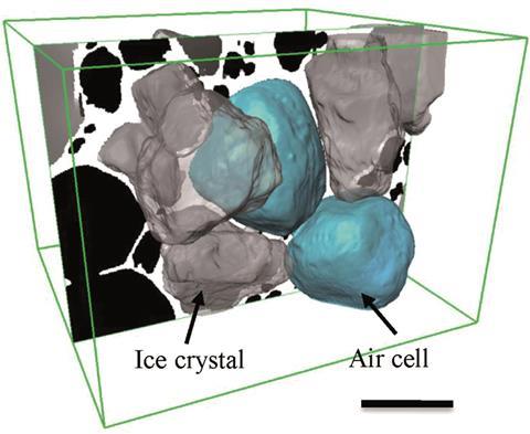
Source: © Royal Society of Chemistry This 3D rendered image shows a central air cell bounded by faceted ice crystals. Scale bar is 100mm
Researchers from the UK have developed a new 3D x-ray tomography (XRT) method to visualise the effects of changing temperature on the microstructure of ice cream.
Ice cream is a mixture of milk, fats, sugars, proteins, emulsifiers, stabilisers and flavours that are aerated and then frozen to form a soft solid comprising about 30% ice, 50% air, and 5–15% fat droplets suspended in a sugar solution. Its quality depends on the size of its ice crystals and air bubbles: smaller crystals and bubbles make it smoother and creamier. And since this complex colloid is unstable above –30˚C, its microstructure will change during shipping and storage (domestic freezers are usually at around –18˚C), which will affect its taste and texture.
Interested? The full article can be read in Chemistry World.
The original RSC Advances article can be read below and is open access:
Synchrotron X-ray tomographic quantification of microstructural evolution in ice cream – a multi-phase soft solid
Enyo Guo et al.,
RSC Adv.,2017, 7, 15561-15573
DOI: 10.1039/C7RA00642J


")