Author Archive

Graphene research on the edge

Electrochemistry of folded graphene edges

Adriano Ambrosi, Alessandra Bonanni and Martin Pumera
Nanoscale
, 2011, C1NR10136F

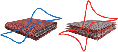
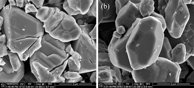
Graphene  has many exciting potential applications, from solar cells to antibacterial sheets. There has been intensive research into the various properties of graphene, and it has been shown to have excellent electronic, electrochemical, optical, mechanical and thermal properties. As graphene has a planar form, it is important to consider the affects of conformational changes of the sheets such as folding and wrinkling, which can alter electrical and electrochemical properties. An important consideration here is the properties of graphene edges, as opposed to on the sheet’s surface.

Pumera and coworkers from Nanyang Technological University, Singapore, have conducted a study of the electrochemistry of folded graphene edges, and compared it to that of open edges. Folded edges have a very different structure compared to closed edges, and therefore it is natural to assume that they should possess different physical, chemical and electronic properties. Pumera et al. conclude in their paper that the electrochemical properties are superior at the open edges, discovering that the electron transfer rate is about 35 times faster at open-edged graphene structures than at folded-edged graphenes. This could be an extremely important consideration when synthesising graphene-based materials for many applications, particularly sensing and bio-sensing, as pointed out by the authors.

To read the full article, click here.

Digg This
Reddit This
Stumble Now!
Share on Facebook
Bookmark this on Delicious
Share on LinkedIn
Bookmark this on Technorati
Post on Twitter
Google Buzz (aka. Google Reader)

Nanoscale junctions for next generation memory

From stochastic single atomic switch to nanoscale resistive memory device

Attila Geresdi, András Halbritter, András Gyenis, Péter Makk and György Mihály

Nanoscale, 2011, DOI:10.1039/C0NR00951B

Scientists from Hungary have published some important work in Nanoscale regarding the use of solid state ionic conductors as atomic-sized junctions in non-volatile computer memory devices. By varying the size of the junctions from single-atom-sized to 10 nm, the group concluded that there is a lower size limit of 3 nm for reliable ionic nano-switches, a size which is well below the resolution of recent lithographic techniques.

In this work, Garesdi et al. created the junctions by gently touching a silver thin film with an electrochemically sharpened tungsten tip. Exposure of the silver film to air established the ionic conductor surface layer, and the nanoscale ‘point-contact’ geometry was sufficient to form a reliable switching device above the 3 nm threshold. Below this value, the switching process was much less reliable. The storage density here, even with the 3 nm limit, would be higher than current NAND flash devices and similar to the proposed bit size threshold of magnetic media which arises due to the superparamagnetic limit.

The authors provide a detailed analysis of the physical properties of the nano-junctions, as well as an explanation of the underlying mechanisms. They conclude that their ionic conductor-based devices are good candidates for non-volatile memory cells.

To read this article, click here.

<!–[if !mso]> <! st1\:*{behavior:url(#ieooui) } –>

From stochastic single atomic switch to nanoscale resistive memory device

Attila Geresdi, András Halbritter, András Gyenis, Péter Makk and György Mihály

Nanoscale

DOI:10.1039/C0NR00951B

Scientists from Hungary have published some important results in Nanoscale regarding the use of solid state ionic conductors as atomic-sized junctions in non-volatile computer memory devices. By varying the size of the junctions from single-atom-sized to 10 nm, the group concluded that there is a lower size limit of 3 nm for reliable ionic nano-switches, a size which is well below the resolution of recent lithographic techniques.

In this work, Garesdi et al. created these junctions by gently touching a silver thin film with an electrochemically sharpened tungsten tip. Exposure of the silver film to air established the ionic conductor surface layer, and the nanoscale ‘point-contact’ geometry was sufficient to form a reliable switching device above the 3 nm threshold. Below this value, the switching process was much less reliable. The storage density here, even with the 3 nm limit, would be higher than current NAND flash devices and similar to the proposed bit size limit of magnetic media which arises due to the superparamagnetic limit.

The authors provide a detailed analysis of the physical properties of the nano-junctions, as well as an explanation of the underlying mechanisms. They conclude that their ionic conductor-based devices are good candidates for non-volatile memory cells.

To read this article, click here.

Digg This
Reddit This
Stumble Now!
Share on Facebook
Bookmark this on Delicious
Share on LinkedIn
Bookmark this on Technorati
Post on Twitter
Google Buzz (aka. Google Reader)

New Nanoprobes: Silica capped green fluorescent protein

Encapsulated enhanced green fluorescence protein in silica nanoparticle for cellular imaging

Zhengwei Cai, Zhangmei Ye, Xiaowei Yang, Yanli Chang, Haifang Wang, Yuanfang Liu and Aoneng Cao
Nanoscale DOI:10.1039/C0NR00956C

Scientists in China have developed a simple method of capping green fluorescent protein (GFP) in silica, which is a vital step in improving the versatility of fluorescent proteins for use as imaging probes.

Cai et al. at Shanghai University developed a covalent attachment route, which means the capping precursors are chemically bonded to the protein, rather than just providing passive encapsulation. The silica shell is then grown from the precursor layer to provide a solid and stable shell. A simple reverse emulsion method was used, and the group achieved a very high encapsulation efficiency and high protein loading. Their characterisation results suggest that encapsulating GFP in silica significantly increases its fluorescence and stability as the capping provides an effective barrier from external interference, such as protease attack, denaturants, and excessive heating.

Fluorescent probes are widely used to image biological structures and processes, both in vivo and in vitro. The main concerns in the design of these probes are their optical properties and the way they interact with their environment. For example, you may have a probe which exhibits excellent optical properties, but is toxic and therefore adversely affects the things you are trying to image. Conversely, you could have a probe which is non-toxic, but is unstable and loses its fluorescence too quickly under excitation. Traditional organic dyes suffered from various problems, including a lack of stability, broad emission profiles and toxicity issues. These have gradually been replaced with modern ‘nanoprobes’ which consist of either fluorescent nanoparticles or nanoparticulate coatings for fluorescent molecules. Of all the nanoprobes developed, fluorescent quantum dots exhibit the best optical properties, however, as they generally contain heavy metals such as cadmium, there are toxicity concerns in many applications.

The silica encapsulated fluorescent protein nanoparticles developed in this work may prove to be an exciting new probe which exhibits excellent optical and stability properties whilst avoiding problems such as toxicity and instability,

To read this article, click here.

Nanoscale DOI:10.1039/C0NR00956C

Digg This
Reddit This
Stumble Now!
Share on Facebook
Bookmark this on Delicious
Share on LinkedIn
Bookmark this on Technorati
Post on Twitter
Google Buzz (aka. Google Reader)

Carbon nanotubes in molecular biology

Effect of surface charge of polyethyleneimine-modified multiwalled carbon nanotubes on the improvement of polymerase chain reaction
Xueyan Cao, Jingjing Chen, Shihui Wen, Chen Peng, Mingwu Shen and Xiangyang Shi
Nanoscale DOI:10.1039/C0NR00833H

There are a vast number of potential applications for carbon nanotubes in many areas of science today, but current uses are mostly associated with their structural properties in bulk quantities. However, there is a vast amount of research being conducted on how the nanoscale properties of carbon nanotubes can be used to perform precise actions at a molecular level. This concept is of particular interest in biomedical science, where fine control of interactions with biomolecules and biological structures is of great importance in developing new diagnostic and therapeutic techniques.

Considering applications of carbon nanotubes in biology, scientists in China have conducted a systematic study of how the surface charge of multi-walled carbon nanotubes affects their performance as additives in polymerase chain reactions (PCR), which are of high importance in molecular biology. Cao et al. at Donghua University, Shanghai, used polyethyleneimine (PEI)-modified multiwalled carbon nanotubes with different surface charge polarities, and showed that positively charged nanotubes could significantly enhance the specificity and efficiency of PCR, even when used at a low concentration.

Polymerase chain reactions are of fundamental importance in molecular biology as a gene amplification technique, where the copying yield of a targeted gene can be increased drastically. However, the technique suffers from low specificity and efficiency, and therefore optimisation procedures are essential. Unfortunately, as the mechanism is complex, this optimisation is not easily achieved. Nanoparticles have been studied as potential solutions to these problems due to their unique physicochemical properties, and indeed carbon nanotubes have been shown to be good additives for PCR optimization. However, this study in China is the first report relating to the optimization of PCR using CNTs with different surface charge polarities, and it represents an exciting development in the field.

To read the whole article, click here.

Digg This
Reddit This
Stumble Now!
Share on Facebook
Bookmark this on Delicious
Share on LinkedIn
Bookmark this on Technorati
Post on Twitter
Google Buzz (aka. Google Reader)

Improving the performance of lithium-ion batteries

New Nanoscale Communication

Enhanced cycleability of LiMn2O4 cathodes by atomic layer deposition of nanosized-thin Al2O3 coatings

Dongsheng Guan, Judith A. Jeevarajan and Ying Wang

Nanoscale, DOI: 10.1039/c0nr00939c

A group of scientists in America have developed a method to put an Al2O3 ‘nano-coating’ on LiMn2O4 cathodes, which results in significantly enhanced performance of the cathode. They claim that this method can be generalised to other electrode materials and a variety of surface coatings in order to significantly improve battery performance.

LiMn2O4 has been widely investigated for use in lithium-ion batteries due to its unique advantages such as high specific capacity and output voltage, and the fact that it is low-cost, abundant and environmentally friendly. However, LiMn2O4 does suffer from a critical problem: it is unstable in the presence of electrolytes and suffers from capacity degradation during cycling. This seriously limits the practical applications of an otherwise very promising material.

Ying Wang and his co-workers used atomic layer deposition (ALD) to deposit the Al2O3 coating on LiMn2O4 cathodes. This method allows fine control over the thickness and conformation of the thin films, and allowed the group to create ultra-thin coatings on the cathodes. Theses ‘nano-coated’ cathodes were then compared with bare cathodes to study differences in electrochemical performance. The group discovered that the Al2O3 coating reduced dissolution of manganese ions from the cathode into the electrolyte, and also reduced decomposition of the electrolyte at the cathode surface. This resulted in a significantly enhanced cycling performance of the LiMn2O4 cathode.

To read more about this study, click here.

Digg This
Reddit This
Stumble Now!
Share on Facebook
Bookmark this on Delicious
Share on LinkedIn
Bookmark this on Technorati
Post on Twitter
Google Buzz (aka. Google Reader)

Carbon Nanotube Vehicles for Cellular Delivery

New Nanoscale Communication

Polyethylenimine–carbon nanotube nanohybrids for siRNA-mediated gene silencing at cellular level

Stéphanie Foillarda, Guy Zuber and Eric Doris

Nanoscale DOI:10.1039/C0NR01005G

Carbon nanotube (CNT) based structures which can act as ‘nanohybrid vehicles’ for the delivery of functional molecules into cells have been developed by scientists in France.

Synthetic interfering RNA (siRNA) is able to inhibit the expression of a targeted gene by triggering enzymatic and sequence-selective degradation of the corresponding mRNAs, which holds great promise for the highly selective treatment of medical disorders at a genetic level. However, nucleic acids require a ‘delivery vehicle’ to take carry them through the cellular membrane and to the sites where they are required. The CNT hybrid nanostructure developed by this group is intended to do exactly this.

Eric Doris and co-workers at CEA, Service de Chimie Bioorganique et de Marquage, covalently modified short CNTs (~200 nm) with the cationic polymer polyethylenimine (PEI), which were then able to bind siRNA. The intention was to build a nanostructure which could enter cells by endocytosis but escape endosomal capture to increase the biological activity of the payload. They appear to be successful in this as they show that their system performs better than a reference lipid carrier.

To read this article, click here.

Digg This
Reddit This
Stumble Now!
Share on Facebook
Bookmark this on Delicious
Share on LinkedIn
Bookmark this on Technorati
Post on Twitter
Google Buzz (aka. Google Reader)

Anisotropic Gold Nanoparticles: Controlling growth with surfactants

New Nanoscale Feature Article

Surfactant-assisted, shape-controlled synthesis of gold nanocrystals

Junyan Xiao and Limin Qi

Nanoscale, DOI: 10.1039/c0nr00814a

This week at Nanoscale we have a published a Feature Article on the shape control of gold nanoparticles using surfactant systems. In this work, the authors Junyan Xiao and Limin Qi provide a comprehensive review of the techniques used to grow anisotropic gold nanoparticles, starting with an overview of the general strategies, before delving deeper into the role of surfactants in the production of some truly remarkable nanostructures. The descriptions of the various growth mechanisms are accompanied by excellent schematic diagrams which provide a valuable insight into the complexities of crystal formation and growth.

It is well known that gold nanoparticles have many unique chemical and physical properties, and there is much interest in applying them in a wide range of exciting applications. For example, they have been studied for use in nanoelectronics, drug delivery, catalysis, sensing, and photothermal therapy, to name but a few. Importantly, they exhibit particularly strong absorption and scattering of light due to localized surface plasmon resonance, a property which will be harnessed in the development of some revolutionary bioimaging devices. However, if these potential applications are to be realized, the growth of gold nanoparticles needs to be highly controlled in order exploit different properties which arise as we change their size, shape and surface chemistry.

Surfactants are vital in nanoparticle synthesis. Not only do they provide a protective capping layer and a means of conjugation, but they play an active role in particle nucleation and growth. Therefore, the choice of surfactant, or the design of a surfactant system, is crucial. In this review, the authors focus on gold nanocrystal synthesis assisted by single surfactants, mixed surfactants, supramolecular surfactants, as well as metal–surfactant complex templates.

To read this article, click here.

Digg This
Reddit This
Stumble Now!
Share on Facebook
Bookmark this on Delicious
Share on LinkedIn
Bookmark this on Technorati
Post on Twitter
Google Buzz (aka. Google Reader)

Interlocking Gold and Silver Nanoparticles with Supramolecular Linkers

New Nanoscale communication

Mechanically interlocked gold and silver nanoparticles using metallosupramolecular catenane chemistry

Carl A. Otter, Philipus J. Patty, Martin A. K. Williams, Mark R. Waterland and Shane G. Telfer

Nanoscale, DOI: 10.1039/c0nr00801j

A specially designed metallosupramolecular linker has been used to ‘interlock’ gold and silver nanoparticles into aggregate assemblies. Waterland, Tefler and coworkers at Massey University in New Zealand used a PEGthiol-functionalised bis(phenanthroline)copper(I) complex which acted to ‘catenate’ the nanoparticles into mechanically interlocked structures, which formed a stable yet chemically modifiable linking mechanism with well defined particle separation.

The mechanism behind this particle interlocking is simple and extremely effective (see figure). The copper(I) centre in the complex arranges two phenanthroline ligands in an orthogonal arrangement., while two phenyl substituents direct the polyethylene glycol (PEG) chains away from each side. The PEG chains, which bestow water solubility on the complex, are terminated with thiol groups which have a strong affinity for the nanoparticle surfaces. Once these thiol groups are attached to the nanoparticles, the system locks two particles together into a physically entwined aggregate. The group employed DLS, SERS and TEM in their analyses, three complementary experimental techniques which allowed them to develop a comprehensive picture of their interlocking nanoparticle systems.

The controlled assembly of nanoparticles into complex structures is extremely important in the quest to design and synthesise complex, efficient and multifunctional nanostructures. Strategies such as those employed in this work, which combines the exciting fields of supramolecular chemistry and nanoparticle design, will be extremely important in the future development of novel nanotechnologies.

To read this communication, click here.

Digg This
Reddit This
Stumble Now!
Share on Facebook
Bookmark this on Delicious
Share on LinkedIn
Bookmark this on Technorati
Post on Twitter
Google Buzz (aka. Google Reader)

Quantum Dot Quality Control

New Nanoscale communication

New integrated elemental and molecular strategies as a diagnostic tool for the quality of water soluble quantum dots and their bioconjugates

Laura Trapiella-Alfonso, Antonio R. Montoro Bustos, Jorge Ruiz Encinar, Jose M. Costa-Fernandez, Rosario Pereiro and Alfredo Sanz-Medel

Nanoscale, DOI: 10.1039/c0nr00822b

Alfredo Sanz-Medel and co-workers at the University of Oviedo, Spain have developed a new strategy for analysing the quality of water soluble and bioconjugated quantum dots.

Their work is a novel concept based on size exclusion chromatography (SEC) coupled with elemental and fluorescence detection. Traditionally, it has been difficult to analyse the quality of quantum dot constructs in a quantitative fashion with methods such as transmission electron microscopy and photoluminescence spectroscopy. The current work aims to provide a general platform which allows a much greater understanding of the quality of quantum dots after water solubilisation and bioconjugation.

Although quantum dots exhibit many excellent properties for use in biomedical imaging, such as bright fluorescence and narrow emission lines, it is vital that these innately hydrophobic particles can be dispersed in water using a reliable capping procedure. Furthermore, for them to be used as functional probes in biological environments they need to be conjugated to other molecules, such as proteins or antibodies. As these steps are vital for development of quantum dot-based biomedical probes, there is an urgent need for advanced methods of purification and characterization of quantum dot bioconjugates, such as the one developed by this group.

Read more about this exciting work here.

Digg This
Reddit This
Stumble Now!
Share on Facebook
Bookmark this on Delicious
Share on LinkedIn
Bookmark this on Technorati
Post on Twitter
Google Buzz (aka. Google Reader)

Improving Cancer Detection with Multimodal Nanoparticles

New Nanoscale Communication

An enzyme-sensitive probe for photoacoustic imaging and fluorescence detection of protease

Xiaohu Xia, Miaoxin Yang, L. Kyle Oetjen, Yu Zhang, Qingge Li, Jingyi Chen and Younan Xia

Nanoscale, 2011, DOI: 10.1039/C0NR00874E

A particularly exciting aspect of research into new nanotechnology is the design and synthesis of multimodal nanomaterials. These are generally nanostructures which incorporate several different functional materials, such as magnetic and fluorescent substances, into a single structure which can then perform several tasks in parallel. It is anticipated that this sort of approach will improve the performance and efficiency of diagnostic and therapeutic nanomaterials in biomedical applications.

In this communication, Xia et al. describe their development of a new imaging probe constructed from gold nanocages functionalised with fluorescent dyes. Gold nanocages are already considered to be good candidates as contrast agents for optical imaging due to their tunable localized surface plasmon resonance features in the near-infrared region, and their compact sizes (<50 nm). They are of particular use in photoacoustic imaging due to their strong light absorption in the near-infrared, which can drastically improve contrast.

The team behind this work has extended the functionality of these gold nanocages by attaching dye molecules which will only fluoresce in the presence of protease (specifically, matrix metalloproteases, MMPs), a common indicator for cancer cell metastasis. In practice, this means that the distribution of the gold nanocages in targeted lesions can be mapped by photoacoustic imaging, while protease activity can be simultaneously mapped using fluorescence spectroscopy or microscopy.

Read this exciting work here.

Digg This
Reddit This
Stumble Now!
Share on Facebook
Bookmark this on Delicious
Share on LinkedIn
Bookmark this on Technorati
Post on Twitter
Google Buzz (aka. Google Reader)