In this HOT article, tumour-specific targeting using superparamagnetic iron oxide nanoparticles (SPIONs) combined with ionising radiation is investigated. Heat shock protein Hsp70 is known to be expressed by cells exposed to stressful conditions or specifically on the membrane of highly aggressive tumour cells. SPIONs, which are negative contrast agents for magnetic resonance imaging (MRI), were synthesised to contain the Hsp70 specific antibody (cmHsp70.1) to increase uptake into various tumour cells.
Conjugation of the cmHsp70.1 was accomplished by introducing amine groups to the dextran polymer coating of SPIONs, allowing covalent attachment to the carboxylic acid functional groups of the antibody through carbodiimide coupling. This was shown to dramatically increase loading of iron oxide into various tumour cells, compared to unconjugated SPIONs. By exposing cells to a non-lethal dose of ionising radiation, additional uptake could be achieved as cells express more of the heat shock mHsp70.
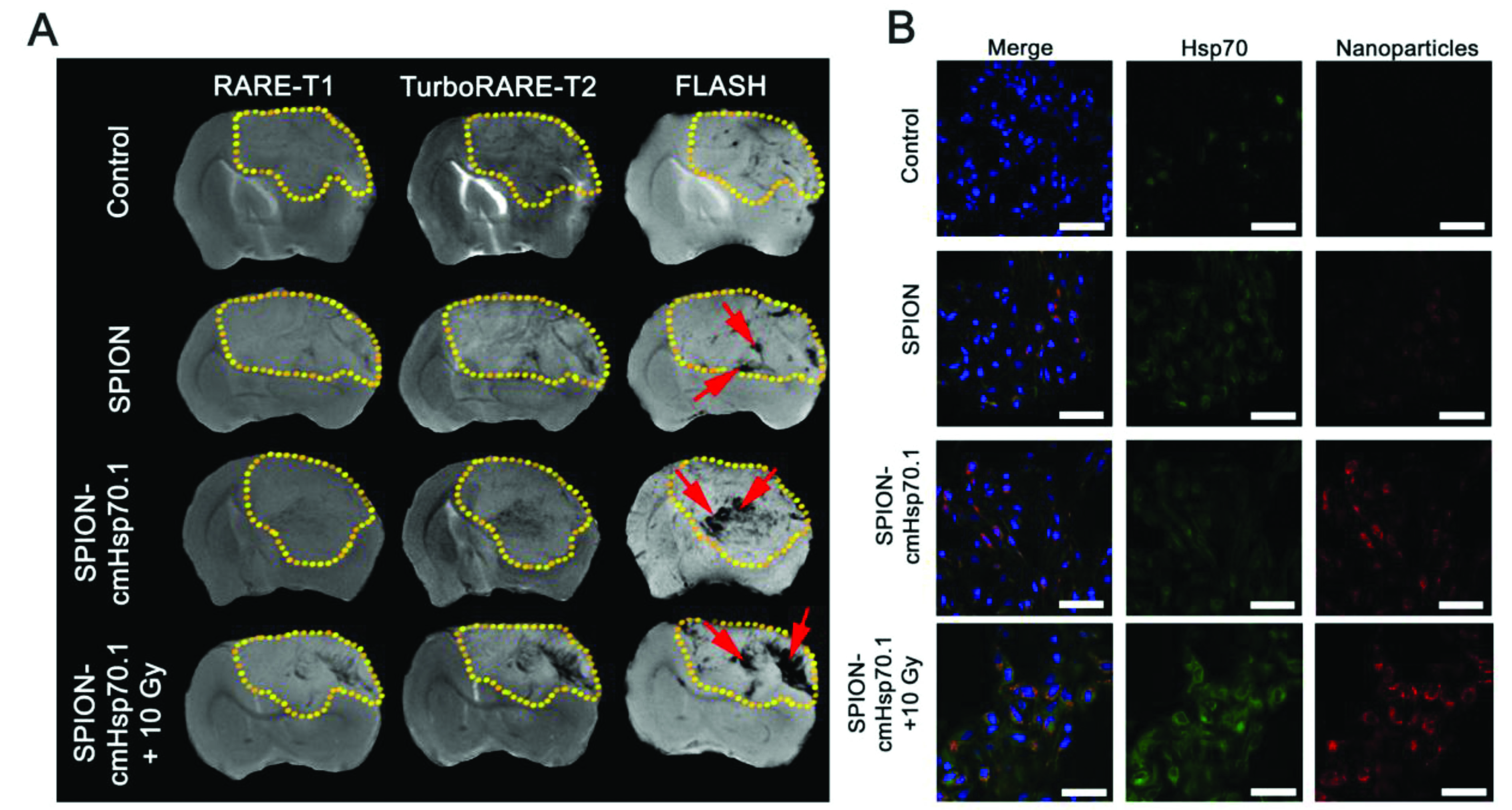
The differences in uptake can be seen in the figure below in both in vivo and in vitro environments using MRI and fluorescence microscopy, respectively. This approach has the potential to be clinically relevant for both diagnosis and therapy of tumours.

Figure 1 Targeting of the orthotopic C6 glioma by SPION–cmHsp70.1 conjugates. (A) Magnetic resonance images for the control, non-irradiated animals treated with SPIONs, SPION–cmHsp70.1 particles and irradiated rats (10 Gy) treated with SPION–cmHsp70.1. Images were obtained in RARE-T1, TurboRARE-T2 and FLASH regimens. Retention of the nanoparticles in the tumor presented as hypotensive zones on T2-weighted and gradient echo images (red arrows). (B) Immunofluorescent images of the brain tumor stained with anti-Hsp70 antibodies (green). Nuclei stained with DAPI (blue). Nanoparticles detected using reflective laser scanning at 488 nm (red). Scale bar, 40 μm.
Ionizing radiation improves glioma-specific targeting of superparamagnetic iron oxide nanoparticles conjugated with cmHsp70.1 monoclonal antibodies (SPION–cmHsp70.1)
Maxim A. Shevtsov, Boris P. Nikolaev, Vyacheslav A. Ryzhov, Ludmila Y. Yakovleva, Yaroslav Y. Marchenko, Marina A. Parr, Valerij I. Rolich, Anastasiya L. Mikhrina, Anatolii V. Dobrodumov, Emil Pitkin and Gabriele Multhoff
Nanoscale, 2015,7, 20652-20664, DOI: 10.1039/C5NR06521F, Paper
Dr Mike Barrow is a guest web writer for the Nanoscale blog, he currently works as a Postdoctoral Researcher at the University of Liverpool. Twitter: @mikesyb


