Archive for February, 2011

Surface nanotechnology for biological applications – themed issue

Issue 2 of Nanoscale out now!

This month’s issue includes a collection of articles on the theme surface nanotechnology for biological applications

This themed issue is Guest Edited by Professor Marcus Textor, Professor Darrell Irvine and Professor Xingyu Jiang. It includes a Review by Antonio Nanci et al. on Nanoscale surface modifications of medically relevant metals: state-of-the art and perspectives and a Communication by Molly Stevens, Kinetic investigation of bioresponsive nanoparticle assembly as a function of ligand design, as well as much, much more!

issue 2 coverCover image

The cover image highlights the paper by Nicholas Melosh and colleagues and shows that the stability of nanoscale hydrophobic bands inside the hydrophobic core of lipid membranes depends on their relative size.

Nanoscale patterning controls inorganic–membrane interface structure
Benjamin D. Almquist, Piyush Verma, Wei Cai and Nicholas A. Melosh
Nanoscale, 2011, 3, 391-400

Want to read more about our themed issues? Please visit the ‘ Themed Issues’ page on our website.

Digg This
Reddit This
Stumble Now!
Share on Facebook
Bookmark this on Delicious
Share on LinkedIn
Bookmark this on Technorati
Post on Twitter
Google Buzz (aka. Google Reader)

Carbon nanotubes in molecular biology

Effect of surface charge of polyethyleneimine-modified multiwalled carbon nanotubes on the improvement of polymerase chain reaction
Xueyan Cao, Jingjing Chen, Shihui Wen, Chen Peng, Mingwu Shen and Xiangyang Shi
Nanoscale DOI:10.1039/C0NR00833H

There are a vast number of potential applications for carbon nanotubes in many areas of science today, but current uses are mostly associated with their structural properties in bulk quantities. However, there is a vast amount of research being conducted on how the nanoscale properties of carbon nanotubes can be used to perform precise actions at a molecular level. This concept is of particular interest in biomedical science, where fine control of interactions with biomolecules and biological structures is of great importance in developing new diagnostic and therapeutic techniques.

Considering applications of carbon nanotubes in biology, scientists in China have conducted a systematic study of how the surface charge of multi-walled carbon nanotubes affects their performance as additives in polymerase chain reactions (PCR), which are of high importance in molecular biology. Cao et al. at Donghua University, Shanghai, used polyethyleneimine (PEI)-modified multiwalled carbon nanotubes with different surface charge polarities, and showed that positively charged nanotubes could significantly enhance the specificity and efficiency of PCR, even when used at a low concentration.

Polymerase chain reactions are of fundamental importance in molecular biology as a gene amplification technique, where the copying yield of a targeted gene can be increased drastically. However, the technique suffers from low specificity and efficiency, and therefore optimisation procedures are essential. Unfortunately, as the mechanism is complex, this optimisation is not easily achieved. Nanoparticles have been studied as potential solutions to these problems due to their unique physicochemical properties, and indeed carbon nanotubes have been shown to be good additives for PCR optimization. However, this study in China is the first report relating to the optimization of PCR using CNTs with different surface charge polarities, and it represents an exciting development in the field.

To read the whole article, click here.

Digg This
Reddit This
Stumble Now!
Share on Facebook
Bookmark this on Delicious
Share on LinkedIn
Bookmark this on Technorati
Post on Twitter
Google Buzz (aka. Google Reader)

Filling the space

Biomineral nanoparticles are space-filling
Li Yang, Christopher E. Killian, Martin Kunz, Nobumichi Tamura and P. U. P. A. Gilbert
Nanoscale, 2011, 3, 603-609

Scientists in the US have tried to answer the question of whether biominerals are mesocrystals or not.

Sea urchin biominerals are known to form from aggregating nanoparticles of amorphous calcium carbonate, which then crystallize into macroscopic single crystals of calcite. The group measured the surface areas of these biominerals, finding them to be comparable to those of space-filling macroscopic geologic calcite crystals. These biominerals are therefore different from synthetic mesocrystals, which are always porous. Based on this results, the group proposes that space-filling amorphous calcium carbonate is the structural precursor for echinoderm biominerals.

Mollusk shells, corals, and echinoderm biominerals have remarkable mechanical properties, making them the object of many studies to shed some light on their formation mechanisms.

Read the whole article now

Article submitted as part of the Themed Issue on Crystallization and Formation Mechanisms of Nanostructures, read the issue here

Digg This
Reddit This
Stumble Now!
Share on Facebook
Bookmark this on Delicious
Share on LinkedIn
Bookmark this on Technorati
Post on Twitter
Google Buzz (aka. Google Reader)

Top Ten most-read Nanoscale articles in January

The latest top ten most accessed Nanoscale articles

See the most-read papers of January 2011 here:

Idalia Bilecka and Markus Niederberger, Nanoscale, 2010, 2, 1358-1374
DOI: 10.1039/B9NR00377K
 
Wolfgang Schärtl, Nanoscale, 2010, 2, 829-843
DOI: 10.1039/C0NR00028K
 
José M. Romo-Herrera, Ramón A. Alvarez-Puebla and Luis M. Liz-Marzán, Nanoscale, 2011, Advance Article
DOI: 10.1039/C0NR00804D
 
Hualan Wang, Qingli Hao, Xujie Yang, Lude Lu and Xin Wang, Nanoscale, 2010, Advance Article
DOI: 10.1039/C0NR00224K
 
Xiaoting Jia, Jessica Campos-Delgado, Mauricio Terrones, Vincent Meunier and Mildred S. Dresselhaus, Nanoscale, 2011, 3, 86-95
DOI: 10.1039/C0NR00600A
 
Wufeng Chen and Lifeng Yan, Nanoscale, 2010, 2, 559-563
DOI: 10.1039/B9NR00191C
 
Rubén Mas-Ballesté, Cristina Gómez-Navarro, Julio Gómez-Herrero and Félix Zamora, Nanoscale, 2011, 3, 20-30
DOI: 10.1039/C0NR00323A
 
Benjamin Weintraub, Zhengzhi Zhou, Yinhua Li and Yulin Deng, Nanoscale, 2010, 2, 1573-1587
DOI: 10.1039/C0NR00047G
 
Jessica M. Rosenholm, Cecilia Sahlgren and Mika Lindén, Nanoscale, 2010, 2, 1870-1883
DOI: 10.1039/C0NR00156B
 
Poulomi Roy, Doohun Kim, Kiyoung Lee, Erdmann Spiecker and Patrik Schmuki, Nanoscale, 2010, 2, 45-59
DOI: 10.1039/B9NR00131J
Digg This
Reddit This
Stumble Now!
Share on Facebook
Bookmark this on Delicious
Share on LinkedIn
Bookmark this on Technorati
Post on Twitter
Google Buzz (aka. Google Reader)

Themed Issue: Modelling for the nanoscale

Nanoscale Themed Issue announcement:

Modelling for the nanoscale

Guest Editors:

Amanda Barnard (CSIRO, Australia)

Changming Li (Nanyang Technological University, Singapore)

Ruhong Zhou (IBM Watson & Columbia University, USA)

Yuliang Zhao (NCNST, China)

Submission deadline: 15 August 2011


The issue will be published in early 2012 and aims to address the recent developments in the field of modelling applied to the nanoscale. This will include studies on CNT-protein and CNT-water interactions (including other nanoparticles and soft matter), confinement and catalysis, DNA-nanopore interactions and sequencing, nanomaterial-environmental interaction modelling, modelling of nanoparticles and nanomaterials (both QM and MM), nucleation, growth and transformations and optical properties of nanostructures as well as modelling of nanotoxicity.

Don’t miss the deadline, submit your contribution before the 15th August 2011.

Digg This
Reddit This
Stumble Now!
Share on Facebook
Bookmark this on Delicious
Share on LinkedIn
Bookmark this on Technorati
Post on Twitter
Google Buzz (aka. Google Reader)

Improving the performance of lithium-ion batteries

New Nanoscale Communication

Enhanced cycleability of LiMn2O4 cathodes by atomic layer deposition of nanosized-thin Al2O3 coatings

Dongsheng Guan, Judith A. Jeevarajan and Ying Wang

Nanoscale, DOI: 10.1039/c0nr00939c

A group of scientists in America have developed a method to put an Al2O3 ‘nano-coating’ on LiMn2O4 cathodes, which results in significantly enhanced performance of the cathode. They claim that this method can be generalised to other electrode materials and a variety of surface coatings in order to significantly improve battery performance.

LiMn2O4 has been widely investigated for use in lithium-ion batteries due to its unique advantages such as high specific capacity and output voltage, and the fact that it is low-cost, abundant and environmentally friendly. However, LiMn2O4 does suffer from a critical problem: it is unstable in the presence of electrolytes and suffers from capacity degradation during cycling. This seriously limits the practical applications of an otherwise very promising material.

Ying Wang and his co-workers used atomic layer deposition (ALD) to deposit the Al2O3 coating on LiMn2O4 cathodes. This method allows fine control over the thickness and conformation of the thin films, and allowed the group to create ultra-thin coatings on the cathodes. Theses ‘nano-coated’ cathodes were then compared with bare cathodes to study differences in electrochemical performance. The group discovered that the Al2O3 coating reduced dissolution of manganese ions from the cathode into the electrolyte, and also reduced decomposition of the electrolyte at the cathode surface. This resulted in a significantly enhanced cycling performance of the LiMn2O4 cathode.

To read more about this study, click here.

Digg This
Reddit This
Stumble Now!
Share on Facebook
Bookmark this on Delicious
Share on LinkedIn
Bookmark this on Technorati
Post on Twitter
Google Buzz (aka. Google Reader)

Nanoscale Hot article: 3D Nano-batteries

Nanoscale Hot Article: just published

Scientists from the US describe the fabrication of 3D multifunctional energy-storage nanoarchitectures.

In these structures, the three critical components of a battery (cathode, separator/electrolyte, and anode) are assembled internally as tri-continuous nanoscopic phases in three steps: First, the formation of a monolithic manganese oxide ambigel nanoarchitecture. Second, the electrodeposition of a conformal ultrathin polymer (separator/electrolyte). And third, the infiltration of a counter insertion electrode (RuO2) within the residual interconnected free volume.

Using Transmission Electron Microscopy, Jeffrey Long and Debra Rolison (US Naval Research Laboratory) and their colleagues have characterized the architectures that contain all three components for a solid-state energy storage device within a void volume of tens of nanometres and have nanometre-thick distances between the opposing electrodes.

Read the article now

Architectural integration of the components necessary for electrical energy storage on the nanoscale and in 3D
Christopher P. Rhodes, Jeffrey W. Long, Katherine A. Pettigrew, Rhonda M. Stroud and Debra R. Rolison
Nanoscale, 2011, DOI: 10.1039/C0NR00731E

Digg This
Reddit This
Stumble Now!
Share on Facebook
Bookmark this on Delicious
Share on LinkedIn
Bookmark this on Technorati
Post on Twitter
Google Buzz (aka. Google Reader)

Carbon Nanotube Vehicles for Cellular Delivery

New Nanoscale Communication

Polyethylenimine–carbon nanotube nanohybrids for siRNA-mediated gene silencing at cellular level

Stéphanie Foillarda, Guy Zuber and Eric Doris

Nanoscale DOI:10.1039/C0NR01005G

Carbon nanotube (CNT) based structures which can act as ‘nanohybrid vehicles’ for the delivery of functional molecules into cells have been developed by scientists in France.

Synthetic interfering RNA (siRNA) is able to inhibit the expression of a targeted gene by triggering enzymatic and sequence-selective degradation of the corresponding mRNAs, which holds great promise for the highly selective treatment of medical disorders at a genetic level. However, nucleic acids require a ‘delivery vehicle’ to take carry them through the cellular membrane and to the sites where they are required. The CNT hybrid nanostructure developed by this group is intended to do exactly this.

Eric Doris and co-workers at CEA, Service de Chimie Bioorganique et de Marquage, covalently modified short CNTs (~200 nm) with the cationic polymer polyethylenimine (PEI), which were then able to bind siRNA. The intention was to build a nanostructure which could enter cells by endocytosis but escape endosomal capture to increase the biological activity of the payload. They appear to be successful in this as they show that their system performs better than a reference lipid carrier.

To read this article, click here.

Digg This
Reddit This
Stumble Now!
Share on Facebook
Bookmark this on Delicious
Share on LinkedIn
Bookmark this on Technorati
Post on Twitter
Google Buzz (aka. Google Reader)

Anisotropic Gold Nanoparticles: Controlling growth with surfactants

New Nanoscale Feature Article

Surfactant-assisted, shape-controlled synthesis of gold nanocrystals

Junyan Xiao and Limin Qi

Nanoscale, DOI: 10.1039/c0nr00814a

This week at Nanoscale we have a published a Feature Article on the shape control of gold nanoparticles using surfactant systems. In this work, the authors Junyan Xiao and Limin Qi provide a comprehensive review of the techniques used to grow anisotropic gold nanoparticles, starting with an overview of the general strategies, before delving deeper into the role of surfactants in the production of some truly remarkable nanostructures. The descriptions of the various growth mechanisms are accompanied by excellent schematic diagrams which provide a valuable insight into the complexities of crystal formation and growth.

It is well known that gold nanoparticles have many unique chemical and physical properties, and there is much interest in applying them in a wide range of exciting applications. For example, they have been studied for use in nanoelectronics, drug delivery, catalysis, sensing, and photothermal therapy, to name but a few. Importantly, they exhibit particularly strong absorption and scattering of light due to localized surface plasmon resonance, a property which will be harnessed in the development of some revolutionary bioimaging devices. However, if these potential applications are to be realized, the growth of gold nanoparticles needs to be highly controlled in order exploit different properties which arise as we change their size, shape and surface chemistry.

Surfactants are vital in nanoparticle synthesis. Not only do they provide a protective capping layer and a means of conjugation, but they play an active role in particle nucleation and growth. Therefore, the choice of surfactant, or the design of a surfactant system, is crucial. In this review, the authors focus on gold nanocrystal synthesis assisted by single surfactants, mixed surfactants, supramolecular surfactants, as well as metal–surfactant complex templates.

To read this article, click here.

Digg This
Reddit This
Stumble Now!
Share on Facebook
Bookmark this on Delicious
Share on LinkedIn
Bookmark this on Technorati
Post on Twitter
Google Buzz (aka. Google Reader)

Transplant tracking

Transplant tracking

Harriet Brewerton

Magnetic nanoparticles could be used to track neural stem cells after a transplant in order to monitor how the cells heal spinal injuries, say UK scientists.

Neural stem cells are a promising treatment for repairing spinal cord injuries as they have the ability to generate tissue, but there is no effective way of monitoring the cells for long periods of time after transplantation.

Transplant tracking

Nguyen TK Thanh at the Davy Faraday Research Laboratory, University College London and the Royal Institution, and colleagues, believe they have the answer. They have developed hollow biocompatible cobalt-platinum nanoparticles and attached them to the stem cells. The nanoparticles are stable for months and have a high magnetic moment – tendency to align with a magnetic field – so that low concentrations can be detected using magnetic resonance imaging (MRI).

‘Magnetic nanoparticles are emerging as novel contrast and tracking agents in medical imaging,’ says Samir Pal at the California Institute of Technology, US, an expert in biological-nanoparticle interactions. ‘When used as a contrast agent for MRI, the nanoparticles allow researchers and clinicians to enhance the tissue contrast of an area of interest by increasing the relaxation rate of water.’

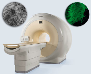
Stem cells attached to biocompatible nanoparticles can be visualised by MRI after transplantation into spinal cord slices

The team labelled stem cells with their nanoparticles, injected them into spinal cord slices and took images of their progress over time. They found that low numbers of the nanoparticle-loaded stem cells could still be detected two weeks after transplantation. ‘The new method demonstrates the feasibility of reliable, noninvasive MRI imaging of nanoparticle-labelled cells,’ says Thanh.

Thanh hopes that her stem cell tracking method will be used during stem cell replacement therapy for many central nervous system diseases. Her team is working towards developing nanoparticles that can be used to diagnose and treat these diseases.

Read the article in Nanoscale:

Magnetic CoPt nanoparticles as MRI contrast agent for transplanted neural stem cells detection

Xiaoting Meng, Hugh C. Seton, Le T. Lu, Ian A. Prior, Nguyen T. K. Thanh and Bing Song
Nanoscale, 2011, DOI: 10.1039/C0NR00846J

Read more Chemistry World News here


Digg This
Reddit This
Stumble Now!
Share on Facebook
Bookmark this on Delicious
Share on LinkedIn
Bookmark this on Technorati
Post on Twitter
Google Buzz (aka. Google Reader)