Posts Tagged ‘PDMS’

How soil-worms allow for realistic human physiology studies

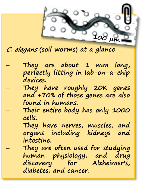
Mice, fruit fly, zebra fish easily come to mind when thinking of animal models for human physiology studies – but one animal is often forgotten, although it is as functional as the others. Have you guessed already? We are talking about soil-dwelling worms, aka C. elegans! These animals combine the simplicity of single-cell systems with the complexity of animal models, therefore they can provide significant insights into human disorders. Please take a moment to look at our note below summarizing the key features of C. elegans.

Muscular strength is a good example of human physiology studies  and it relies on calcium-initiated muscle contraction, sarcomere composition and organization, and translocation of actin and myosin molecules. Analysis of such parameters can reveal the formation of muscular dystrophy, a muscle degenerative disorder. However, the measurement of these parameters has been a challenge due to the dependence on random animal-behavior that yields irreproducible results. Recently, researchers from Texas Tech University collaborated with Rutgers University and University of Nottingham to study muscular strength in C. elegans. They achieved to obtain results independent of animal behavior and gait in a miniature system consisting of an elastic micropillar forest (Figure 1a and 1b).

The microfluidic system is made of PDMS, and contains bendable micropillars hanging from the microchannel ceiling. The pillars are bent upon the action of the body muscles when C. elegans crawls through the pillar array. Individual pillar bending events can be quantified using a microscope-camera system and image analysis (Figure 1c). The pillar density is designed to create high mechanical resistance to locomotion, therefore maximum exertable force can be measured independent of animal behavior. Here, maximum exertable force corresponds to the peak force exerted by human quadriceps muscle in a standardized knee extension resistance test.

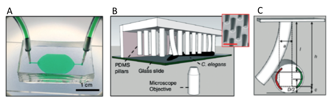
Figure 1. (a) Image of the microfluidic device with the pillar forest and the ports. (b) Schematic demonstration of the C. elegans strength measurement apparatus. Inset shows a scanning electron microscope picture of the pillars. (c) A sketch of interaction with a pillar by the worm body. The pillar is bent due to the action of the body muscles (shown in red and green). Image from Rahman et al.

The authors of this study explain that animals produce strong forces in highly resistive areas and demonstrate different locomotion regimes based on the body size relative to gap between pillars. Besides the body size, body configuration and behavioral characteristics can be the sources to the magnitude of the force exerted on the pillars. Thanks to the probabilistic nature of the parameters sourcing of the force exerted, a reproducible algorithm can be defined for quantifying muscle strength. Using this strategy, researchers showed for the first time that locomotion between microfluidic pillars comprises of three regimes: non-resistive (worm contacts with 1-2 pillars and doesn’t adjust body posture), moderately resistive (worm contacts with >2 pillars and minimally adjust body posture), and highly resistive (worm contacts with multiple pillars and body posture adjustment is disabled). When operated at highly-resistive regime, the microfluidic system suppresses the animal behavior. This system allows for (1) discriminating between the muscle strength or weakness levels of individual worms of different ages, (2) determining body length decrease and muscular contraction levels led by levamisole treatment, (3) comparing the muscular strength in the wild and mutant C. elegans types. According to the researchers, the future studies can help us to obtain deeper understanding in molecular and cellular circuits of neuromuscular function as well as dissection of degenerative processes in disuse, aging, and disease.

To download the full article for free* click the link below:

NemaFlex: a microfluidics-based technology for standardized measurement of muscular strength of C. elegans
Mizanur Rahman, Jennifer E. Hewitt, Frank Van-Bussel, Hunter Edwards, Jerzy Blawzdziewicz, Nathaniel J. Szewczyk, Monica Driscolld, and Siva A. Vanapalli
Lab Chip, 2018, Lab on a Chip Recent Hot Articles
DOI: 10.1039/c8lc00103k


About the Webwriter

Burcu Gumuscu is a postdoctoral fellow in Herr Lab at UC Berkeley in the United States. Her research interests include development of microfluidic devices for quantitative analysis of proteins from single-cells, next generation sequencing, compartmentalized organ-on-chip studies, and desalination of water on the microscale.

Digg This
Reddit This
Stumble Now!
Share on Facebook
Bookmark this on Delicious
Share on LinkedIn
Bookmark this on Technorati
Post on Twitter
Google Buzz (aka. Google Reader)

Zenith in “artery”

When cutting a finger, thrombocytes and fibrin in the blood make up the blood-clotting mechanism, aka. haemostasis, to stop the blood loss. Another way to trigger this mechanism is having an artery damaged by atherosclerosis, which is often caused by several genetic or acquired factors. In the latter case, thrombosis develops within a vein or artery, obstructing or stopping the blood flow to major organs like the heart and eventually causing heart attack. Considering every year over 14 million lives worldwide are lost to heart attacks, more investigation on this topic is needed without any doubt.

Recently, a research team led by Andries van der Meer published a research article in Lab on a Chip on mimicking arterial thrombosis in 3D vascular structures, representing a major step forward in the development of accurate and faster methods of studying arterial thrombosis without using animals. The authors highlighted the inconvenience of using animal models to predict arterial thrombosis in humans. This is mainly due to fundamental differences between human and animal physiology, the researchers explain. For instance, rodent platelet biology, coagulation dynamics, and shear stress in mice arteries significantly vary between humans and mice.

thrombosis on chip

Figure 1. Three-dimensional models of a healthy and stenotic vessels and thrombosis formation upon blood perfusion through the channels.

The paper uses miniaturized vascular structures mimicking 3D architectures found in both healthy and stenotic blood vessels in-vitro (Figure 1). They combined stereolithography and 3D printing of computed tomography angiography data to construct 3D-printed templates of vessels in PDMS microchips. The 3D printed vessels are then coated with human umbilical vein endothelial cells, forming a monolayer fully covering the surface. In the next step, the artificial vessels are perfused with blood at normal arterial shear rates, allowing a blood clot to form as it would happen in the human body. The 3D printed vessel is clinically more relevant when compared to 2D vessel models, since the realistic flow profiles of blood and even distribution of shear stress across the vessel are of great importance when researching arterial thrombosis. Hugo Albers, the co-first author of the paper explains what led the team to try 3D models: “Other groups have worked on thrombosis-on-a-chip before, but we wanted to incorporate flow profiles that are similar to what one would find in-vivo. So we opt for a round and thus 3D shape. Since the stenotic geometry is an important part of this work, we wanted to find a technique that allowed us to make almost any shape we could come up with. Thus 3D-printing seemed to be the way to go.”

When it comes to defining the challenges in 3D organ-on-chip modeling and fabrication, “we needed to replicate the cellular environment using human endothelial cells and human whole blood to fully mimic the nature of vasculature” says Albers. “Incorporating the shape of vasculature to recreate the flow profiles found in-vivo and recreating the shape of vasculature on a small scale was quite challenging, since the resolution of 3D-printing quickly started to be the limiting factor. Furthermore, we ran into problems related to working with whole blood. We had to figure out how to perfuse small channels with blood without instigating thrombosis outside of the microfluidic channel.” The researchers successfully overcame the challenges mentioned by Albers and mimicked the formation of thrombosis in a stenotic vessel model as seen in Figure 1 (bottom).

The researchers note that the next step involves co-culturing arterial endothelial cells and smooth muscle cells with human umbilical vein endothelial cells or moving to different cell lines such as differentiated human induced pluripotent stem cells. “I think we can also apply the 3D-printing technique to create thrombosis-on-a-chip devices with different geometries, e.g. aneurysms or bifurcated geometries”, says Albers.

To download the full article for free* click the link below:

Mimicking arterial thrombosis in a 3D-printed microfluidic in vitro vascular model based on computed tomography angiography data

Pedro F. Costa, Hugo J. Albers, John E. A. Linssen, Heleen H. T. Middelkamp, Linda van der Hout, Robert Passier, Albert van den Berg, Jos Malda and Andries D. van der Meer

Lab Chip, 2017, Paper

DOI: 10.1039/C7LC00202E

This paper is included in our Organ-, Body- and Disease-on-a-Chip Thematic Collection. To read other articles in the collection, visit – rsc.li/organonachip

About the Webwriter

Burcu Gumuscu is a postdoctoral fellow in Herr Lab at UC Berkeley in the United States. Her research interests include development of microfluidic devices for quantitative analysis of proteins from single-cells, next generation sequencing, compartmentalized organ-on-chip studies, and desalination of water on the microscale.

Digg This
Reddit This
Stumble Now!
Share on Facebook
Bookmark this on Delicious
Share on LinkedIn
Bookmark this on Technorati
Post on Twitter
Google Buzz (aka. Google Reader)