Emerging Investigators Series – Ye Ai


Dr. Ye Ai is currently an Associate Professor at Singapore University of Technology and Design (SUTD). He obtained his B.S. in Mechanical Engineering from Huazhong University of Science and Technology (China) in 2005 and his Ph.D. in Mechanical and Aerospace Engineering from Old Dominion University (USA) in 2011. Prior to joining SUTD as an assistant professor in 2013, he worked as a postdoctoral researcher at the Bioscience Division of Los Alamos National Laboratory from June 2011 to January 2013. He was a visiting scholar at Massachusetts Institute of Technology (MIT) from August 2014 to July 2015. He was promoted to associate professor with tenure in September 2019. Dr. Ai’s research interest focuses on developing novel microfluidic technologies for particle/cell manipulation and single cell analysis. His research team is also striving to translate their innovative microfluidic technologies to commercial market through collaborations with industry.

Read Dr Ai’s recent Emerging Investigator Series paper: Microfluidic impedance cytometry device with N-shaped electrodes for lateral position measurement of single cells/particles in the most recent issue of Lab on a Chip, and find out more about him and his work below.


  1. Your recent Emerging Investigator Series paper focuses on measuring the lateral position of single cells or particles. How has your research evolved from your first article to this most recent article?

My first research article when I was a PhD student was to develop a finite element model with dynamically deformed mesh that can simulate the transient motion of finite-size particles in microscale fluid flows. My PhD research mainly focused on electrokinetics for manipulating particles, cells and ions in micro/nanoscale. My postdoctoral training at the Bioscience Division of Los Alamos National Laboratory exposed me to a lot of biological problems, in particular the need in high-throughput cellular analysis at the single cell level. My previous research experience has somehow shaped my current research focus into single cell manipulation and analysis using novel microfluidic technologies when I become an independent principal investigator in Singapore.

  1. What aspect of your work are you most excited about at the moment?

I am most excited to apply our developed microfluidic technologies for solving real biomedical problems and enabling new biological studies. As an example, in 2017 my team published our single cell sorting technology using a highly focused acoustic beam in Lab Chip (DOI: 10.1039/c7lc00678k). Later, I was approached by quite a number of research teams worldwide who wanted to try our sorting technology. These email communications have encouraged me to apply our developed prototype for real biomedical problems. Right now, I have established collaboration with a few biomedical research institutes in Singapore and we have found that our sorting technology is not causing any cell damage, which is however challenging for conventional FACS machine. We currently have the idea to commercialize this single cell sorting technology. Let us see what is going to happen in the next few years.

  1. In your opinion, what are the key considerations when designing a microfluidic platform for real-time measurements?

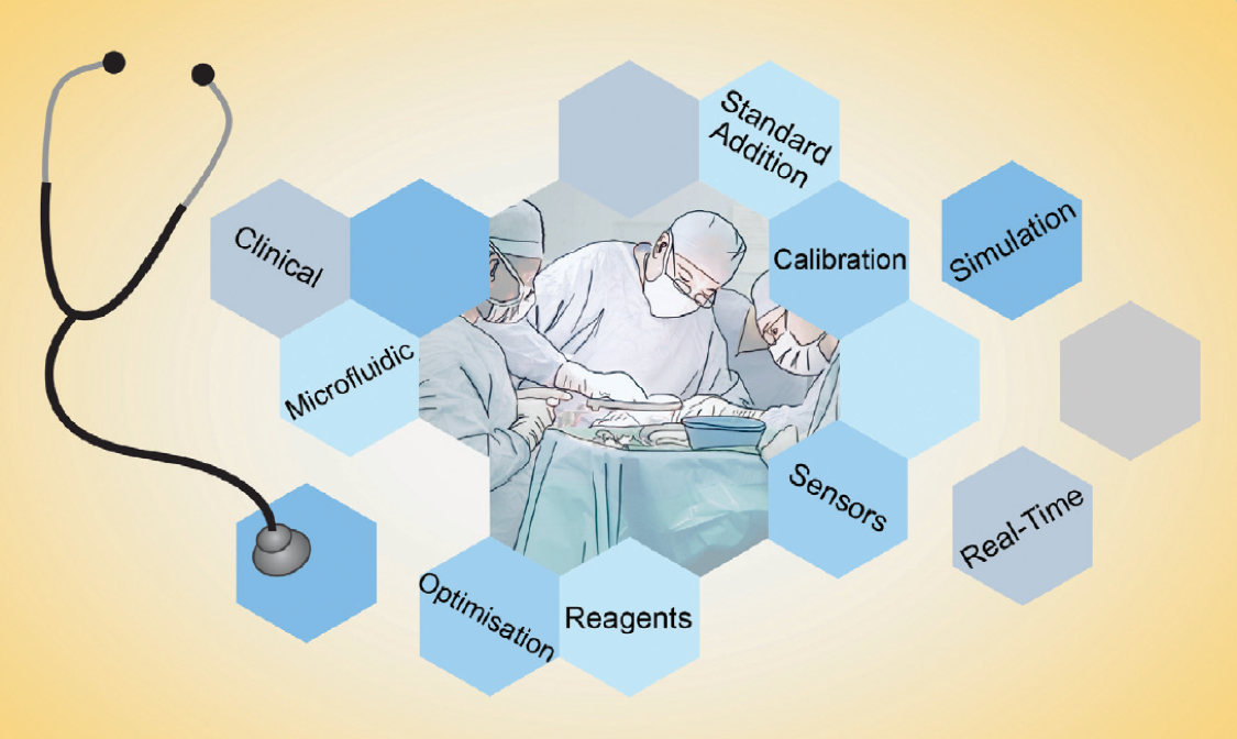
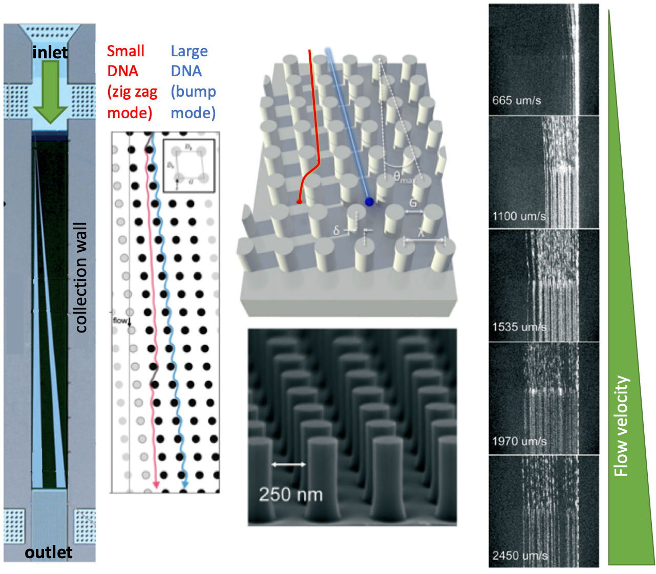
My research team is currently developing both hydrodynamic and acoustic cell sorting platforms. The conventional way to quantify the sorting performance (e.g. purity and recovery) is to run additional cell analysis of collected samples, typically using a flow cytometer. In this work (DOI: 10.1039/c9lc00819e), we designed and validated a new impedance cytometry device that enables the measurement of the lateral positions and physical properties of individual particles and cells. The integration of this new device with any cell sorting platform will allow the evaluation of the sorting performance to be implemented in the same system.

The key consideration of integrating these real-time measurements really depends on whether there is a critical need. But I do see a lot of sorting applications need these real-time, in-line measurements for the purposes of quality control and workflow simplification. And any integration will somehow complicate the system and increase the cost, so the other key consideration is the ease of integration. Integration of electronics is generally easier compared to optics, and we are measuring intrinsic biophysical properties rather than labelling approaches; therefore, I do see great opportunities to integrate our new impedance cytometry device with a variety of cell sorting platforms.

  1. What do you find most challenging about your research?

The microfluidics and Lab on a chip research area is interdisciplinary in nature. My challenge is always to find the right people (e.g. students, postdoctoral fellows, collaborators) and secure sufficient resources to work on real impactful research problems.

  1. How do you spend your spare time?

I am trying to make a balance between work and personal life, so I mainly spend my spare time with my family members, especially my second kid is only 8 months old. I also spend some of my spare time to do physical exercise, which can help relax and leave some time for free thinking.

  1. Which profession would you choose if you were not a scientist?

I rarely thought about this before. Perhaps I would choose to be a doctor.

  1. Can you share one piece of career-related advice or wisdom with other early career scientists?

Based on my own experience, it is important to define a unique research domain based on your own expertise and the surrounding research ecosystem when early career scientists start their independent research. It is also wise to have a clear vision about what you want to achieve in the next five years.

Digg This
Reddit This
Stumble Now!
Share on Facebook
Bookmark this on Delicious
Share on LinkedIn
Bookmark this on Technorati
Post on Twitter
Google Buzz (aka. Google Reader)

New thematic collection open for submissions – Single Cell Analysis

We are delighted to announce a new thematic collection in Lab on a Chip, focusing on multimodal single cell analysis, with Professors Daniel T. Chiu and Pratip K. Chattopadhyay as thought leaders.

Daniel Chiu

Professors Chiu and Chattopadhyay describe the current challenges in the field in their recent editorial in Lab on a Chip on “The Next Frontier in Single Cell Analysis: MultiModal Studies and Clinical Translation”:

Biological processes are inherently complex. Stochasticity, redundancy, plasticity, and noise are built into fundamental cellular activities from gene transcription to protein expression. A major challenge in biomedical research is to untangle this complexity. Microarray technology influenced biological research because it demonstrated clearly the wide selection of cellular molecules available for measurement and provided an efficient means to query them. However, microarrays require a large amount of material and assay large numbers of cells together in bulk.

Single cell analysis overcomes the problems of bulk measurements, but for many years the only available technology—flow cytometry—was incapable of highly multiplexed measurements. The current movement in single cell analysis is multimodal characterization. These approaches, which are rapidly replacing one-dimensional single cell analysis in biomedical research, simultaneously combine measurements of transcription with post-transcriptional regulation, epigenetic modifications, and surface protein expression. It is possible that lipid and metabolite composition, and/or cellular morphology may also be analyzed with the transcriptome or proteome.

We now have a dizzying array of tools that provide us with the potential to comprehensively and accurately characterize the cells involved in a biological process. We are a step away from using these tools widely and efficiently to impact clinical care, but there are large obstacles we must break down first. With a better understanding of the complexity ingrained in cellular systems, how do we smartly choose subsets of markers and cell types to survey, remembering that samples from patients are often limited as are research budgets? Once we know what to measure, there is the critical question of how to measure it, since there are a myriad of technical platforms and data analysis tools from which to choose. As we make measurements, how do we ensure that they are robust—are there general validation and quality control principles we can establish, or are such measures wholly platform-specific? Finally, are highly multiplexed, single cell technologies valuable only as a screening tool to identify simple biomarkers, or can these highly complex technologies (and their associated data analysis algorithms) be used directly for clinical diagnostics?

We invite review and research manuscripts that suggest answers to these questions and related issues for inclusion in a thematic collection focused on multimodal single cell analysis. If you are interested in submitting to the collection please contact the Editorial Office.

This collection open for submissions now, and into 2020.


If you’re interested in this topic, you can read our previous thematic collection on droplet-based single-cell sequencing here. The articles are free to read until November 15th 2019.


Digg This
Reddit This
Stumble Now!
Share on Facebook
Bookmark this on Delicious
Share on LinkedIn
Bookmark this on Technorati
Post on Twitter
Google Buzz (aka. Google Reader)

What are the challenges to sample-in-answer-out technologies in clinical settings?

The boom in microfluidic total analysis systems is spurred by the use of microscale fabrication techniques. In 1999, Agilent Technologies introduced a coin-size device called “LabChip” to the market for the analysis of DNA. The timing of the market release of this device was remarkable as it benefited from the hype of the Human Genome Project. The LabChip has drawn considerable attention from both academia and industry. As a result, the shrink of chemistry labs into coin-sized microfluidic devices has evolved more from technological push than from market pull. Technology-driven development pathway brought challenges, especially for the clinical integration of microfluidic devices due to the lack of standards, focus, and communication between academia and clinics.

A recent Lab on a Chip paper from Martyn Boutelle’s Lab addresses this issue. The authors identify the problem from a well-addressed microdialysis perspective. They state that “taking a microfluidic system into clinical environment brings lots of challenges, not least that during setups and developments the very low the flow rates used in combination with microdialysis means that leaks and misdirection of flows are very hard to see.” The authors define the most significant challenge as the development of a device that’s robust enough to be used by and provide enough information to, the clinical team without micromanagement by experts.

The authors attack the problem by creating a sensor-based online system associated with electrochemical measurement, which would be able to analyze the sample in a miniaturized platform continuously. They developed the microfluidic sensors and chip, but they wanted to increase the use of their technology in real-world scenarios by non-experts, so they looked into ways to introduce more precision and control to the platform. The authors combined their technology with LabSmith microfluidic components and constructed breadboard like layouts for typical lab protocols. The authors add that “the main surprise was the ability to bring the rigor of an analytical laboratory into unusual places such as abattoirs, surgical theatres and public transport!”

In this work and the previous work of the authors, the performance of 3-D printed chips was compared to the PDMS ones. The authors found that PDMS material is much more vulnerable than the 3D print outs when frequently handled. The sensor attached to the chip is programmed to calibrate in regular intervals (e.g., every three hours) in single or multiphase flow conditions. Authors describe the advantage of the system that ”remote access to the scripts allows interaction with the system without the requirement of a highly skilled person being right next to it, which in the context of surgical theatres and hospital wards is a distinct advantage. If the codes and scripts are available to less skilled personnel, they are still able to interact and use the system and by making the system more user-friendly a wider audience and more enthusiasm is generated for the product, increasing interest, uptake, and use.”

The authors would like to improve the platform further by making it wearable since it already has grounds for such an operation with wireless sensors. The next thing to be improved in the system is the feed. At this moment, the syringes have to be regularly refilled. This might not be a problem in the laboratory; however, monitoring can last for days in a clinical setting, and periodically refilling the syringes may lead to noise artifacts. Another improvement could be the ease of operation and troubleshooting when, e.g., a tubing becomes blocked in the middle of the measurement when the user is short on space and time.

Lastly, the authors think that this pioneering platform can help shape the future in the market by giving more people access to an area of science that was previously highly skilled, whilst maintaining analytical robustness. This will be the start to break the barrier between academia-made devices and clinical settings.


To download the full article for free* click the link below:

Clinical translation of microfluidic sensor devices: focus on calibration and analytical robustness

Sally A. N. Gowers, Michelle L. Rogers, Marsilea A. Booth, Chi L. Leong, Isabelle C. Samper, Tonghathai Phairatana, Sharon L. Jewell, Clemens Pahl, Anthony J. Strong and Martyn G. Boutelle, Lab Chip, 2019, Lab on a Chip Hot Articles

DOI: 10.1039/C9LC00400A

*access is free with an RSC account (free to register)


About the Webwriter

Burcu Gumuscu is a researcher in Mesoscale Chemical Systems Group at the University of Twente in the Netherlands. Her research interests include the development of microfluidic devices for quantitative analysis of proteins from single-cells, next-generation sequencing, compartmentalized organ-on-chip studies, and desalination of water on the microscale.

Digg This
Reddit This
Stumble Now!
Share on Facebook
Bookmark this on Delicious
Share on LinkedIn
Bookmark this on Technorati
Post on Twitter
Google Buzz (aka. Google Reader)

Emerging Investigators Series – Jacqueline Linnes

Dr. Jacqueline Linnes is an Assistant Professor in the Weldon School of Biomedical Engineering at Purdue University. She received her B.S. in Engineering from the Purdue University and Ph.D. in Biongineering from the University of Washington. She was a Fogarty Postdoctoral Fellow at the Division of Global Health Equity within Brigham and Women’s Hospital/Harvard Medical School and continued her postdoctoral training at Boston University in Biomedical Engineering. She has received numerous awards including the Mandela Fellows Global Innovation Challenge Award (2017), Fast Company’s World Changing Ideas Finalist (2018), and Marta E. Gross Assistant Professorship of Biomedical Engineering (2018).

Dr. Linnes’s research lab develops real-time detection technologies to prevent, diagnose, and better understand the pathogenesis of diseases. This work emphasizes the translation of fundamental microfluidics and biological assays into point-of-care diagnostics using human-centered design principles. Her extensive experience in translational research includes co-founding and managing early-stage field-testing and user feedback for two startup companies. She has co-developed point-of-care health diagnostics, wearable devices, and water purification technologies for use in the US, Bolivia, Nicaragua, Kenya, Zambia, and Haiti.

Read Dr Linnes’s Emerging Investigator article “Microfluidic Rapid and Autonomous Analytical Device (microRAAD) to Detect HIV from Whole Blood Samples” and find out more about her in the interview below.


Your recent Emerging Investigator Series paper focuses on detecting HIV using a microRAAD. How has your research evolved from your first article to this most recent article?

As a PhD student, I published on the causes of bacterial adhesion to proteins adsorbed to medical devices (in 2012). These infections are incredibly difficult to detect and I found that I didn’t want to just study the causes of infections but to develop the diagnostic tools themselves. I now use the molecular biology and surface analysis skills that I developed in my PhD to create point-of-care diagnostic devices in my own lab. A critical shift in my thinking came when I realized that both the technical skills and the problem solving mindset that I learned in my PhD research were transferable to entirely different fields. In my two postdocs, I worked on Global Health projects ranging from infection control to point-of-care diagnostic devices.  Now in my own research lab, we focus on developing, integrating and automating real-time detection technologies including point-of-care diagnostics and wearable devices, to meet the needs of underserved populations. This article is an example of a sample-to-answer test we developed to automate molecular detection of a pathogen (HIV) from whole blood sample at the point-of-care.


What aspect of your work are you most excited about at the moment?

I started my lab in 2015 with the vision that we could automate molecular detection in point-of-care diagnostics, so that the dvices could be used by anyone, anywhere in the world. I love that we have pulled together individuals with expertise in so many different fields from materials science, to electrical engineering, to molecular biology in order to make this technology work. A huge contingent of my lab and Dr. Stanciu’s lab, and at all levels, from undergraduate researchers to PhD’s have contributed to this project. Now we are bringing in more expertise in translational clinical research. I am currently in Kenya and just handed over a batch of these microRAADs to my colleague, Dr. Eddy Odari at Jomo Kenyatta University of Agriculture and Technology. Dr. Odari will be testing the MicroRAADs using real patient samples and I can’t wait to find out the results.


In your opinion, what is the biggest advantage to using your microRAAD compared to other methods of detecting HIV?

I know there’s still a ways to go, but I believe that the microRAAD platform will ultimately bridge the gap between laboratory-based molecular detection instruments and truly point-of-care diagnosis of HIV in the field.


What do you find most challenging about your research?

Designing technologies sample-to-answer molecular diagnostics that are both highly sensitive and remain robust and accessible to the clinicians, technicians, and patients who need them is incredibly challenging. In my lab, we find it critical to test out our ideas and prototypes via formal and informal usability studies to understand what can be done practically in the field settings that they are designed for. We redesign anything that isn’t actually usable in the real world.


At which upcoming conferences or events may our readers meet you?

I am at the 4th Africa International Biotechnology and Biomedical Conference in Nairobi and Mombasa, Kenya, and this October I will be attending the 2019 Biomedical Engineering Society Annual Meeting in Philadelphia, USA, and the 2019 MicroTAS conference in Basel, Switzerland.


How do you spend your spare time?

I have a 5 year old and a 3 year old so “spare time” is perhaps an overstatement, but we spend a lot of time outdoors at parks and playgrounds and my husband and I built a tree house in our backyard this summer.


Which profession would you choose if you were not a scientist?

That’s a tough one. I love my job as a biomedical engineering faculty member. I do think it would be fantastic to work at a science museum developing and building exhibits and outreach activities.


Can you share one piece of career-related advice or wisdom with other early career scientists?

Don’t underestimate the power that people play in your research. Play well with others, find a place that supports you in your efforts, seek out excellent employees and mentees, and make sure to invest in their development and in your own. Whenever possible, work directly with the people that you ultimately want to use your technology. It is both incredibly motivating and absolutely critical to making an impact that reaches beyond the confines of your own lab.

Dr Jacqueline Linnes

Dr Jacqueline Linnes (Picture credit: Purdue University photo/Rebecca Wilcox)

Digg This
Reddit This
Stumble Now!
Share on Facebook
Bookmark this on Delicious
Share on LinkedIn
Bookmark this on Technorati
Post on Twitter
Google Buzz (aka. Google Reader)

Lab on a Chip Thematic collection on cancer immunotherapy-open for submissions

Professor James R. Heath, Institute for Systems Biology, Seattle, USA

We are very happy to announce a new thematic collection in Lab on a Chip on cancer immunotherapy with Professor James R. Heath, President and Professor at Institute for Systems Biology in Seattle, acting as Thought leader.

In his recent Editorial published in Lab on a Chip, Professor James R. Heath wrote “The rapid development of cancer immunotherapies over the past 5-10 years is not only revolutionizing clinical cancer care, but it is also making the immunotherapy field a proving ground for many new measurement and computational technologies.  An understanding of the technological needs of this field can be gleaned by placing those needs within the context of state-of-the-art treatments. […]

The level of personalisation that is now being tested in the clinic hardly was unthinkable just a decade ago.  The newness of personalized cancer immunotherapies means that, as a rule, they are still extremely expensive.  An urgent and unmet need is to develop technologies that can assist in the democratization of such treatments.[…]

A unique characteristic of the biology of immuno-oncology is that it can invariably be mined to generate new hypotheses for how to improve treatments.  Such hypotheses might include approaches for improved bioengineering of T cells, or the potential identification of new immune checkpoints, etc.  While this characteristic gives cancer immunotherapy a very bright future, it also means that finding technologies that can rapidly and inexpensively validate or negate such hypotheses is an urgent and rapidly expanding need

We welcome primary research and review content relating to how lab-on-a-chip technologies can be developed to address these and related challenges for inclusion in a thematic collection in Lab on a Chip focused on immuno-engineering and immuno-therapy. This collection is now open for submissions and we are looking for submissions into 2020.

Please note that all submitted manuscripts will be subject to peer review in accordance with the journal’s normal standards.

Lab on a Chip publishes significant and original work related to miniaturisation, at the micro- and nano-scale, of interest to a multidisciplinary readership. The journal seeks to publish work at the interface between physical technological advancements and high impact applications that are of direct interest to a broad audience.

We have compiled a collection of recent papers and reviews published in Lab on a Chip on this topic. These articles can be read at rsc.li/immunotherapy and are available free to access* until the 15th November 2019. A couple of highlights from this collections are shown below.

Graphical Abstract from Segaliny, Zhao, et al., 2018 (DOI: 10.1039/C8LC00818C)


Functional TCR T cell screening using single-cell droplet microfluidics

Aude I. Segaliny, Weian Zhao, et al.


MATE-Seq: microfluidic antigen-TCR engagement sequencing

Alphonsus H. C. Ng, James R. Heath et al.


If you’re interested in contributing to this collection,

please contact the Lab on a Chip Editorial Office.


Digg This
Reddit This
Stumble Now!
Share on Facebook
Bookmark this on Delicious
Share on LinkedIn
Bookmark this on Technorati
Post on Twitter
Google Buzz (aka. Google Reader)

Wearable-Implantable Sensors Thematic Collection-open for submissions

We are very pleased to announce a new Thematic Collection on Wearable and Implantable sensors!

Cover Image from 10.1039/C7LC00914C

Cover image for 10.1039/C7LC00914C

A ‘super-team’ of Lab on a Chip authors (10.1039/c7lc00914c) recently wrote, “Wearable sensing technology has recently and rapidly moved from largely a vision of science fiction to a wide array of established consumer and medical products. This explosion of wearable sensors can be attributed to several factors, such as affordability and ergonomics provided by advances in miniaturized electronics, the proliferation of smart-phones and connected devices, a growing consumer desire for health awareness, and the unmet need for doctors to continuously obtain medical quality data from their patients.”

Following this, we at Lab on a Chip have been inspired to create an Editors’ Choice collection highlighting some of our favourite recent papers in the area and to also seek more contributions in this area. The collection will feature a series of papers that address aspects of the issues involved in creating wearable or implantable sensors and their applications for diagnostics, medicine and therapeutics, health awareness and other novel applications.

Below is a selection of content highlights featured in the collection so far. In addition, all papers are free to read until 31st October 2019*.

Wearable sensors: modalities, challenges, and prospects

J. Heikenfeld, A. Jajack, J. Rogers, P. Gutruf, L. Tian, T. Pan, R. Li, M. Khine, J. Kim, J. Wang and J. Kim


Flexible plastic, paper and textile lab-on-a chip platforms for electrochemical biosensing

Anastasios Economou, Christos Kokkinos and Mamas Prodromidis


Microfluidic neural probes: in vivo tools for advancing neuroscience

Joo Yong Sim, Jae-Woong Jeong, et al.


Passive sweat collection and colorimetric analysis of biomarkers relevant to kidney disorders using a soft microfluidic system

Yi Zhang, John A. Rogers, et al.


Complete validation of a continuous and blood-correlated sweat biosensing device with integrated sweat stimulation

A Hauke, J. Heikenfeld, et al.


Interested in submitting to the collection?

We are interested in contributions of review and research articles in this area and this collection is now open for submissions into 2020. If you’re interested in contributing to this collection, please contact the Editorial Office.

*Access is free through an RSC account (free to register)

Digg This
Reddit This
Stumble Now!
Share on Facebook
Bookmark this on Delicious
Share on LinkedIn
Bookmark this on Technorati
Post on Twitter
Google Buzz (aka. Google Reader)

We are delighted to announce that Hang Lu is the 2019 winner of the “Pioneers of Miniaturization” Lectureship!

The 14th “Pioneers of Miniaturization” Lectureship, sponsored by Dolomite and Lab on a Chip , is for early to mid-career scientists who have made extraordinary or outstanding contributions to the understanding or development of miniaturised systems.

The 2019 “Pioneers of Miniaturization” Lectureship will be presented to Professor Lu at the µTAS 2019 Conference in Basel, Switzerland, being held on 27-31 October 2019. Professor Lu will receive a certificate, a monetary award and will give a short lecture during the conference.

Many congratulations to Professor Hang Lu on this achievement from the Lab on a Chip Team!

About the Winner

Professor Hang Lu is the Love Family Professor, School of Chemical and Biomolecular Engineering, Georgia Institute of Technology, USA.

Professor Lu earned her PhD in Chemical Engineering from Massachusetts Institute of Technology, USA in 2003. After a postdoctoral fellowship with Professor Cornelia I. Bargmann, at University of California San Francisco and the Rockefeller University, she was appointed as an Assistant Professor at School of Chemical and Biomolecular Engineering, Georgia Institute of Technology.

In recognition of her outstanding achievements, Professor Lu has received numerous awards and international recognition, including being invited to join Board of Directors, Chemical and Biological Microsystems Society, invited to present at the Nobel Symposium on Microfluidics (2017) and the National Academy of Sciences’ Kavli Frontiers of Science Symposia (2014, 2012, 2009), awarded the ACS Analytical Chemistry Young Innovator Award, Chemical and Biological Microsystems Society (2013), Council of Systems Biology in Boston (CSB2) Prize in Systems Biology (2011), a National Science Foundation CAREER award (2010), an Alfred P. Sloan Foundation Research Fellowship (2009), a DARPA Young Faculty Award (2007), a DuPont Young Professor Award (2006), the Saville Lectureship of Princeton University (2013), the H. C. Van Ness Award Lectures of Rensselaer Polytechnic Institute (2011), and is a fellow of the American Institute for Medical and Biological Engineering (AIMBE) and  a fellow of the American Association for the Advancement of Science (AAAS). She has authored more than 140 peer-reviewed publications and has served on the Editorial Board of Lab on a Chip as Associate Editor since 2017. She is currently the director of the Interdisciplinary Bioengineering Program, and the associate director of the NSF-Simons Foundation supported Southeast Center for Mathematics and Biology, Georgia Institute of Technology.

Professor Lu has pioneered the use of microfluidic systems for imaging and performing genetic studies with small organisms, primarily the nematode C. elegans. In a series of studies published since 2008 she established a set of technologies to streamline imaging, phenotyping, and sorting of C. elegans based on features that are difficult to distinguish and discern by human eyes. The throughput of these technologies were often 1,000 times that of conventional approaches. Professor Lu’s technology has enable faster and more accurate experiments and revolutionized how genetic screens and high-content imaging experiments are done currently in other scientists’ labs. In parallel, her lab has also engineered micro systems for high-content experiments with cells, aggregates, organoids, and embryos to extract high-dimensional information for systems biology studies.

The Lu group performs research at the interface of engineering and biology. They engineer automated microfluidic systems, microscopy tools, and image imformatic technologies to address questions in neuroscience, cell biology, and biotechnology that are difficult to answer using conventional techniques. Applied to the study of fundamental biological questions, these new techniques allow the Lu group to gather large-scale quantitative data about complex systems.

Learn about the Lu group online

Digg This
Reddit This
Stumble Now!
Share on Facebook
Bookmark this on Delicious
Share on LinkedIn
Bookmark this on Technorati
Post on Twitter
Google Buzz (aka. Google Reader)

Emerging Investigator Series – Joo H. Kang

Dr. Joo H. Kang is currently an Assistant Professor of the Department of Biomedical Engineering, School of Life Sciences at Ulsan National Institute of Science and Technology (UNIST), Korea. He received double Bachelor’s degrees in Chemical Engineering and Life Science from Sogang University in 2002 and his M.S. and Ph.D. in Bioengineering from Korea Advanced Institute of Science and Technology (KAIST) in 2004 and 2008, respectively. He joined Children’s Hospital Boston, Harvard Medical School as a research fellow in 2009, and he continued his work at the Wyss Institute, Harvard University as a Wyss Technology Development Fellow from 2012-2016. He received several awards in his early career, including Postdoctoral Award for Professional Development from Harvard University, Wyss Technology Development Fellowship from Harvard University, Baxter Young Investigator Award from Baxter Inc., and Young Frontiers in Bio and Braining Engineering from KAIST. His research interests include multiscale biofluidic approaches for tackling infectious diseases and cancer, and miniaturized organ-mimicking microsystems.

Read Joo H. Kang’s Emerging Investigator article “Measurement of the magnetic susceptibility of subtle paramagnetic solutions using the diamagnetic repulsion of polymer microparticles” and find out more about him in the interview below:


Your recent Emerging Investigator Series paper focuses on measuring magnetic susceptibility of subtle paramagnetic solution using diamagnetic repulsion of polymer microparticles. How has your research evolved from your first article to this most recent article?

One of the research topics that interested me was to discriminate the subtle differences in the magnetic susceptibility of materials in a microfluidic regime. The first paper I published in regards to this (Kang,JH, et al., JACS, 2009) demonstrated the capability of discriminating the magnetic susceptibility of “solid microparticles” where they are diamagnetically forced to be located at a quasi-isomagnetic position in a microfluidic channel (a position where the differences of the magnetic susceptibility between the solid particles and surrounding media become nearly zero). When I was invited to make contribution to the Emerging Investigator Series of Lab on a Chip last year, I wanted to revisit this, and this time I aimed to assess the subtle magnetic susceptibility of “surrounding paramagnetic solutions”.

What aspect of your work are you most excited about at the moment?

As for the paper, I was surprised of the sensitivity of the device that can discriminate the magnetic susceptibility. We compared our results with those assessed by a conventional superconducting quantum interference device (SQUID), and found that our approach is even more sensitive than the conventional one. Likewise, we can unveil various scientific approaches when exploring fluidic regimes at the micro and nanoscale, and this is the most exciting aspect as being a part of the research community in this field.

In your opinion, what applications can your current approach be used for?

Because this is a platform technology, various applications are possible where we need to measure the magnetic susceptibility of paramagnetic solutions. Assessment of residual magnetic nanoparticles in biological samples, for examples, would be one of the potential uses. We could also use this platform to evaluate metal contamination of drinking water, such as chromium or iron oxide, which alters the magnetic susceptibility of water.

What do you find most challenging about your research?

Taking research from the “bench to products”. Since I started my independent research career, I realized that I have to make considerable efforts to get this happen while playing multiple roles at the same time. But I am enjoying it.

In which upcoming conferences or events may our readers meet you?

I am planning to attend microTAS 2019 that will be held in Basel, Switzerland this year.

How do you spend your spare time?

I am spending my time with my family, hiking, swimming, and playing soccer or games with my little son and daughter.

Which profession would you choose if you were not a scientist?

Probably an architect. This was one of the paths I was thinking of when I was a high school student.

Can you share one piece of career-related advice or wisdom with other early career scientists?

A clear vision on your own research and collaborators who you can share your vision with.

Digg This
Reddit This
Stumble Now!
Share on Facebook
Bookmark this on Delicious
Share on LinkedIn
Bookmark this on Technorati
Post on Twitter
Google Buzz (aka. Google Reader)

Emerging Investigator Series – Mei He

Dr. He is a tenure-track assistant professor at the University of Kansas. She received her PhD degree from the University of Alberta with professor Jed Harrison, and postdoctoral training from the University of California, Berkeley with professor Amy Herr. She is the vice chair of the ASABE Biosensor program and the Councilor of the American Electrophoresis Society. Dr. He is also the founder of Clara Biotech Inc. and the founder committee for the MidWest 3D technology society. Dr. He Received NIH Maximizing Investgator’s Research Award for Early Stage Investigators in 2019. She also received the Lab on Chip Outstanding Reviewer for the year of 2018. One of her publications also received the 2018 SLAS Technology Readers Choice Award. Her research interests include biomedical microfluidic devices and sensing approaches, 3D biomaterials, and nanodelivery, employed in programming and monitoring biomimetic immunity associated with extracellular vesicles.

Read Mei He’s Emerging Investigator article “3D-printing enabled micro-assembly of a microfluidic electroporation system for 3D tissue engineering” and find out more about her in the interview below:


Your recent Emerging Investigator Series paper focuses on “3D-printing Enabled Micro-assembly of Microfluidic Electroporation System for 3D Tissue Engineering”. How has your research evolved from your first article to this most recent article?

My first article observed the microscale evolution of porous polymer materials in the microfluidic channel when I was a PhD student. I found very interesting phenomena in the microfluidic device which actually inspired me to explore more surrounding dimensions, surface chemistry, and scales. Till to my recent article focusing on 3D geometric influence on cellular behavior and their extracellular vesicles secretion dynamics, the 3D dimension in microscale is intriguing in the biological system.

What aspect of your work are you most excited about at the moment?

I am very excited to take the microfluidic technology and phenomena into the biology world, as it will bring new investigation and discovery. Biology is still in its infancy stage, I am very excited to see how microfluidic technology could advance this growth.

In your opinion, what is the biggest impact your microfluidic electroporation system will have in tissue engineering?

Intracellular delivery of regulatory or therapeutic targets into the cell is very crucial in the field of tissue engineering and regenerative medicine. Current existing electro-transfection systems, including microfluidic platforms and commercial benchtop systems, are only able to study monolayer cell suspensions in vitro, which is incapable of clinical translation within in vivo tissue microenvironment. So developing a 3D, in vivo like tissue microenvironment with effective electro-transfection is very important to move to the clinical study in the future. We actually are more interested in downstream, precise control and manipulation of cellular machinery for secreting exosomes and extracellular vesicles under the transfection-induced stimulus, such technology is not existing yet but very important for understanding the interconnection of cargo internalization with cellular level responses elicited by exosomes delivery pathway.

What do you find most challenging about your research?

Building up an in vivo like tissue system with precise control is not straightforward. The environment in a controlled lab setting is totally different than in an in vivo biological system. So the analyzed information actually is not representative of the real situation in the human in vivo system. There are huge heterogeneities present in the cell population as well as human individuals, which poses the challenges for correctly understanding cellular system regulation, such as immunity, in our human body. Mimicking in vivo living system is very challenging, but crucial for understanding quite a few of mechanism and disease pathogenesis. Our research introduces new microfluidic technology and material solutions to solve such challenges.

In which upcoming conferences or events may our readers meet you?

I will attend next year Gorden Research Conference in Bioanalytical Sensors as well as the MicroTAS annual meeting.

How do you spend your spare time?

I have a 7-year-old boy and expecting a new baby girl this year. My spare time definitely is occupied by kids and watching them growing.

Which profession would you choose if you were not a scientist?

I always like to discover new things since I was a child. If I am not a scientist, I would like to be a greeting card designer or paleontologist.

Can you share one piece of career-related advice or wisdom with other early career scientists?

Being a life-long learner and always keeping strong scientific curiosity will definitely help with your research development. Get good mentors around you and you will appreciate their advice.

Digg This
Reddit This
Stumble Now!
Share on Facebook
Bookmark this on Delicious
Share on LinkedIn
Bookmark this on Technorati
Post on Twitter
Google Buzz (aka. Google Reader)

Through a cheaper ‘book of life’

Mapping the human genome project has been one of the world’s largest scientific collaborations. Completing the full genome sequencing for “the book of life” took more than 10 years with the efforts of 1000’s scientists and a budget of $3 billion. About 20 years after the finalization of this enormous project, it is now possible to complete a full human genome sequencing within 8 days for about $1,000 thanks to more advanced sequencing tools. Further improvements in genome sequencing tools are still warranted today because the genome sequencing field has been embraced by many more applications, including forensics, disease modeling & identification, and personalized medicine (e.g., identifying the genes that cause a medicine to work in one patient but not in another).

Initial sequencing technologies relied on standard DNA electrophoresis techniques such as slab gels and capillaries, allowing for the preparation of only small numbers of samples at a time. The sample preparation limitation was the primary reason for the increased costs and processing duration during the human genome project. Many efforts have been directed towards improving sample preparation techniques in the last decades. As the first step, electrophoresis techniques have been optimized to boost the sample throughput with user-friendly, smaller, and functional platforms. Traditional DNA separation gels, which have been used as the golden standard for many decades, have been replaced by microfabricated post arrays and nanometer-scale deterministic lateral displacement arrays.

A nice example of nanometer-scale deterministic lateral displacement arrays has been demonstrated recently by researchers from IBM T.J. Watson Research Center and Icahn School of Medicine at Mount Sinai in New York, USA. In this work, the researchers fractioned DNA in the range of 100-10.000 base pairs with a size-selective resolution of 200 base pairs. To achieve that, four different microchip configurations were fabricated on silicon wafers, where the array and nanopillar sizes were tuned for each configuration to obtain the optimum separation performance for the selected range of DNA fragments. Each configuration contained several separate arrays to conduct independent runs in a single chip. Instead of applying an electric field, the researchers applied pressure-driven force to separate the fragments. This strategy is particularly useful for separating non-charged species without being affected by buffer conditions (e.g., ionic strength).

The separation mechanism in the nanometer-scale deterministic lateral displacement array is simple: If the size of a DNA fragment is larger than the diameter of the pillars, the fragment is deflected towards the collection wall at a large angle, also called the bump mode. If the size of a DNA fragment is smaller than the diameter of the pillars, the fragment migrates at an angle nominally zero, termed as the zig zag mode. In such a system, diffusion of DNA fragments lead to intermediate migration angles, termed as the partial-bump mode. Different sizes of DNA fragments could be separated in the array since the fragments will follow distinct trajectories thanks to the existence of different modes. Figure 1 summarizes the separation mechanisms and gives an outline for the nanometer-scale deterministic lateral displacement array.

In the nanometer-scale deterministic lateral displacement array, the gap sizes were tuned from microscale to nanoscale only, without application of any other molecules that could change the DNA diffusion behavior, ionic strength (changing the effective gap distances). In such a setting, the researchers identified, for the first time, the flow velocity-dependence of different fragment lengths. Mainly, changing flow velocity caused a transition between bump and zig zag modes for the given size range of different DNA fragments: Slow speeds lead to partial-bump mode, and high speeds lead to the collapse of all DNA fragments to zigzag mode. The nanometer-scale deterministic lateral displacement array could also be used as a purification tool with 75% recovery and 3-fold concentration enhancement of DNA fragments. This tool could be used effectively for preparing next-generation sequencing libraries, on-chip DNA characterization, and circulating DNA characterization applications.


Figure 1. The nanometer-scale deterministic lateral displacement array and DNA separation mechanism at different flow velocities.

To download the full article for free* click the link below:

Gel-on-a-chip: continuous, velocity-dependent DNA separation using nanoscale lateral displacement

Benjamin H. Wunsch, Sung-Cheol Kim, Stacey M. Gifford, Yann Astier, Chao Wang, Robert L. Bruce, Jyotica V. Patel, Elizabeth A. Duch, Simon Dawes, Gustavo Stolovitzky and  Joshua T. Smith, Lab Chip, 2018, Lab on a Chip Articles

DOI: 10.1039/c8lc01053f


About the Webwriter

Burcu Gumuscu is a researcher in Mesoscale Chemical Systems Group at the University of Twente in the Netherlands. Her research interests include the development of microfluidic devices for quantitative analysis of proteins from single-cells, next-generation sequencing, compartmentalized organ-on-chip studies, and desalination of water on the microscale.


Digg This
Reddit This
Stumble Now!
Share on Facebook
Bookmark this on Delicious
Share on LinkedIn
Bookmark this on Technorati
Post on Twitter
Google Buzz (aka. Google Reader)