Archive for the ‘Chemistry World Highlights’ Category

Moving microrobots with bubbles

Microrobots smaller than the width of a human hair have been directed to assemble patterns made of single yeast cells and cell-laden agarose microgels using cavitation bubbles by a team from Hawaii. The robots could be used to push cells together to grow artificial tissue.

The microbot manipulating agarose gel blocks, some containing cells, into a 3x4 array

The microbot manipulating agarose gel blocks, some containing cells, into a 3x4 array

There have been a number of different methods used to manipulate single cells into patterns; including micromanipulators, which physically trap and hold cells but need skilled technicians to use them; and optical tweezers, which can be automated but usually need strong lasers or electrical fields that can affect the cells.

Read the full article in Chemistry World.

Or read the Lab on a Chip paper:
Hydrogel microrobots actuated by optically generated vapour bubbles
Wenqi Hu, Kelly S. Ishii, Qihui Fan and Aaron T. Ohta
Lab Chip, 2012,12, 3821-3826
DOI: 10.1039/C2LC40483D

Digg This
Reddit This
Stumble Now!
Share on Facebook
Bookmark this on Delicious
Share on LinkedIn
Bookmark this on Technorati
Post on Twitter
Google Buzz (aka. Google Reader)

3D-printed miniaturised fluidic devices

milli- and microfluidic devices

A variety of milli- and microfluidic devices printed in polypropylene using the 3D Touch printer. Image credit: Geoffrey J T Cooper/Lee Cronin/University of Glasgow

UK scientists have developed 3D printing technology for making miniaturised fluidic reactionware devices that can be used for chemical syntheses, in just a few hours.

Having recently built a 3D printer in his laboratory, Leroy Cronin and his colleagues from the University of Glasgow have now shown that intricate micro- and milli-scale reactionware can be printed. This technology offers scientists the freedom to design bespoke reactors using low cost materials, with a quick production turn-around. Initial design to a functional reactor is completed within a matter of hours and chemical reactions using the device can be completed in the same day.

.

See the full article in Chemistry World

Or read the Lab on a Chip paper:

Configurable 3D-Printed millifluidic and microfluidic ‘lab on a chip’ reactionware devices
Philip J. Kitson, Mali H. Rosnes, Victor Sans, Vincenza Dragone and Leroy Cronin
DOI: 10.1039/C2LC40761B

Digg This
Reddit This
Stumble Now!
Share on Facebook
Bookmark this on Delicious
Share on LinkedIn
Bookmark this on Technorati
Post on Twitter
Google Buzz (aka. Google Reader)

Paper and plastic HIV test

African mother and child holding hands

If untreated, the mortality rate of HIV‐infected infants may reach 35% by one year of age, and 53% by two years of age If untreated, the mortality rate of HIV‐infected infants may reach 35% by one year of age, and 53% by two years of age. Sourced from www.shutterstock.com

A DNA test to detect HIV in infants in resource-poor countries is a step closer, thanks to a technique to amplify DNA samples developed by US scientists.

Currently, there are no suitable methods to test for HIV in infants in resource-poor areas. The rapid anti-HIV antibody tests for adults can’t be used for early diagnosis in infants. This is because maternal anti-HIV antibodies persist in infant blood for 18 months after birth, even in HIV-negative infants, resulting in false positives.

Efforts to improve infant HIV tests include analysing DNA extracted from dried blood spot samples using the polymerase chain reaction to amplify the DNA, but this requires expensive equipment and trained technicians. It can also take four weeks to get a results, so for the many patients who aren’t able to return to the clinic, an instant result is more practical.

Now, Brittany Rohrman and Rebecca Richards-Kortum at Rice University, Houston, have made a paper and plastic-based device that can amplify 10 strands of HIV DNA to detectable levels in just 15 minutes using dried blood samples.

See the full article in Chemistry World


Or read the Lab on a Chip paper:
A paper and plastic device for performing recombinase polymerase amplification of HIV DNA
Brittany Rohrman and Rebecca Richards-Kortum
Lab Chip, 2012, DOI: 10.1039/C2LC40423K

Digg This
Reddit This
Stumble Now!
Share on Facebook
Bookmark this on Delicious
Share on LinkedIn
Bookmark this on Technorati
Post on Twitter
Google Buzz (aka. Google Reader)

Genetic testing? We’ve got an app for that

US scientists have developed a device dubbed Gene-Z for point-of-care genetic testing using a smartphone interface that has realistic commercial potential.

The device, made by Robert Stedtfeld at Michigan State University and colleagues, is cheap, with an estimated manufacture cost of less than $350. It’s small, about the size of a tablet computer, and weighs less than a bag of sugar. And, you can operate it via an app on a smartphone.
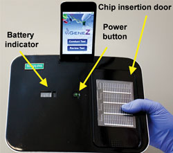
The Gene-Z Device

The Gene-Z device

The team’s prototype consists of a disposable plastic chip with four arrays containing the dried primers required for a genetic assay, allowing parallel processing of four samples with multiple genetic markers for accurate disease diagnosis. The chip sits in an aluminium heater bed, through which LEDs are fed for real-time optical detection, and the whole thing is encased in sleek black packaging with a dock for an iPod Touch or iPhone.

Holger Becker, co-founder of microfluidic ChipShop, Germany, is impressed. ‘I like it for a couple of reasons,’ he says, ‘it’s not over-engineered, it addresses a couple of very typical problems in a point-of-care diagnostic device and there is a simplicity in the approach, especially in the detection system.’

The team made two major changes to existing designs, explains team member Syed Hashsham. First, they made the disposable microfluidic chip from polyester, rather than the more traditional silicon or polydimethylsiloxane. Hashsham says he was inspired by ‘kitchen chemistry’. If plastic food bags can be sealed to keep your sandwiches fresh, he reasoned, then plastic membranes in the chip would stop samples escaping during loading. This avoids the need for mechanical valves and the plastic chips are easy to design and fabricate. You can think one up in the morning and use it in the afternoon, he says, whereas silicon chips take weeks to develop.

Second, the team rejected polymerase chain reactions (PCR) in favour of the loop-mediated isothermal amplification (LAMP) assay. The LAMP assay requires no thermal cycling, requires cheaper optics for detection and is robust – as opposed to PCR, which Hashsham describes as ‘finicky’. Finally, they used WiFi and a smartphone with a custom-built app as the interface to manage data from the device.

Using WiFi does throw up a couple of problems, however: the safe transmission of potentially sensitive patient data via a wireless network, use in hospital settings where signals can interfere with other equipment and viability in areas without an internet connection. Stedtfeld and his team are working with other groups to address the encryption and interference issues. They say that their next step is to make the Gene-Z device compatible with an ordinary mobile phone, so that information can be transmitted via SMS and, by adding solar cells, the device could be used in rural settings.

Gene-Z: a device for point of care genetic testing using a smartphone
Robert D. Stedtfeld, Dieter M. Tourlousse, Gregoire Seyrig, Tiffany M. Stedtfeld, Maggie Kronlein, Scott Price, Farhan Ahmad, Erdogan Gulari, James M. Tiedje and Syed A. Hashsham
DOI: 10.1039/C2LC21226A

Read the original article at Chemistry World

Digg This
Reddit This
Stumble Now!
Share on Facebook
Bookmark this on Delicious
Share on LinkedIn
Bookmark this on Technorati
Post on Twitter
Google Buzz (aka. Google Reader)

Pressurising red blood cells for information

Scientists in Canada have developed a method to study the changes in red blood cells caused by the most common malaria parasite Plasmodium falciparum.

Mosquito

The malaria parasite is spread by mosquitos. © Shutterstock

Malaria causes approximately one million deaths each year and there is a lot of research surrounding the disease’s biomechanics, aimed at diagnosing patients and developing treatments. The parasite, spread by mosquitoes, infects red blood cells and reduces their ability to deform. Measuring the deformability of an infected red blood cell can provide vital information about the disease’s mechanism and response to treatment.

Current methods to measure deformability are complicated or not sensitive enough, but Hongshen Ma at the University of British Columbia, Vancouver, and colleagues, have designed an accurate and simple microfluidic device for this purpose. The device consists of two layers that control the cells so that only a single cell is introduced into a funnel containing a series of different sized constrictions. The pressure required to push the cell through a constriction is measured precisely and used to calculate the deformability.

The team used their device to show that the deformability of uninfected red blood cells can be distinguished from cells at various stages of infection.

Abhishek Jain at Boston University, US, is an expert in biomedical devices and comments that ‘the device is elegant in the sense that it can be easily scaled up for diagnosis and high throughput drug testing for not only malaria but other pathologies like sickle cell anaemia’.

‘We hope our technique will provide a useful biomechanical assay for the development of new drugs,’ says Ma, who adds that ‘the ability to easily measure the deformability of red blood cells will help researchers study the mechanism of the disease and investigate complex challenges, such as drug resistance.’

Ma’s team plans to further test their device and use it to study the mechanisms of drug resistance.

Microfluidic biomechanical assay for red blood cells parasitized by Plasmodium falciparum
Quan Guo , Sarah J. Reiling , Petra Rohrbach and Hongshen Ma
Lab Chip, 2012, Advance Article
DOI: 10.1039/C2LC20857A

Original article published at Chemistry World

Digg This
Reddit This
Stumble Now!
Share on Facebook
Bookmark this on Delicious
Share on LinkedIn
Bookmark this on Technorati
Post on Twitter
Google Buzz (aka. Google Reader)

Staining tissue samples at the microscale

A vertical microfluidic probe developed by researchers in Switzerland can create a range of immunohistochemistry staining conditions on a single tissue sample.

Immunohistochemistry is a process of detecting antibody biomarkers and is regularly used to reveal abnormal cells in tissue sections. Pathologists conducting immunohistochemistry tests often work with very limited samples for which they don’t know the optimal staining conditions. Under-staining can give a false negative, but over-staining causes loss of contrast and can generate false positives.

Diamond-shaped probe used to analyse tissue samples

The diamond-shaped probe is positioned above the tissue sample and scans horizontally across it. The microchannels inject and aspirate liquids at a distance of 1-30µm from the tissue section

Now, a microfluidic device developed by Emmanuel Delamarche and colleagues at the IBM Zurich Research Laboratory, Rüschlikon, offers local staining of tissue sections, which allows a range of staining conditions to be used on one section. The microfluidic probe is positioned vertically above the tissue section and scans horizontally across it. The head of the probe has two apertures at its apex. The liquid is injected onto the tissue section from one aperture and aspirated at the second. ‘Instead of incubating the entire tissue section, the probe can scan it with a variable speed so as to vary locally the incubation time. This is a simple trick, but it should give an optimal staining contrast at least on one spot of the sample,’ explains Delamarche. Tissue sections are prepared for staining in the conventional fashion and post-staining processing is also unchanged.

Delamarche and the team have shown proof of concept with their current research, but in the future, they hope it will receive clinical validation. In addition, the probe could be used for fundamental research. ‘Developing a novel tissue staining method to detect various biomarkers is critical to obtain a more accurate and sophisticated understanding of drug discovery and clinical pathology,’ explains Je-Kyun Park, an expert in bioengineering at the Korea Advanced Institute of Science and Technology, South Korea. Using the microfluidic probe to scan multiple locations with different conditions could help analyse tissue microarrays.

Micro-immunohistochemistry using a microfluidic probe
Robert D. Lovchik, Govind V. Kaigala, Marios Georgiadis and Emmanuel Delamarche
Lab Chip, 2012, Advance Article
DOI: 10.1039/C2LC21016A

Original article published at Chemistry World

Digg This
Reddit This
Stumble Now!
Share on Facebook
Bookmark this on Delicious
Share on LinkedIn
Bookmark this on Technorati
Post on Twitter
Google Buzz (aka. Google Reader)

Lab on a Chip Issue 1 just published!

Welcome to the first 2012 issue of Lab on a Chip

On the front cover of our first issue of Volume 12 an the article from Neus Sabaté et al. on their fuel cell-powered microfluidic platform for lab-on-a-chip applications.  This hot article was recently highlighted in Chemistry World.

Fuel cell-powered microfluidic platform for lab-on-a-chip applications
Juan Pablo Esquivel, Marc Castellarnau, Tobias Senn, Bernd Löchel, Josep Samitier and Neus Sabaté
DOI: 10.1039/C1LC20426B

On the inside front cover we have an image from Eric Stava et al. showing their work on the mechanical actuation of ion channels using a piezoelectric planar patch clamp system.

Mechanical actuation of ion channels using a piezoelectric planar patch clamp system
Eric Stava, Minrui Yu, Hyun Cheol Shin, Hyuncheol Shin, Jonathan Rodriguez and Robert H. Blick
DOI: 10.1039/C1LC20636B

In this issue we also have the editorial introduction from Editor Harp Minhas – Meeting the challenge – discussing our new developments and plans for the coming year, we think it’s going to be an exciting one!

Take a look at the issue

Digg This
Reddit This
Stumble Now!
Share on Facebook
Bookmark this on Delicious
Share on LinkedIn
Bookmark this on Technorati
Post on Twitter
Google Buzz (aka. Google Reader)

Automated micro particle image velocimetry and a heart on a chip on the cover of Issue 24

Welcome to the final issue of 2011!

On the front cover of Issue 24 we have an article from Per Augustsson et al., who have developed a platform for micro particle image velocimetry (μPIV) for analyzing two-dimensional acoustophoresis.  The device is automated, temperature-stable and has uncertainties below 5% and is therefore able to conduct high-precision measurement of the acoustophoretic velocity field in microchannels.

Automated and temperature-controlled micro-PIV measurements enabling long-term-stable microchannel acoustophoresis characterization
Per Augustsson, Rune Barnkob, Steven T. Wereley, Henrik Bruus and Thomas Laurell
DOI: 10.1039/C1LC20637K


The inside front cover highlights the article from Kevin Kit Parker and colleagues that recently featured in Chemistry World.  The article describes a ‘heart on a chip’, exploiting muscular thin film technology to measure contractility and the effect of cell architecture on tissue contraction.

Ensembles of engineered cardiac tissues for physiological and pharmacological study: Heart on a chip
Anna Grosberg, Patrick W. Alford, Megan L. McCain and Kevin Kit Parker
DOI: 10.1039/C1LC20557A

Also in this issue is the latest Research Highlights article from Ali Khademhosseini, and a Focus article on droplet microfluidics for protein engineering and analysis from Helene Andersson Svahn and Haakan Joensson.

Digg This
Reddit This
Stumble Now!
Share on Facebook
Bookmark this on Delicious
Share on LinkedIn
Bookmark this on Technorati
Post on Twitter
Google Buzz (aka. Google Reader)

A lab you can wear?

Malaysian scientists have created a flexible and environmentally friendly microfluidic device using a cloth decorating technique for printing wax onto cotton.

Dedy Wicaksono at the University of Technology Malaysia was inspired by his batik-patterned clothes to create the device. ‘Batik processing is wax patterning to create regions of differing hydrophilicity and hydrophobicity on cloth,’ says Dedy. The technique is traditionally used to prevent dye spreading from one area of cloth to another, creating coloured patterns. Using wax printing methods for paper and silk based microfluidic devices is known, but these have required specialist equipment or expensive materials.

Wicaksono’s method needs neither of these things. First, his team prepared the cotton by scouring it with sodium hydroxide and anhydrous sodium carbonate solutions. The treatment removes the outer layer, exposing underlying cellulose fibres. Both the chemical composition (increased oxygen content) and physical structure (increased surface roughness) of the fibre surface were altered, increasing the wettability and wicking rate.

Microfluidic device in different stages

(a)-(f) are photographs showing two hydrophilic channels with different dye solutions that cross each other vertically and horizontally in three layers without mixing. (b)-(e) are front views of the device before, 5 seconds, 2 minutes and 5 minutes after dropping the dyes into different channels. (f) is the bottom layer 5 minutes after adding the dyes.

Then, they printed the pattern for the microfluidic device onto paper. The paper was dipped into hot batik wax, then dried, before the pattern was cut from the sheet and attached to the scoured cloth using a few pins. Heat treatment melted the wax again, and it spread onto the surface and into the cloth, filling the gaps in the weave and within the fibres. The fatty acids in the wax increase the hydrophobicity of the fibres where they are applied, creating barriers to liquid flow.

Dedy’s group made both 2D and 3D devices; the latter was made by folding layers of the patterned cloth. A test using ink solutions revealed the ink moving along the cloth’s hydrophilic channels, filling a device in minutes. In a further test, the team was able to detect the protein bovine serum albumin colorimetrically using the devices, with the result visible to the naked eye.

‘A key innovation here is the batik-inspired method of transferring patterned wax on paper to cotton cloth,’ comments Shashi Murthy, an expert in microfluidic devices at Northeastern University, US. He adds that the technology ‘has the potential to provide a rapid and low cost readout for analytes characterised by relatively simple colorimetric assays’.

Dedy is now investigating ways to exert more control over the liquid flow so that more complex microfluidic devices can be developed. ‘By making the channels inside a flexible cloth, we are envisioning an embeddable wearable lab in the very near future,’ he says.

Flexible microfluidic cloth-based analytical devices using a low-cost wax patterning technique
Azadeh Nilghaz, Dedy H. B. Wicaksono, Dwi Gustiono, Fadzilah Adibah Abdul Majid, Eko Supriyanto and Mohammed Rafiq Abdul Kadir
Lab Chip, 2012, Advance Article
DOI: 10.1039/C1LC20764D

Original article published at Chemistry World

Digg This
Reddit This
Stumble Now!
Share on Facebook
Bookmark this on Delicious
Share on LinkedIn
Bookmark this on Technorati
Post on Twitter
Google Buzz (aka. Google Reader)

Heart-on-a-chip

A heart-on-a-chip device could help detect drugs that limit heart tissue contraction, say US scientists.

The device was created using small thin strips of tissue made from heart muscle cells that are connected to electrodes to stimulate contraction. Observing the contraction response of the tissue allows scientists to study the effect of physiological factors or test drugs for cardiotoxicity. Replicating segments of heart tissue makes it possible to rapidly measure contraction data at the tissue level, rather than just studying individual cells.

Heart on a chip

The researchers, led by Kevin Parker from Harvard University, Cambridge, created up to eight separate strips of tissue on one chip by growing a sheet of heart muscle cells in a flat film and then cutting away sections to leave isolated strips that were only connected to the film by one edge. Creating several strips on one chip allowed the team to conduct multiple experiments at once. The contraction experiments were observed by looking vertically down onto the chip and monitoring the change in length as the strips contracted and bent up. ‘The heart-on-a-chip allows us to capture in 2D what a healthy or diseased heart might look like and look at how much force the tissue will generate,’ says Anna Grosberg, a member of the research team.

The team then used the heart-on-a-chip to investigate the effects of different environments on contraction response. Experiments with epinephrine (adrenaline) showed the device can be used to measure the dose-dependent effect of drugs on contraction response. The team also showed that the heart-on-a-chip can be used as a platform to investigate the effect of cell architecture on tissue contraction.

‘The heart-on-a-chip technology is the first scalable technique that recapitulates in vitro the anisotropic cell organisation of native cardiac muscle, while simultaneously allowing coupled electrical and mechanical function to be quantitatively assayed,’ says Andrew McCulloch, an expert in cardiac bioengineering at the University of California, San Diego, US. ‘Once this technology can be deployed using human stem cell-derived cardiomyocytes, we will have a powerful new platform for screening new drugs for heart diseases like arrhythmia and heart failure.’

Ensembles of engineered cardiac tissues for physiological and pharmacological study: Heart on a chip
Anna Grosberg, Patrick W. Alford, Megan L. McCain and Kevin Kit Parker
DOI: 10.1039/C1LC20557A

Original article published at Chemistry World

Digg This
Reddit This
Stumble Now!
Share on Facebook
Bookmark this on Delicious
Share on LinkedIn
Bookmark this on Technorati
Post on Twitter
Google Buzz (aka. Google Reader)