Author Archive

Emerging Investigator Series: Scott Tsai

Dr. Scott Tsai is an Associate Professor in the Department of Mechanical and Industrial Engineering at Ryerson University, and an Affiliate Scientist at the Li Ka Shing Knowledge Institute of St. Michael’s Hospital. He obtained his BASc degree (2007) in Mechanical Engineering from the University of Toronto, and his SM (2009) and PhD (2012) degrees in Engineering Sciences from Harvard University. After a brief NSERC Postdoctoral Fellowship at the University of Toronto, Dr. Tsai joined Ryerson University at the end of 2012. He leads the Laboratory of Fields, Flows, and Interfaces (LoFFI), which is currently developing innovative microfluidic approaches to generate and use water-in-water droplets and microbubbles in a variety of biomedical applications. Dr. Tsai is a recipient of the United States’ Fulbright Visiting Research Chair Award (2018), Ontario’s Early Career Researcher Award (2016), Canadian Society for Mechanical Engineering’s I. W. Smith Award (2015), and Ryerson University’s Deans’ Teaching Award (2015).

Read Dr. Tsai’s Emerging Investigator Series article “Microfluidic diamagnetic water-in-water droplets: a biocompatible cell encapsulation and manipulation platform” and find out more about him in the interview below:

Your recent Emerging Investigator Series paper focuses on microfluidic diamagnetic water-in-water droplets. How has your research evolved from your first article to this most recent article

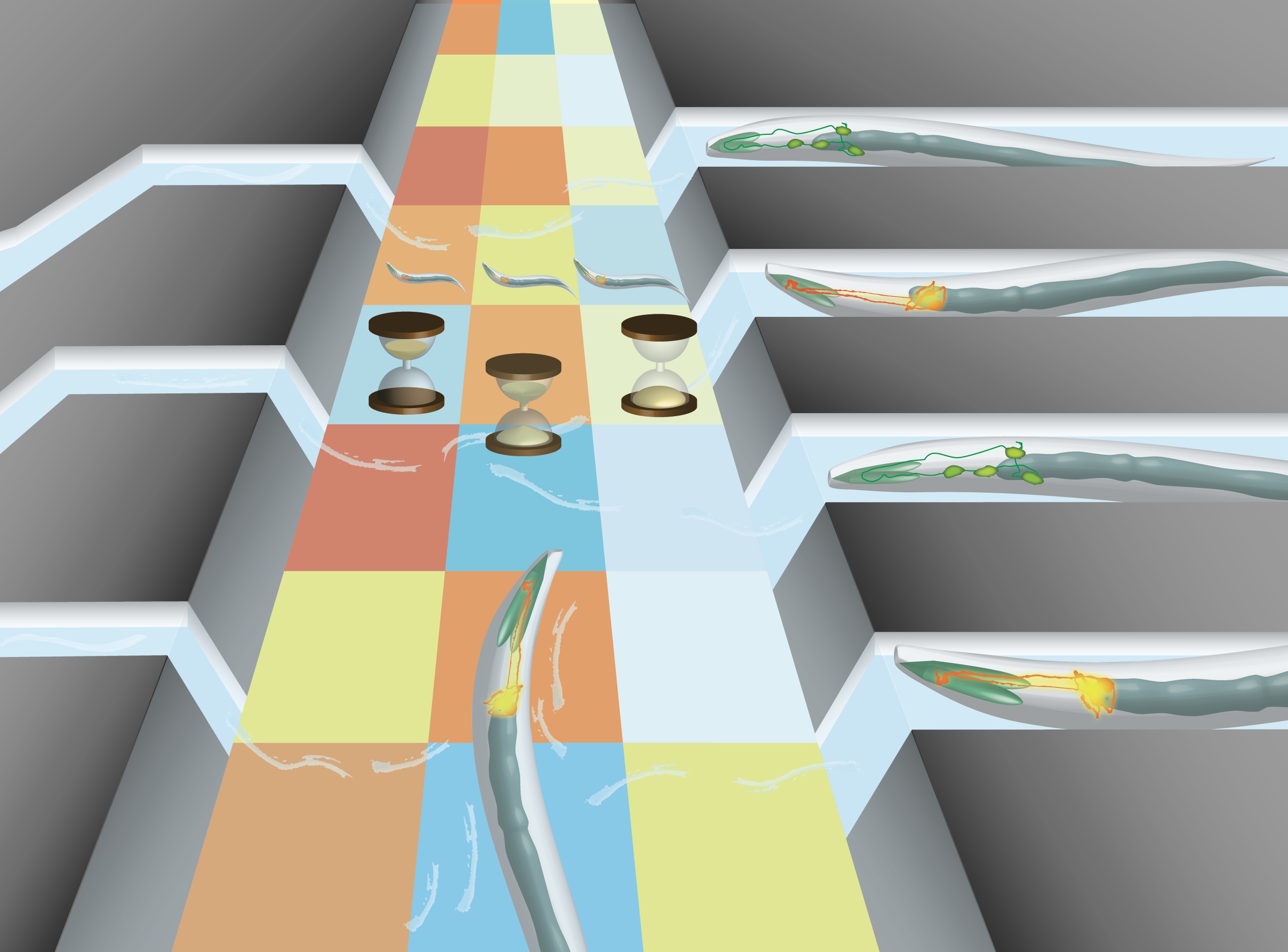
My group’s research is contributing to the exciting and emerging theme of all-aqueous and biocompatible water-in-water droplet microfluidics, which was started just a few years ago. Following the pioneering work of others, such as Anderson Shum (University of Hong Kong), we demonstrated that pulsating inlet pressures could be used to break-up all-aqueous threads into water-in-water droplets (Moon et al.Lab on a Chip, 2015). We subsequently simplified the approach by using weak hydrostatic pressure to passively create the water-in-water droplets (Moon et al.Analytical Chemistry, 2016), which enabled other applications, such as cellular encapsulation and release (Moon et al.Lab on a Chip, 2016), Pickering-style stabilization (Abbasi et al.Langmuir, 2018), and diamagnetic manipulation, as described in this paper. I am really excited to see what other tools emerge in the coming years that leverage water-in-water droplet microfluidics.

What aspect of your work are you most excited about at the moment?

I think right now, we as a community are still in the phase of developing the functionality and performance of water-in-water droplet microfluidics platforms. However, I have not seen many papers that leverage the unique characteristics of water-in-water droplets, which are based in aqueous two phase systems (ATPS). For example, ATPS fluids can selectively partition molecules and cells into or out of the droplet phase. I am most excited to see what we and other researchers can create that exploits unique features such as this.

In your opinion, what do you think is the future of droplet encapsulation technologies for single cell analysis?  

Predicting the future is always difficult, but I would say that at some point there will start to be some kind of standardization of encapsulation technologies, so that the entire field can move further into single cell analysis applications.

What do you find most challenging about your research?

I find that the most challenging aspect is related to selecting the right problems to work on. Water-in-water droplet microfluidics is emerging and there are many possibilities for research in this area. However, we have limited resources like time and funding, so we have to be selective about which problems to work on, and try to pick the ones that we think will have the greatest impact on advancing the field.

In which upcoming conferences or events may our readers meet you?

I always enjoy learning more about fluid mechanics at the American Physics Society’s Division of Fluid Dynamics (APS-DFD) meeting. I am also a huge fan of the Ontario on a Chip symposium, which was sponsored by Lab on a Chip this year. I was the co-host with Edmond Young and Milicia Radisic (both from the University of Toronto) of Ontario on a Chip for a few years, and my entire group attends the symposium every year. The keynote talks by international leaders in our field draws many attendees, but I am also always amazed by the quality of the student presentations.

How do you spend your spare time?

These days, my spare time is mostly spent with my young family—my wife Edlyn, and my two daughters, Abigail (4) and Chloe (1). I also do some volunteer work for my church. Additionally, as kind of an avid cyclist, I enjoy mountain biking on weekends, and road biking for my commute to work, and for charity rides for example Ride for Heart. Interestingly, I also bike commute sometimes with my Ontario on a Chip co-host, and LOC Emerging Investigator Edmond Young – so perhaps cycling contributes to one’s ability to be a good investigator!

Which profession would you choose if you were not a scientist?

This seems to change at various points in my life. (If you had asked me this question when I was a teenager, I would have probably said that I wanted to be a professional basketball player.) Right now, I think if I was not a scientist I would probably be a bicycle mechanic. Indeed, if I ever retire from science, I might actually attempt to get a job as a mechanic at my local bike shop.

Can you share one piece of career-related advice or wisdom with other early career scientists?

Find a mentor that is well-established, willing to help you, and understands the intricacies of academia, funding, and other aspects of science careers. I was fortunate to receive this kind of mentorship from one of my collaborators from the start of my independent research career. I regard this mentorship as probably the most important factor that has helped me advance in my career.

Digg This
Reddit This
Stumble Now!
Share on Facebook
Bookmark this on Delicious
Share on LinkedIn
Bookmark this on Technorati
Post on Twitter
Google Buzz (aka. Google Reader)

Emerging Investigator Series: Yi-Chin Toh

Dr Yi-Chin Toh obtained her B.Eng in Chemical Engineering and PhD in Bioengineering from the National University of Singapore in 2001 and 2008 respectively. She did her post-doctoral training at the Massachusetts Institute of Technology in 2008 before joining the Institute of Bioengineering and Nanotechnology, A*STAR as a research scientist in 2010. Since 2014, she is an Assistant Professor at the Department of Biomedical Engineering, National University of Singapore.  Yi-Chin’s research interest is in engineering multi-tissue in vitro models to mimic complex biological interactions during human development and diseases, and translate them into scalable platforms for disease modeling and drug testing applications. Dr Toh is a recipient of the National University of Singapore Research Scholarship, A*STAR Graduate Scholarship and A*STAR International Fellowship.

Read her full Emerging Investigator Series article A liver-immune coculture array for predicting systemic drug-induced skin sensitization”  and find out more about her in the video interview below:


Digg This
Reddit This
Stumble Now!
Share on Facebook
Bookmark this on Delicious
Share on LinkedIn
Bookmark this on Technorati
Post on Twitter
Google Buzz (aka. Google Reader)

Emerging Investigator Series: Weiqiang Chen

Dr. Weiqiang Chen is an Assistant Professor in the Departments of Mechanical and Aerospace Engineering and Biomedical Engineering at New York University. He received his B.S. in Physics from Nanjing University in 2005 and M.S. degrees from Shanghai Jiao Tong University in 2008 and Purdue University in 2009, both in Electrical Engineering. He earned his Ph.D. degree in Mechanical Engineering from the University of Michigan in 2014.  He is the receipt of the National Institute of Biomedical Imaging and Bioengineering Trailblazer Award, the American Heart Association Scientist Development Award,  the NYU Whitehead Fellowship, the 2013 Baxter Young Investigator Award, the University of Michigan Richard F. & Eleanor A. Towner Prize for Outstanding PhD Research, and the ProQuest Distinguished Dissertation Award. Dr. Chen’s research interests focus on Lab-on-a-Chip, biomaterials, mechanobiology, stem cell biology, caner biology, and immune engineering.

Read his latest Emerging Investigator Series article An integrated adipose-tissue-on-chip nanoplasmonic biosensing platform for investigating obesity-associated inflammation and read more about him in the interview below:

Your recent Emerging Investigator Series paper focuses on “An Integrated Adipose-Tissue-On-Chip Nanoplasmonic Biosensing Platform for Investigating Obesity-associated Inflammation”. How has your research evolved from your first article to this most recent article?

We have previously been interested in developing microfluidic biosensing systems for immune cell cytokine detections. Quantitative measurements of the cytokine-producing capacity of different immune cells are critically important for characterizing the immune cell functions and defining the “immune phenotypes” of patients for clinical diagnosis and interventions. Although many advanced biosensing techniques have been purposed for cytokine profiling, there are no clinically available methods that integrate high-resolution immune cell monitoring and multiplexed cytokine detection together in its native tissue microenvironment, which is a big hurdle to understand the cellular immunophenotype behaviors during disease progression. This has inspired us to consider of combining the label-free nanoplasmonic biosensors with an Adipose-Tissue-on-Chip system reported in the recent article to quantitatively characterize obesity-associated dynamic cytokine secretion behaviors in a complex biomimetic adipose tissue microenvironment.

What aspect of your work are you most excited about at the moment?

I am most excited about the potential of our research for future applications in personalized medicine.

In your opinion, what do you think the contribution of disease-on-a-chip models for personalised medicine is likely to be?

Traditional medicine, which mostly relies on test of treatment on animal models, has many limitations. This is because the traditional animal models cannot mimic human pathophysiology accurately. Alternatively, the disease-on-a-chip platform can mimic an ideal microenvironment of human pathologic tissue, which enables a more accurate way to test the medicine activity under molecular- and cellular-scale in human organ tissue in vitro. Particularly, the cellular immunophenotype behavior is highly correlated with the tissue microenvironment. Unquestionably, integrating in situ cellular and molecular immune profiling in an organotypic setting can provide a reliable and accurate screening to characterize cell functions in its native tumor microenvironment. More importantly, due to the heterogeneity between individual human being, even for patients who are diagnosed with the same disease, their response to a same medicine treatment can be diversified. The minimized microfluidic disease-on-a-chip model can be derived from the specific patient’s cells or explanted pathophysiology tissue, allows parallel screening of multiple medical treatments in personalized manner. Therefore, this technology will provide the most efficient way to identify the most effective personalized medical options before applying to the patient’s own self.

What do you find most challenging about your research?

The most challenging part about our research is to accelerate translational technologies from the benchtop to the clinic to make a real impact on patients. A number of obstacles, including differences in culture between basic and clinical researchers, difficulty in developing and sustaining interdisciplinary collaborations, inadequate training and knowledge in medical sciences, and lack of funding, resources, or infrastructure, have limited the opportunities for basic bioengineering investigators, including myself, to conduct translational research. Therefore, my lab has established strong collaboration with many clinical colleagues at NYU Langone Health. Their expertise will contribute most significantly to our research from the biology and clinical aspects. Through this, I believe our engineering methods can introduce real impact to translational clinical research.

In which upcoming conferences or events may our readers meet you?

We will present our research in the upcoming Biomedical Engineering Society (BMES) Annual Meeting and the International Conference on Miniaturized Systems for Chemistry and Life Sciences (µTAS 2018).

How do you spend your spare time?

I enjoy watching movies, spending time with family and friends, and traveling to different cities around the world.

Which profession would you choose if you were not a scientist?

I would probably choose a career in social science.

Can you share one piece of career-related advice or wisdom with other early career scientists?

Something I could suggest is to be patient in testing your new ideas. It always take some time, normally years, for a scientist to prove a new scientific idea and develop it into practical methods or solutions.

Digg This
Reddit This
Stumble Now!
Share on Facebook
Bookmark this on Delicious
Share on LinkedIn
Bookmark this on Technorati
Post on Twitter
Google Buzz (aka. Google Reader)

Quantitative and multiplex microRNA assays from unprocessed cells in isolated nanoliter well arrays

MIT researchers develop a simple new chip that simplifies sample preparation and reduces sample volume for micro-RNA analysis

In recent years, microRNAs (miRNAs) have emerged as potential biomarkers for a range of diseases. These short (~22 nucleotide) sequences of non-coding RNA function by silencing messenger RNA and thus, provide a form of post-transcriptional regulation in the cell. Consequently, their regulation can have a significant impact on cell function. While they are implicated in many diseases, their role in cancer is of particular interest. It is known that multiple miRNAs are dysregulated in tumours compared to healthy tissues and they can be used as biomarkers in cancer diagnosis. Testing for multiple miRNAs provides a better diagnosis, so it is important to be able to test them as a panel. There are many techniques available to test for miRNAs such as gene chips, quantitative RT-PCR, and RNA-seq, but these technologies all have their own shortcomings. What is common to all them is the need for sample preparation, and mainly RNA extraction from the tissue or cells to be tested. In their recent HOT article, Tentori and researchers from the Doyle Group at MIT describe a chip that could overcome this problem and perform multiplexed testing of miRNA all within a minimum volume of just a few nanoliters.

The new chip comprises two glass slides which can be sandwiched together. The bottom slide has an array of 300 µm diameter wells which contain the miRNA sensors and serve to isolate samples into a small reaction volume. The top slide is then used to deliver lysis reagents to the sample. The authors tried a variety of designs for the top plate, but ultimately settled on an array of 30 µm diameter wells. This has two advantages; 1) Reagents are precisely metered and 2) the top and bottom plates can be sandwiched robustly without any need for precision handling or alignment. This last point is really important to Augusto Tentori, a postdoc in the Doyle Group and the lead author of the paper. Tentori wants “to make devices that are simple and robust, so translation is easier.” The miRNA sensors are polyethylene glycol diacrylate (PEGDA) hydrogels that contain complementary DNA probes.  The posts are photopolymerized in the wells and various sizes, shapes, and patterns of posts can be made. Further, a single well can contain a variety of posts, each functionalized with a different DNA probe targeting a different miRNA. In this way, multiplexed assays can be performed with spatial separation. The chip format of the assay is more sensitive than previous formats and can detect miRNA from less than 20 cells.

Tentori is really excited about the prospects of this new chip. His co-authors include pathologists who have been guiding the project to make sure it is clinically useful, and he really wants to see this technology get into the hands of pathologists and diagnostic technicians.

Read the full article by Tentori et al. here “Quantitative and multiplex microRNA assays from unprocessed cells in isolated nanoliter well arrays” that features in Lab on a Chip’HOT article collection

About the webwriter

Darius Rackus (right) is a postdoctoral researcher in the Dittrich Bionalytics Group at ETH Zürich. His research interests are in developing integrated microfluidic tools for healthcare and bioanalysis

Digg This
Reddit This
Stumble Now!
Share on Facebook
Bookmark this on Delicious
Share on LinkedIn
Bookmark this on Technorati
Post on Twitter
Google Buzz (aka. Google Reader)

Emerging Investigator Series: Kiana Aran

Dr. Aran received her undergraduate degree in electrical engineering at the City University of New York in 2007 and her Ph.D in biomedical engineering at Rutgers University in 2012. She then continued her postdoctoral studies in bioengineering at the University of California Berkeley and was a recipient of National Institutes of Health (NIH) postdoctoral training fellowship at the Buck Institute for Age Research in 2015. She joined Keck Graduate Institute in 2017 as an Assistant Professor and is a currently a visiting scientist at the University of California Berkeley. She is also a consultant for Bill and Melinda Gate Foundation and cofounder of NanoSens innovations. Since starting her faculty position in 2017, Dr. Aran work has been supported by various funds including one RO1, industry sponsored research and a 3 years subaward from University of California Berkeley.

Read her Emerging Investigator Series article “Graphene-based biosensor for on-chip detection of Bio-orthogonally Labeled Proteins to Identify the Circulating Biomarkers of Aging during Heterochronic Parabiosis” and find out more about her in the interview below:


Your recent Emerging Investigator Series paper focuses on digital detection of tagged proteins in vivo to identify the circulating biomarkers in aging. How has your research evolved from your first article to this most recent article?

I was a recipient of an NIH T32 award during my last years of postdoc training at UC Berkeley (2015-2017) which provided me with the opportunity to work with Dr. Irina Conboy, a pioneer in utilizing bio-orthogonally labeled proteins for research on aging. This approach enabled the detection of several rejuvenating protein candidates from the young parabiont, which were transferred to the old mammalian tissue through their shared circulation. Although this method was very powerful, there were a lot of challenges with the detection of these tagged proteins including very time consuming, complex and expensive assays, large sample requirement and false positive. I started working on this project as a side project along with other projects I had with Dr. Conboy just to make the process more facile so we can detect these protein faster and more continuously. This work is a great example of utilizing a lab on a chip technology for a real application. I have also started collaboration with nanomedical diagnostics, a startup company in San Diego, to enable mass production of our chips with high reproducibility. With my academic and industrial collaborators, we are planning to utilize this technology for at least two different applications in the near future to better understand the biology of aging.

What aspect of your work are you most excited about at the moment?

I am very excited to see my technologies go beyond publications. Two of my inventions have been licensed toward developing medical devices for drug delivery and medical diagnostics and the joy from that have been the biggest drive for me to work harder.

In your opinion, what is the future of portable devices for biosensing?

Even though still in its emerging stage, digital diagnostics and 2D materials will shape the future of biosensors.

What do you find most challenging about your research?

As a junior faculty, it is difficult to recruit highly motivated postdoctoral researchers. Unfortunately, the majority of postdoctoral candidates often look for a well-established laboratory headed by a leading scientist. Students and postdoc should know that working in a new lab can be challenging but can provide researcher with opportunities to strengthen their scientific and management skills.

In which upcoming conferences or events may our readers meet you?

I will be presenting some of my biosensor work in the upcoming IEEE EMBS MNMC Conference

Above: Dr Aran supports a community in Africa by building a school

How do you spend your spare time? 

I love what I do so I can not define my spare time from my work time. I have recently co-founded a startup in digital diagnostics so that takes my weekend but I love it. I also paint when I feel like painting.  But I do run, swim and work out routinely after work and enjoy a variety of sports.  I have also started traveling to small villages around the world with my friends where we help in building schools.

Which profession would you choose if you were not a scientist?

I love painting and would have explored a career in art.

Can you share one piece of career-related advice or wisdom with other early career scientists?

Have a vision. Sometimes we get lost in small career goals such as publishing but if you have a vision of what you are trying to accomplish you can define your career path, you will start working with the right people who support your vision, you will attend the right conferences and workshops for your career development and you will definitely be successful.

Lab on a Chip Issue 21

Digg This
Reddit This
Stumble Now!
Share on Facebook
Bookmark this on Delicious
Share on LinkedIn
Bookmark this on Technorati
Post on Twitter
Google Buzz (aka. Google Reader)

Emerging Investigator Series: Adriana San Miguel

Adriana San Miguel is an Assistant Professor in the Department of Chemical & Biomolecular Engineering at NC State University. She is part of the Synthetic and Systems Biology Chancellor´s Faculty Excellence Program. Her work combines engineering and biology and focuses on developing tools to perform high-throughput automated experiments with the model organism C. elegans. Her team uses these tools and this organism to better understand aging, stress, and the nervous system.

Adriana is originally from San Luis Potosi, Mexico. After receiving a BS in Chemical Engineering at the Monterrey Institute of Technology (ITESM) in 2005, she worked in the water treatment and cement industries for 2 years. She obtained a Ph.D. in Chemical Engineering from the Georgia Institute of Technology in 2011. She then took on a Postdoctoral Research position at Georgia Tech, and a second one at the Dana-Farber Cancer Institute in Boston. In 2013, Adriana was awarded the NIH K99 Pathway to Independence Award to study the mechanisms regulating synaptic plasticity and aging in the nematode C. elegans.

Read her Emerging Investigator Series article “A microfluidic platform for lifelong high-resolution and high throughput imaging of subtle aging phenotypes in C. elegansand find out more about her in the interview below:

Your recent Emerging Investigator Series paper focuses on a microfluidic platform for high-resolution and high throughput imaging of aging phenotypes in C. elegans. How has your research evolved from your first article to this most recent article? 

We have previously been interested in developing systems for high-throughput imaging and analysis of subcellular features in C. elegans, and gained interest in quantitatively assessing how these change during aging. Aging studies pose several technical challenges that could not be addressed with previous microfluidic devices, thus our interest in developing a system capable of performing high-throughput analysis of aging phenotypes.

What aspect of your work are you most excited about at the moment?

I am excited about the possibility of doing systems biology enabled by microfluidics. Lab on chip approaches provide an excellent level of precision in experimental conditions which are unfeasible with traditional C. elegans techniques. Microfluidics also allows high-throughput studies that result in large data sets, and as a consequence, a need to perform unsupervised quantitative data analysis. When combined, these powerful approaches enable better understanding of biological processes from a systems perspective.

In your opinion, what are the challenges with using microfluidics for whole organism behaviour?

Behavioral studies are challenging, particularly because there is a large degree of variability within a population. Microfluidics can help by facilitating animal handling and ensuring the stimuli used is precise (in time, concentration, and localization). Although it is difficult to determine how representative a microfluidic environment (where animals navigate in 2D) resembles their natural 3D environment, microfluidic chips enable single animal tracking, thus increasing the confidence of noisy behavioral readouts.

What do you find most challenging about your research?

Our research requires the integration of several different fields. We develop microfluidic tools, and use them to answer fundamental biological questions. In addition, we use automation, image processing, machine learning, and analysis tools for the large data sets we acquire. Integrating all of these can certainly be challenging, but achieving fully integrated systems is what we find very motivating.

In which upcoming conferences or events may our readers meet you?

I typically attend the American Institute of Chemical Engineers Annual Meetings, as well as the International C. elegans meeting, among others.

How do you spend your spare time?

I enjoy working out every day, it is a necessary part of my routine. I also enjoy baseball, live music, reading, and spending time with family and friends.

Which profession would you choose if you were not a scientist?

I’d probably still be an Engineer, or maybe an artist of some sort.

Can you share one piece of career-related advice or wisdom with other early career scientists?

Something I could suggest is to not be afraid of testing new ideas and methods or venturing into different fields. Although it can be challenging to develop something that is completely new, if it sparks your interest, give it a try. It is very rewarding and motivating to get something new working.


San Miguel Journal Front Cover

Digg This
Reddit This
Stumble Now!
Share on Facebook
Bookmark this on Delicious
Share on LinkedIn
Bookmark this on Technorati
Post on Twitter
Google Buzz (aka. Google Reader)

Acoustofluidics 2018


Acoustofluidics-18-Web-banner_date

Acoustofluidics 2018 is a three-day conference that will take place this year in Lille, France between 29th – 31st August. 

The meeting is dedicated to exploring the science, engineering, and use of micro to nanoscale acoustofluidics. The full list of invited speakers has now been confirmed and published, as well as information on registration fees and the cost of the conference dinner. Please see the conference website for details on abstract submission and how to register.

Digg This
Reddit This
Stumble Now!
Share on Facebook
Bookmark this on Delicious
Share on LinkedIn
Bookmark this on Technorati
Post on Twitter
Google Buzz (aka. Google Reader)

2018 Art in Science Competition

Get your entries in before the deadline on 15th October 2018 (23:59 Honolulu, Hawaii, USA time)


The µTAS 2018 Conference will feature the 11th Art in Science competition entitled ‘Under the Looking Glass: Art from the World of Small Science‘, sponsored and supported by National Institute of Standards and TechnologyLab on a ChipMicroTAS and the Chemical and Biological Microsystems Society.

Since the earliest publications of the scientific world, the aesthetic value of scientific illustrations and images has been critical to many researchers. The illustrations and diagrams of earlier scientists such as Galileo and Da Vinci have become iconic symbols of science and the scientific thought process.

In current scientific literature, many scientists consider the selection of a publication as a “cover article” in a prestigious journal to be very complimentary.

Deadline 15th October 2018 at 23:59 Honolulu, Hawaii, USA time—please note this is a month before the conference!


Are you attending the µTAS 2018 Conference?

Would you like your image to be featured on the cover of Lab on a Chip?

To draw attention to the aesthetic value in scientific illustration while still conveying scientific merit, NIST, LOC and CBMS are sponsoring this annual competition. Applications are encouraged from authors in attendance of the µTAS Conference and the winner will be selected by a panel of judges and presented at the Royal Society of Chemistry/Lab on a Chip booth during the last poster session of the 2018 MicroTAS conference.

Applications must show a photograph, micrograph or other accurate representation of a system that would be of interest to the µTAS community and be represented in the final manuscript or presentation given at the Conference.

They must also contain a brief caption that describes the illustration’s content and its scientific merit. The winner will be selected on the basis of aesthetic eye appeal, artistic allure and scientific merit. In addition to having the image featured on the cover of Lab on a Chip, the winner will also receive a financial prize at the Conference.


Art in Science Competition Submission Process

Step 1. Sign-In to the Electronic Form Using Your Abstract/Manuscript Number

Step 2. Fill in Remaining Information on Electronic Submission Form

Step 3. Upload Your Image

Good Luck!

You can also take a look at the winners from last year on our blog.

Digg This
Reddit This
Stumble Now!
Share on Facebook
Bookmark this on Delicious
Share on LinkedIn
Bookmark this on Technorati
Post on Twitter
Google Buzz (aka. Google Reader)

Emerging Investigator Series – Rebecca Pompano

We are delighted to introduce our latest Lab on a Chip Emerging Investigator, Rebecca Pompano.

Dr. Rebecca Pompano is an Assistant Professor in the Departments of Chemistry and Biomedical Engineering at the University of Virginia, and a member of the Beirne B. Carter Center for Immunology Research.  She completed a BS in Chemistry at the University of Richmond in 2005, and a PhD in 2011 at the University of Chicago, working in the laboratory of Dr. Rustem Ismagilov.  She completed a postdoc in the University of Chicago Department of Surgery, leading a collaboration between Dr. Joel Collier, a tissue engineer, and Dr. Anita Chong, an immunologist.  Since 2014, she has been a faculty member at UVA, where her research interests center on developing microfluidic and chemical assays to unravel the complexity of the immune response.  She received an Individual Biomedical Research Award from The Hartwell Foundation and the national 2016 Starter Grant Award from the Society of Analytical Chemists of Pittsburgh.  Recently, her lab was awarded an NIH R01 to develop hybrids of microfluidics and lymph node tissue to study inflammation.  In addition to her research, she is active in advocating for continued funding for education and biomedical research on Capitol Hill.

Read her Emerging Investigator Series article “User-defined local stimulation of live tissue through a movable microfluidic port” and find out more about her in the interview below:

Your recent Emerging Investigator Series paper focuses on stimulation of live tissue through a movable microfluidic port. How has your research evolved from your first article to this most recent article?

My current research combines some seemingly disparate themes from my prior work.  My first article in graduate school used droplet microfluidics to study blood clotting, and I became fascinated with how spatial organization affects the function of complex biological systems. Later, I also worked on the physics of fluid flow in a reconfigurable SlipChip device… and both of these ideas make a comeback in this current paper!  Then in my postdoc, I had the fabulous opportunity to work in both a bioengineering lab and an immunology lab, studying the mechanism of action of a new non-inflammatory vaccine. The research in my lab now is really at the intersection of bioanalytical chemistry, bioengineering, and immunology.  We develop new tools to study the immune system and how it is organized. This particular paper offers a new technology to pick and choose where to deliver a drug or stimulant to a piece of live tissue, and we demonstrated it for lymph nodes, our favorite immune organ.

What aspect of your work are you most excited about at the moment?

I am very excited about the ideas we are pursuing, specifically that our tools to control and detect how tissue is organized might prove useful for other researchers.  As a chemist by training, I’m thrilled to collaborate with creative bioengineers and immunologists like Jennifer Munson (Virginia Tech) and Melanie Rutkowski (U Virginia) to work on inflammatory diseases and tumor immunology.  Seeing our chips at work in their labs is very rewarding.

In your opinion, what is the biggest advantage of using local stimulation over global stimulation for measuring tissue responses?

Local stimulation, by which I mean delivering fluid or a drug to one region of tissue, rather than bathing the entire sample in media, gives you the chance to ask unique questions about spatial organization.  For example, I envision using this microfluidic technique to determine whether a drug is more effective when delivered to one area of tissue than another, and then developing a nanoparticle that targets just the right region.  It can also be used to mimic local biological events, like diffusion of signals from a blood vessel, to determine how inflammation initiates and propagates through live tissue.

What do you find most challenging about your research?

Studying the immune system – its complexity is what I love about it, but it is challenging when the cells and tissue do exactly the opposite of what you expected!  This happens over and over when we ask a real biological question. I suppose it shows how much there is still to learn, and why new tools are so desperately needed to predict and control immunity.

In which upcoming conferences or events may our readers meet you?

I’m looking forward to MicroTAS in Taiwan this year. I also bounce around between Pittcon (analytical chemistry), the Society for Biomaterials annual meeting, and the annual AAI Immunology conference.  This fall I’ll be attending the BMES annual meeting (Biomedical Engineering Society) for the first time!  There is not yet a focused conference for immunoanalysis and immunoengineering, but I’m hoping one will form soon.

How do you spend your spare time?

A few years ago I would have said knitting… I had a great group of friends in graduate school who would get together to knit every week.  I still wear those socks and sweaters!  Now though, my husband Drew and I spend most of our free time playing together with our 2-year old, Jasper.  Sometimes I also go to Drew’s gigs to be a rock star’s spouse instead of a chemistry professor for a while.  He’s a bassist in several great bands in Charlottesville (check a few of them out – Pale Blue Dot and 7th Grade Girl Fight).

Which profession would you choose if you were not a scientist?

I almost went into science policy instead of academia. I seriously entertained the idea of working at USAID helping promote vaccines internationally, or working in a think tank to help guide health-related policies.  I’m still very passionate about the need for scientists to inform the public and our elected officials about the science underlying issues like health, education, and care of the environment.

Can you share one piece of career-related advice or wisdom with other early career scientists?

A former mentor recommended me the book, Ask For It, by Linda Babcock and Sara Laschever, and it completely changed how I operate.  I think many early career scientists could benefit from this book, which is about overcoming self-doubt to ask for what you really need. Although ostensibly written for women, in science I see so many men and women who could achieve something great with just a little confidence booster.

Digg This
Reddit This
Stumble Now!
Share on Facebook
Bookmark this on Delicious
Share on LinkedIn
Bookmark this on Technorati
Post on Twitter
Google Buzz (aka. Google Reader)

Roll-to-roll PDMS-chips for the masses in molecular diagnostics

PDMS microfluidic devices for molecular diagnostics are now produced at scale using roll-to-roll manufacturing

If there is one material that has enabled microfluidic research in academia, poly(dimethylsiloxane) (PDMS) is surely it. PDMS is cheap and easy to prototype with, and its elastomeric properties have led to complicated structures (e.g. valving) in microfluidic channels. Although it is great for rapid prototyping, there is often a disconnect between the prototype and high throughput manufacturing due to a lack of scalable production methods. Researchers at VTT-Technical Research Centre of Finland and the University of California Berkeley have recently reported a roll-to-roll method for fabricating PDMS microfluidic chips.

In roll-to-roll (R2R) processing—common to the paper industry—long sheets of materials are continuously processed, feeding through rollers and modules with different functionalities. To form R2R microfluidic devices, PDMS was applied to an aluminized paper substrate and then embossed by a heated nickel imprinting cylinder which also cured the PDMS. The devices had good reproducibility and channel depths around 100 µm were achieved. Replication from the nickel master was automated and performed at high throughput of 1.5 m/min. Olli-Heikki Huttunen, one of the authors on the paper, said that “although the process required a lot of fine tuning, it was surprisingly simple.” Like other high-throughput manufacturing techniques (e.g. injection moulding), the nickel tool is quite expensive, but these costs can be overcome by the volume of production.

As a proof-of-principle application, the authors demonstrated nucleic acid detection by loop-mediated isothermal amplification (LAMP). Reagents were spotted and dried in the microchannels using a roll-to-roll compatible dispensing machine, and PDMS lids with vias for fluidic and vacuum connections were formed by a roll-to-roll process (though vias were manually punched) and then bonded manually. Huttunen said that the next steps are to figure out how to manufacture the entire device roll-to-roll, but that it should not be too challenging.

Using aluminized paper as the base substrate for the devices offered a couple advantages. One is that the aluminium dramatically reduced the paper’s autofluorescence. Another advantage was the aluminum reflected back both excitation and emission light, resulting in stronger signals. Results from the test could be read within 20 minutes, suggesting that these devices would be useful for low-cost point-of-care testing.

The challenge for the future, says corresponding author Luke Lee, will be “to learn what the new rules of thinking and design are for roll-to-roll microfluidics in order to solve the problem of mass production in integrated molecular diagnostics for all.” This is an exciting new prospect for both PDMS and the microfluidics community.

To read the full paper for free*, click the link below:

PDMS microfluidic devices for molecular diagnostics are now produced at scale using roll-to-roll manufacturing

*article free to read from 06/06/2018 – 06/07/2018

About the Webwriters

Darius Rackus (Right) is a postdoctoral researcher at the University of Toronto working in the Wheeler Lab. His research interests are in combining sensors with digital microfluidics for healthcare applications.

Digg This
Reddit This
Stumble Now!
Share on Facebook
Bookmark this on Delicious
Share on LinkedIn
Bookmark this on Technorati
Post on Twitter
Google Buzz (aka. Google Reader)