Author Archive

A closer look at erythrocytes in motion

Blood analysis is usually the first step involved in the diagnosis of various diseases, such as typhoid and malaria. The biochemical and cellular equilibrium of blood is very sensitive to physiological variations occurring in the body at various disease stages. Thus, a fast and accurate examination of blood properties is essential. The morphological and biochemical changes in erythrocytes are used as  the pathological signatures of various diseases.

Flow cytometry is used  to examine blood cells, which requires hydrodynamic sheath flow alignment and fluorescence antibody labelling, making it time-consuming and expensive. Advanced light scattering techniques (such as digital holography) are often seen as suitable alternatives, as they provide fast and label-free measurements.

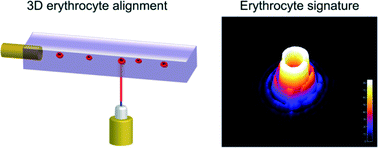
In a recent Lab On A Chip articleNetti et al. from the Italian Institute of Technology, in collaboration with scientists from Germany and Russia, presented a camera-based light scattering approach, coupled with a viscoelasticity -induced cell migration technique. This new system is used to characterise the morphological properties of erythrocytes in microfluidic flows.

They obtained light scattering profiles (LSPs) of individual living cells in microfluidic flows over a wide angular range and matched them with scattering simulations to characterise their morphological properties. A healthy erythrocyte diameter lies between 6 and 9 µm. The diameter values obtained from the experiment lie between 7 and 8.3 µm, which is in good agreement with the existing literature.

‘The results demonstrate the ability of a rapid and cost effective way to measure the average dimensions of an erythrocyte population which can be easily related to the health of a patient,’ concludes Netti.


To gain deeper insight into LSP acquisition and simulation, you can read the full article for free* by following the link below.
Optical signature of erythrocytes by light scattering in microfluidic flows
D. Dannhauser, D. Rossi, F. Causa, P. Memmolo, A. Finizio, T. Wriedt, J. Hellmers, Y. Eremin, P. Ferraro and   P. A. Netti
Lab Chip, 2015,15, 3278-3285
DOI: 10.1039/C5LC00525F

*Access is free until 27/09/2015 through a registered RSC account.
Digg This
Reddit This
Stumble Now!
Share on Facebook
Bookmark this on Delicious
Share on LinkedIn
Bookmark this on Technorati
Post on Twitter
Google Buzz (aka. Google Reader)

µ-Med A 2015 workshop

µ-Med A 2015, an international workshop on microsystems technologies for African health. This interesting workshop will be held during 16-19 September 2015 at Protea hotel, Stellenbosch, South Africa.

The event will bring together researchers, technologists, entrepreneurs, non-governmental organizations and funding bodies to interact on the latest developments and future trends in the multidisciplinary field of microsystems technology.

The workshop will focus on the following themes:

  • Burden of disease in Africa
  • Microfluidic diagnostic technologies
  • Point of care diagnostics
  • Paper based diagnostics

Feel free to read more about the success of the first workshop held in 2011.


Register now and contribute to the efforts to improve health in Africa!


For additional information, please visit µ-Med A website.

Digg This
Reddit This
Stumble Now!
Share on Facebook
Bookmark this on Delicious
Share on LinkedIn
Bookmark this on Technorati
Post on Twitter
Google Buzz (aka. Google Reader)

3D Bio-etching is here!

3D technology has revolutionised the entertainment industry by offering viewers the experience of being part of the action, going on in a movie rather than simply watching it. Thanks to 3D technology, we can sky walk hand in hand with George Clooney in ‘Gravity’.

Microfluidics setup for 3D bioetching

The history of 3D technology can be drawn all the way back to the invention of the stereoscope by David Brewster in 1844. Last two decades have seen 3D technology replacing 2D in all walks of life, ranging from entertainment, physics, microelectronics, tissue engineering and regenerative medicine. For e.g., microelectromechanosensors (MEMS) are 3D devices produced by using soft lithography techniques. MEMS installed in air-bags in the cars have saved thousands of lives by sensing pressure levels during accidents.

Can we use 3D technology to have a better look at the complex events happening at cellular level? One of the major challenges in tissue engineering is that the conventional approaches are mainly limited to 2D monolayers systems and do not allow manipulation of complex multilayer tissue. Cells grown on 2D substrates may respond and differentiate distinctly than those in more physiologically relevant 3D environments. The emergence of 3D technology has enabled scientists to mimic the exact cellular environments and helped to provide better insights into the cell signalling, migration and differentiation in cells.

One of the ways of mimicking the cellular architectures is bio-etching which involves subtractive manufacturing. Bioetching of monolayers of cells in response to laser cuts or scratch assays is achieved by using 2D cell culture studies. But the actual biological systems such as tissues and organs are much more complex and cannot be mimicked using simple monolayers. For long time, scientists have been working on developing better technologies to address this problem. One of the ways to achieve this is 3D bio-etching.

William C. Messner et al. from Tufts University in a recent article in Lab on a Chip explain the utility of 3D bioetching technique to create and shape 3D composite tissues using a microfluidics based approach. The ability to shape the 3D form of multicellular tissues and to control 3D stimulation will have a high impact on tissue engineering and regeneration applications in bioengineering and medicine as well as provide significant improvements of highly complex 3D integrated multicellular biosystems.


Can 3D bio-etching help us to design tissue architecture of our choice mimicking different biological events? Find out by reading the full paper for free* using the link below:

3D bio-etching of a complex composite-like embryonic tissue
Melis Hazar, Yong Tae Kim, Jiho Song, Philip R. LeDuc, Lance A. Davidson and William C. Messner
Lab Chip, 2015, Advance Article
DOI: 10.1039/C5LC00530B


*Access is free through a registered RSC account.

Digg This
Reddit This
Stumble Now!
Share on Facebook
Bookmark this on Delicious
Share on LinkedIn
Bookmark this on Technorati
Post on Twitter
Google Buzz (aka. Google Reader)