Archive for February, 2015

Hooked on a Feeling: measuring cell-substrate adhesion with ISFET devices

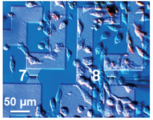
By developing an ion-sensitive field-effect transistor with small gate dimensions, scientists at the University of Applied Sciences Kaiserslautern in Germany were able to measure cell-substrate adhesion on the single cell scale.

To survive, most mammalian cells attach to other cells and the extracellular environment in order to regulate their growth, proliferation, and migration. Electrical impedance spectroscopy is one way to quantitatively monitor cell-substrate interactions. The strength of cellular adhesion to a substrate with integrated electrodes can be measured by comparing the ratio of the readout voltage to the applied alternating current. Yet this method is limited groups of many cells as the size of the microelectrode must be larger than 100 μm in diameter. Smaller features are subject to greater interface impedance between the electrode and liquid media and this background impedance overwhelms the desired cell-substrate measurements. Suslorapova and colleagues thus used an ion-sensitive field-effect transistor (ISFET) with small gate dimensions to overcome this limitation. The group was able measure the effects of enzymatic digestion with trypsin and an apoptosis-inducing drug on single cell detachment using the ISFET devices with a 16 by 2 square micron gate.

The authors create an equivalent circuit model to interpret recorded impedance spectra from their single cell and small cell groups grown in contact with the field-effect transistor devices. The seal resistance and membrane capacitance parameters which can be extracted from the measured transistor transfer function (TTF) provide measures of cell shape and adhesion to the substrate. Changes in TTF correspond to adhesion of individual cells on top of the ISFET gates. This platform and the model developed to interpret TTF signal opens exciting avenues to monitoring cell adhesion in high throughput yet still at single cell resolution.

Download the full research paper paper for free* for a limited time only!

Electrical cell-substrate impedance sensing with field-effect transistors is able to unravel cellular adhesion and detachment processes on a single cell level
A. Susloparova , D. Koppenhöfer , J. K. Y. Law , X. T. Vu and S. Ingebrandt. Lab Chip, 2015, 15, 668-679. DOI: 10.1039/C4LC00593G

*Access is free until 27.03.15 through a publishing personal account. It’s quick, easy and free to register!

Digg This
Reddit This
Stumble Now!
Share on Facebook
Bookmark this on Delicious
Share on LinkedIn
Bookmark this on Technorati
Post on Twitter
Google Buzz (aka. Google Reader)

Google Glass to monitor plant health

‘Okay Glass, image a leaf’

Scientists in the US have developed their very own pair of rose-tinted spectacles by adapting Google Glass to measure the chlorophyll concentration of leaves.

Aydogan Ozcan and his research group at the University of California are passionate about creating new technologies through innovative, photonic methods and are well acquainted with the possibilities of wearable technology in scientific research. Chlorophyll concentration is a handy metric for monitoring plant health and the system devised by Ozcan’s team combines Google Glass with a custom made leaf holder and bespoke software to determine just that.

To read the full article visit Chemistry World.

Quantification of plant chlorophyll content using Google Glass
Bingen Cortazar, Hatice Ceylan Koydemir, Derek Tseng, Steve Feng and Aydogan Ozcan  
Lab Chip, 2015, Advance Article
DOI: 10.1039/C4LC01279H, Paper

Digg This
Reddit This
Stumble Now!
Share on Facebook
Bookmark this on Delicious
Share on LinkedIn
Bookmark this on Technorati
Post on Twitter
Google Buzz (aka. Google Reader)

New YouTube Videos

Functional colloidal micro-sieves assembled and guided above a channel-free magnetic striped film


Non-equilibrium electrokinetic micromixer with 3D nanochannel networks 


Fabrication of 3D High Aspect Ratio PDMS Microfluidic Networks with A Hybrid Stamp  

Digg This
Reddit This
Stumble Now!
Share on Facebook
Bookmark this on Delicious
Share on LinkedIn
Bookmark this on Technorati
Post on Twitter
Google Buzz (aka. Google Reader)

Silver lining for paper Ebola test

Ebola, yellow fever and dengue can be tested for in one go

Researchers in the US have developed a silver nanoparticle-based paper test to simultaneously detect dengue, yellow fever and Ebola. This could provide a cheap and reliable diagnosis for all three diseases, that’s as quick as a home pregnancy test.

The Ebola epidemic in West Africa underscores an urgent need for rapid diagnostics; quick identification and patient isolation can benefit the sick and the healthy. However, dengue, yellow fever and Ebola all initially manifest as a fever and headache, so are easily mixed up.

To read the full article please visit Chemistry World.

Multicolored silver nanoparticles for multiplexed disease diagnostics: distinguishing dengue, yellow fever, and Ebola viruses
Chun-Wan Yen, Helena de Puig, Justina O. Tam, José Gómez-Márquez, Irene Bosch, Kimberly Hamad-Schifferli and Lee Gehrke  
Lab Chip, 2015, Advance Article
DOI: 10.1039/C5LC00055F, Communication

Digg This
Reddit This
Stumble Now!
Share on Facebook
Bookmark this on Delicious
Share on LinkedIn
Bookmark this on Technorati
Post on Twitter
Google Buzz (aka. Google Reader)

New YouTube Videos

PDMS-based turbulent microfluidic mixer  


NAIL: Nucleic Acid detection using Isotachophoresis and Loop-mediated isothermal amplification 


Triboelectric effect as a new strategy for sealing and controlling the flow in paper-based devices 

Digg This
Reddit This
Stumble Now!
Share on Facebook
Bookmark this on Delicious
Share on LinkedIn
Bookmark this on Technorati
Post on Twitter
Google Buzz (aka. Google Reader)

New YouTube Videos

Ultrasonic assisted bonding method for heterogeneous microstructures using self-balanced zig 


Protein Footprinting by Pyrite Shrink-Wrap Laminate 


Micro Propulsion by Acoustic Bubble for Navigating Microfluidic Spaces 

Digg This
Reddit This
Stumble Now!
Share on Facebook
Bookmark this on Delicious
Share on LinkedIn
Bookmark this on Technorati
Post on Twitter
Google Buzz (aka. Google Reader)

Introducing Editorial Board Member Xudong Fan

We are delighted to welcome Xudong (Sherman) Fan to the Lab on a Chip Editorial Board.

Dr Fan is currently a Professor in the Department of Biomedical Engineering at the University of Michigan.

Having completed his B.S and M.S at Peking University, Xudong moved to the USA to complete his PhD at the University of Oregon in the Oregon Center for Optics. From 2000 to 2004, Xudong worked at Research Corporate Lab at 3M Company. In 2004 he took up a position as an Assistant Professor at the University of Missouri where he became a member of Christopher S. Bond Life Sciences Center and the International Center for Nano/Micro Systems and Nanotechnology. In 2010 Xudong moved to the University of Michigan where he is currently a Professor of Biomedical Engineering, a member of Michigan Center for Integrative Research in Critical Care and Wireless Integrated Microsensing and Systems.

My Research Goal:
“My research goal is to use the state-of-the-art photonics, nanotechnology, microfluidics, and other engineering tools to detect and analyse bio/chemical species in both liquid and gas phases.”
Professor Xudong (Sherman) Fan, Lab on a Chip Editorial Board Member

Research in The Fan Lab focuses on the development of novel bio/chemical sensor platforms for analytes in either liquid or gas phase using optofluidic technology and multi-dimensional micro-gas chromatography technology. The groups most recent publication in Lab on a Chip ‘Optofluidic lasers with a single molecular layer of gain’ was added to our Lab on a Chip 2014 HOT Articles collection as it received particularly high scores during peer review.

Last year Xudong received a Departmental Award for Outstanding Accomplishment and become a fellow of Optical Society of America. Congratulations Xudong!

Digg This
Reddit This
Stumble Now!
Share on Facebook
Bookmark this on Delicious
Share on LinkedIn
Bookmark this on Technorati
Post on Twitter
Google Buzz (aka. Google Reader)

New YouTube Videos

A versatile valving toolkit for automating fluidic operations in paper microfluidic devices 


Ship-in-a-bottle femtosecond laser integration of optofluidic microlens arrays with center-pass units enabling coupling-free parallel cell counting with 100% success rate 


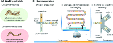
Hydrogel-droplet microfluidic platform for high-resolution imaging and sorting of early larval Caenorhabditis elegans 

Digg This
Reddit This
Stumble Now!
Share on Facebook
Bookmark this on Delicious
Share on LinkedIn
Bookmark this on Technorati
Post on Twitter
Google Buzz (aka. Google Reader)

January’s Free HOT Articles

These HOT articles, published in January 2015 were recommended by our referees and are free* to access for 4 weeks

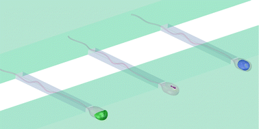
Microfluidic single sperm entrapment and analysis
B. de Wagenaar, J. T. W. Berendsen, J. G. Bomer, W. Olthuis, A. van den Berg and L. I. Segerink
Lab Chip, 2015, Advance Article
DOI: 10.1039/C4LC01425A, Paper

Graphical abstract: Microfluidic single sperm entrapment and analysis

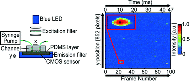
Optofluidic ultrahigh-throughput detection of fluorescent drops
Minkyu Kim, Ming Pan, Ya Gai, Shuo Pang, Chao Han, Changhuei Yang and Sindy K. Y. Tang
Lab Chip, 2015, Advance Article
DOI: 10.1039/C4LC01465K, Paper

Graphical abstract: Optofluidic ultrahigh-throughput detection of fluorescent drops

Hydrogel-droplet microfluidic platform for high-resolution imaging and sorting of early larval Caenorhabditis elegans
Guillaume Aubry, Mei Zhan and Hang Lu
Lab Chip, 2015, Advance Article
DOI: 10.1039/C4LC01384K, Paper

Graphical abstract: Hydrogel-droplet microfluidic platform for high-resolution imaging and sorting of early larval Caenorhabditis elegans

Take a look at our Lab on a Chip 2015 HOT Articles Collection!

*Access is free until 11.03.15 through a publishing personal account. It’s quick, easy and free to register!

Digg This
Reddit This
Stumble Now!
Share on Facebook
Bookmark this on Delicious
Share on LinkedIn
Bookmark this on Technorati
Post on Twitter
Google Buzz (aka. Google Reader)

Culturing Cells on Druggable Bubbles

When testing the effects of drugs on cells and tissues, laboratory scientists generally use a pretty crude approach. They simply mix the desired concentration of the drug with the culture medium and then add it to separate plastic wells in which cells or tissues have been cultured. Of course, this approach has its limitations: when testing many different concentrations and mixtures of drugs, the amount of wells needed for an experiment grows very quickly to overwhelming numbers. Moreover, all of the drug is generally added at once, not taking into account the gradual pharmacokinetic profiles that you would normally find in the human body.

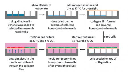
In a paper in Lab on a Chip, scientists from Corning, Inc. and Massachusetts General Hospital demonstrate a new, microengineered approach for treating cultured cells with drugs. Instead of growing the cells on flat surfaces, cells are grown on micro-modified wells plates that contain regular patterns of tiny holes. The microholes can be filled with dried-up drug and then sealed off with a semi-permeable layer of collagen. When cells are grown on the collagen, culture medium starts seeping into the air-filled microholes. The dried-up drug then dissolves and diffuses through the collagen layer, exposing the microscopic patch of cells around the microhole to the drug (see also the figure below).

A schematic overview of how to will microwells with drug and culture hepatocytes on them

The authors only show the results of a simple proof-of-principle experiment with Nefazodone-filled holes leading to local toxicity for cultured liver cells. The technique looks promising however, and it will be interesting to see further development. How easy will it be to load holes with different concentrations and mixtures of drugs? Can the process of drugs diffusing into the medium be controlled? Can we control time-release profiles by changing the shape of the holes, by changing the surface properties of the holes, or maybe by changing the gas content of the medium?

It will also be interesting to see whether the technique can be combined with high-throughput imaging, like fluorescence microscopy or scanning electrochemical microscopy as was recently shown for cells in microscopic wells by Sridhar, et al.

Make sure to check out the paper by Goral, et al. in which they outline their technique while it is still free* to access.

*Access is free until 9.03.15 through a publishing personal account. It’s quick, easy and free to register!

Digg This
Reddit This
Stumble Now!
Share on Facebook
Bookmark this on Delicious
Share on LinkedIn
Bookmark this on Technorati
Post on Twitter
Google Buzz (aka. Google Reader)