Archive for the ‘Hot Article’ Category

STAT3/IL-6 dependent induction of inflammatory response in osteoblast and osteoclast formation in nanoscale wear particle-induced aseptic prosthesis loosening

Total hip arthroplasty (THA) is required in orthopaedic surgery mostly for treating end stage joint disease. A good number of patients (9.1%) require revision surgeries due to aseptic loosening, which means osteolysis around the prostheses caused by the generation of wear particles. Now, there are two established mechanisms to understand wear induced osteolysis: wear particles produce inflammatory cytokines and activated osteoclasts; and wear particles disturb the differentiation, survival and function of osteoblasts and osteoclasts. However, the mechanism of interaction between osteoblasts and osteoclasts and the influence of wear particles is not clearly understood.

The schematic illustration of TiAl6V4 nanoparticle induced activation of the STAT3/IL-6 pathway resulting in the activation of osteoclast and osteolysis, which can be inhibited by CP690,550

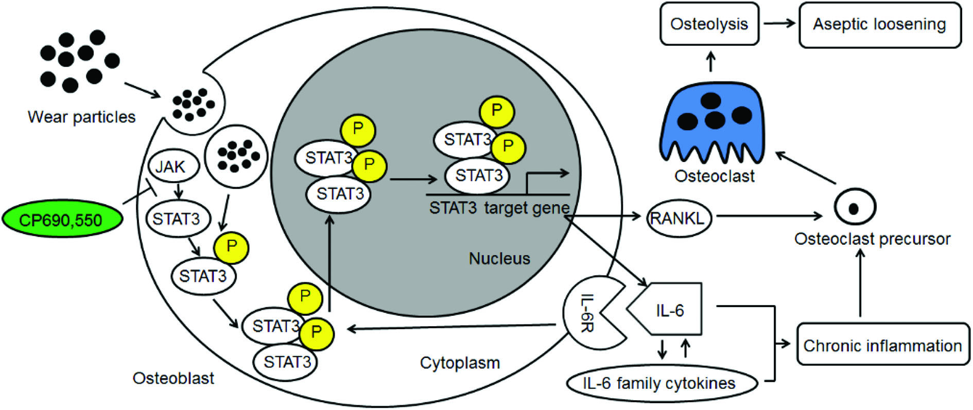
Researchers from China sought to analyze the role of interleukin-6 (IL-6) dependent inflammatory response in osteoblasts treated with TiAl6V4 nanoparticles (TiPs) and also the protective action of IL-6/STST3 (an important transcription factor) inhibition. TiPs obtained from the prosthesis of a patient with aseptic loosening were characterized for shape, size distribution, chemical composition, etc.

As measured by real time-PCR, the expression of IL-6, IL-11, LIF and OSM all increased when MC3T3-E1 cells were stimulated by TiPs in vitro as well as in the periosteum of mice. It was also found via western blotting that the protein levels of activated STAT3 were upregulated following treatment of cells with TiPs in a time and dose dependent fashion. This corroborated the immunofluorescence staining results both in vitro and in vivo, thereby confirming that TiPs activate STAT3 expression in osteoblasts. By using CP690,550 as an inhibitor of STAT3 activation, it was found that upregulation of IL-6- and IL-6-dependent inflammatory cytokine expression was reduced.

Next, using real-time PCR, it was shown that mRNA expression of RANKL, an indicator of osteoclastogenesis increased 13 fold in osteoblasts stimulated by TiPs. Using micro-CT with 3-dimensional reconstruction and quantitative analysis of bone parameters, it was confirmed that inhibition of the STAT3/ IL-6 pathway by inhibitor CP690,550 resulted in suppression of TiP induced osteolysis.

All these results taken together suggest that TiPs induced activation of STAT3 which led to osteolysis by promoting inflammation in osteoblasts and activating osteoclasts and that the inhibition of STAT3 activation by CP690,550 (tofacitinib) significantly reduced the activation of osteoclasts and protected against osteolysis via the STAT3/IL-6 signalling pathway. This suggests use of tofacitinib as a potential therapy for aseptic loosening.

To find out more please read:

STAT3/IL-6 dependent induction of inflammatory response in osteoblast and osteoclast formation in nanoscale wear particle-induced aseptic prosthesis loosening

Biomaterials Science, 2020, DOI: 10.1039/D0BM01256D


About the web writer:

Saswat Choudhury is a graduate student at the Indian Institute of Science Bangalore pursuing research on biomaterials and tissue engineering. He studies bioabsorbable polymers, design and characterization for biomedical applications. Besides research, he is also interested in science communication. You can find him on Twitter @saswatchoudhur1

Digg This
Reddit This
Stumble Now!
Share on Facebook
Bookmark this on Delicious
Share on LinkedIn
Bookmark this on Technorati
Post on Twitter
Google Buzz (aka. Google Reader)

Oversized composite braided biodegradable stents with post-dilatation for pediatric applications: mid-term results of a porcine study

Biodegradable stents (BDSs) have proven to be better compared to permanently implanted metallic stents for the treatment of endovascular diseases in children.  Currently, BDSs that are made out of polylactic acid (PLA) with degradation times of 2–3 years are not suitable for infants, where the ideal healing time for an artery is 3 to 6 months. Poly p-dioxanone (PPDO) is an ideal alternative owing to its availability, FDA approval in clinics and suitable degradation time of 6 months.  But, braided PPDO fiber stents still have lower stiffness than standard self-expanding metal ones.

Researchers from China have come up with a novel design strategy to reinforce the mechanical properties of PPDO fibers by using an elastomeric polycaprolactone (PCL) coating which can serve as a binder at to improve the compression performance. This self-expandable, fiber-based, composite braided biodegradable stent (CBBS) made of PPDO and PCL was then assessed for its physical properties, changes in mechanical properties during degradation, etc and compared with the control, cobalt–chromium-based alloy self-expanding stents (WALLSTENTs/WSs). CBBSs delivered in sheaths post dilation exhibited similar mechanical properties as WSs.

In vitro degradation studies showed that CBBSs post-dilation retained effective mechanical support and stent weight (almost 90%) for at least 16 weeks, which is adequate for arterial healing. These results corroborate with the hydrolysis mechanisms involved in degradation of PDDO, the main component and with in vivo histopathological evaluation.

Lastly, the stents were implanted in porcine models without resulting in any evidence of complications such as implant migration, thrombosis, dissection or aneurism. The mechanical performance of CBBS was also not worse than metallic stents in vivo. Angiographic analysis revealed vessel stenosis and an inflammatory response (intima proliferation) at 4 months due to hydrolysis induced degradation of the stent. But this inflammation was resolved at 12 months due to the complete degradation of CBBSs unlike the WSs. When different diameters of WSs were compared, the ones in oversized common iliac arteries exhibited higher luminal gain initially but there was stenosis and vascular injury compared to normal-sized abdominal aortas in the mid-term follow-up period

All the results combined demonstrate the advantages of these novel composite braided degradable stents over the standard metallic ones in terms of mechanical strength and appropriate degradation rate.

To find out more please read:

Oversized composite braided biodegradable stents with post-dilatation for pediatric applications: mid-term results of a porcine study

Jing Sun, Kun Sun,  Kai Bai, Sun Chen, Fan Zhao, Fujun Wang, Nanchao Honga and Hanbo Hu

Biomaterials Science, 2020, DOI: 10.1039/d0bm00567c


About the web writer:

Saswat Choudhury is a graduate student at the Indian Institute of Science Bangalore pursuing research on biomaterials and tissue engineering. He studies bioabsorbable polymers, design and characterization for biomedical applications. Besides research, he is also interested in science communication. You can find him on Twitter @saswatchoudhur1

Digg This
Reddit This
Stumble Now!
Share on Facebook
Bookmark this on Delicious
Share on LinkedIn
Bookmark this on Technorati
Post on Twitter
Google Buzz (aka. Google Reader)

3D in vitro modeling of Alzheimer’s disease using electrospun microfiber scaffolds

Alzheimer’s disease (AD) presently occupies the topmost position among the most commonly diagnosed neurodegenerative diseases worldwide with the number of affected people forecasted to reach 100 million by 2050. It is characterized by progressive memory loss, impairment of cognitive function, and inability to perform activities of daily life. The key to understanding AD lies in developing effective models which should ideally recapitulate all aspects of the disease. Furthermore, high inaccessibility to the human brain makes it desirable to study neuronal function and degeneration using appropriate in vivo or in vitro model systems of brain cultures. Increasing evidence indicates the superiority of three-dimensional (3D) in vitro cell culture platforms over conventional two-dimensional (2D) monolayer cultures in mimicking native in vivo microenvironments.

Researchers from Singapore have recently developed a novel 3D in vitro model of AD by encapsulating patient induced pluripotent stem cell (iPSC) derived neural progenitors in poly(lactic-co-glycolic acid) (PLGA) microtopographic scaffolds fabricated using wet electrospinning. They demonstrate that 3D culture robustly recapitulates and accelerates early-stage AD pathogenesis compared with Petri dish-based 2D monolayer controls.

Schematic showing fabrication of PLGA 3D scaffold

First, they achieved deep cellular infiltration and uniform distribution inside the 3D microfibrous scaffold by optimizing various parameters such as fiber diameter, pore size, porosity and hydrophilicity. The stiffness of the microfiber scaffold was found to be comparable to the elasticity of native brain tissue, indicating its capability to promote realistic physiological responses.

Next, they compared key neural stem cell features including viability, proliferation and differentiation in 3D culture with 2D monolayer controls. The 3D microfibrous substrate reduced cell proliferation and significantly accelerated neuronal differentiation within just seven days of culture.

Finally, they demonstrated that 3D scaffold-based culture spontaneously enhanced pathogenic amyloid-beta 42 (Aβ42) and phospho-tau levels in differentiated neurons carrying familial AD (FAD) mutations compared with age-matched healthy controls. More importantly, recapitulation of both pathologies was more pronounced and consistent in 3D culture compared with the same cell lines in 2D monolayer culture conditions.

Taken together, the results indicate that the tunable scaffold-based 3D neuronal culture platform serves as a suitable in vitro model that robustly recapitulates and accelerates pathogenic characteristics of FAD-iPSC derived neurons. It can also be extended to model other complex neurodegenerative diseases and to evaluate prospective therapeutic candidates.

To find out more please read:

A microfiber scaffold-based 3D in vitro human neuronal culture model of Alzheimer’s disease

Vivek Damodar Ranjan, Lifeng Qiu, Jolene Wei-Ling Lee, Xuelong Chen, Se Eun Jang, Chou Chai, Kah-Leong Lim, Eng-King Tan, Yilei Zhang, Wei Min Huang and Li Zeng

Biomaterials Science, 2020, DOI: 10.1039/D0BM00833H

Digg This
Reddit This
Stumble Now!
Share on Facebook
Bookmark this on Delicious
Share on LinkedIn
Bookmark this on Technorati
Post on Twitter
Google Buzz (aka. Google Reader)

A novel biomaterial implant for repair of spinal cord injury

Spinal cord injury (SCI) can be categorized as traumatic (90% of cases) or non-traumatic based on its origin. Traumatic SCI occurs when the primary injury is an external mechanical force (arising from traffic accidents, sports, violence etc.,), which damages the spinal cord and initiates a cascade of multiple secondary complications including neuronal/glial death with very slim chances of recovery. Current treatments for SCI are mainly palliative; however, studies involving surgical interventions for reconstructing injured sites via cell implantation have shown promise. Moreover, incorporating cells within engineered biomaterial substrates which act as extracellular matrix (ECM) substitutes not only lowers cell density requirements but also enables more accurate localised transplantation. Both natural and synthetic biomaterials are being investigated in this regard.

Researchers from the UK have recently developed Proliferate®, a macroporous and biodegradable polymer based on cross-linked poly-ε-lysine (pεK) as a biomaterial candidate for SCI implantation. They demonstrate the biocompatibility of the material with CNS cells via in vitro and in vivo studies, both in the original form and on incorporating functional ECM peptides.

First, they synthesized the polymer in two formats: (i) as inserts suspended in 24-well plate culture wells for in vitro studies and (ii) in tubular form with parallel channels facilitating cell guidance for in vivo studies. The material exhibited a beaded, heterogeneous 3D topography with the porosity capable of being tuned by varying the degree of cross-linking.

Next, they cultured astrocytes on the Proliferate® inserts in vitro and compared  cell morphology with controls grown on PLL-coated coverslips. Staining results showed that the astrocytes adopt a fibrous, ramified morphology typical of in vivo conditions when cultured on the inserts. In addition, the polymer supported differentiation, neuronal survival as well as neurite extension in myelinating cultures; however, myelination was slightly delayed in comparison with coverslip-based controls.

Finally, they implanted the tubular form of the biomaterial into adult rat contusion SCI for in vivo assessment at two timepoints i.e. 7 weeks and 6 months post-implantation. The Proliferate®  implants induced extensive vascularisation and cellular infiltration with no significant difference being observed in microglial response surrounding non-implanted injury cavities and construct-implanted injuries. Although, construct-tissue borders were permissive to astrocyte growth and migration, most cell guidance channels were observed to disintegrate with time and organized axonal growth seen only in intact channels.

Taken together, the results indicate the potential of this novel material, both as a solo implant as well as a substrate for delivery of essential biomolecules to the injury site for facilitation of axonal regeneration following SCI.

To find out more please read:

A novel poly-ε-lysine based implant, Proliferate®, for promotion of CNS repair following spinal cord injury

Sara Hosseinzadeh, Susan L. Lindsay, Andrew G. Gallagher, Donald A. Wellings, Mathis O. Riehle, John S. Riddell and Susan C. Barnett

Biomater. Sci., 2020, 8, 3611-3627, DOI: 10.1039/D0BM00097C

Digg This
Reddit This
Stumble Now!
Share on Facebook
Bookmark this on Delicious
Share on LinkedIn
Bookmark this on Technorati
Post on Twitter
Google Buzz (aka. Google Reader)

Culturing stem-cell derived neurons on a “bed-of-nails” substrate

Interfacing living cells with inorganic nanowire (NW) array substrates is one of the latest areas of exploration in life sciences with potential applications in electrical stimulation, biosensors, cell injection, axonal guidance and so on. A growing body of evidence has identified the role of substrate nanotopography in regulating various cellular phenotypes including cell morphology, adhesion, proliferation, differentiation and intracellular signaling. However, cellular interactions with high surface area vertical nanowires are relatively unexplored and further studies are necessary to fully reveal the correlations between NW array geometry and stem cell behavior.

Researchers from Germany have recently interfaced human induced pluripotent stem cell (hiPSC)-derived neurons with tailor-made silicon nitride NW array substrates, achieving highly efficient neuronal differentiation and generating electrophysiologically mature neurons within 4 weeks of culture.

Figurative demonstration of the interface between stem cells and a person

First, they fabricated NW arrays using a top-down dry reactive ion etching (RIE) approach in 3 different arrangements – random, hexagonal and rectangular. The NW lengths were fixed to 1.2 μm with pitches of 1.8 μm and 4 μm, resulting in low density (LD) and high density (HD) arrangements respectively. The cells were transferred onto the NW substrates after 14 days in vitro (DIV) and cultured for another 14-16 DIV before performing functional characterization.

Next, they assessed viability of cells cultured on NW substrates and found that both material used as well as substrate topology had no negative impact on cell viability compared with controls cultured on glass coverslips. Furthermore, on studying cellular outgrowth and morphology, they observed that cells rested on NW tips in the case of HD arrays whereas they encapsulated the NWs in LD arrays, thus indicating the effect of NW density on regulating the settling regime of the cells.

Finally, they tested the electrophysiological integrity of the hiPSC-derived neurons via patch clamping and observed that the neurons cultured on the NW substrates fired characteristic action potentials and demonstrated no significant differences in electrophysiological parameters compared with controls.

Taken together, the results indicate the potential of this platform in stem cell research and regenerative medicine for interfacing human stem cell-derived neurons with tailor-made nanostructured substrates to achieve desired cell behaviors.

To find out more please read:

Interfacing human induced pluripotent stem cell-derived neurons with designed nanowire arrays as a future platform for medical applications

Jann Harberts, Undine Haferkamp, Stefanie Haugg, Cornelius Fendler, Dennis Lam, Robert Zierold, Ole Pless and Robert H. Blick

Biomater. Sci., 2020, 8, 2434-2446, DOI: 10.1039/D0BM00182A

Digg This
Reddit This
Stumble Now!
Share on Facebook
Bookmark this on Delicious
Share on LinkedIn
Bookmark this on Technorati
Post on Twitter
Google Buzz (aka. Google Reader)

Camouflaging tumor targeting nanoparticles with red blood cell membrane for pretargeted multimodal imaging of cancer

Managing cancer requires visualization of tumors using a plethora of imaging modalities such as positron-emission tomography (PET), magnetic resonance imaging (MRI), computed tomography (CT), photoacoustic tomography and optical imaging. Upconversion nanoparticles (UCNPs), a new generation of optical nanomaterials which convert near-infrared (NIR) radiation to visible light by a process called “upconversion luminescence” (UCL), are garnering a lot of attention in cancer diagnostics due to their ability to selectively label cancer cells.

Researchers from Suzhou, China have recently coated tumor targeting UCNPs with red blood cell (RBC) membranes to render them stealthy, effectively preventing them from immune attack and clearance by the host system. Subsequently, they assessed the utility of these RBC-UCNPs for targeted multimodality imaging of 4T1 breast cancer, a triple-negative breast cancer.

First, they isolated cell membranes from the RBCs, reconstructing them into vesicles which were used to encapsulate UCNPs via extrusion. Folic acid (FA) molecules were inserted into the surface of these RBC-UCNPs to assess the tumor-targeting ability of nanoparticles. Upconversion fluorescence imaging revealed that RBC-FA-UCNPs intravenously injected into mice bearing 4T1 subcutaneously transplanted tumors exhibited quick accumulation, long-term retention and reduced uptake by the immune system.

Next, they investigated the feasibility of using these biomimetic nanoparticles in MRI and PET imaging for the detection of tumors in vivo. They found that the MR signal was significantly enhanced by the FA-RBC-UCNPs, indicating the increased circulation time of particles at the tumor site. Furthermore, a combination of pre-targeting strategy and in vivo click chemistry was utilized to mediate PET imaging, which indicated that the biomimetic nanoparticles displayed a higher tumor uptake of the tracer compared with controls, on application of a short half-life radionuclide.

Finally, they conducted in vivo toxicity studies in mice over a span of 30 days, to assess cytotoxicity of the nanoparticles. Blood chemistry, hematology, and histological analyses indicated non-significant induction of toxicity and organ damage, in turn demonstrating the biocompatibility of the biomimetic nanoparticles and their suitability for clinical utilization.

Taken together, the results indicate the potential of this platform for further applications in realizing early diagnosis, bioimaging and treatment of tumors, especially for deep-seated lesions.

To find out more please read:

Red blood cell membrane-coated upconversion nanoparticles for pretargeted multimodality imaging of triple-negative breast cancer

Mengting Li, Hanyi Fang, Qingyao Liu, Yongkang Gai, Lujie Yuan, Sheng Wang, Huiling Li, Yi Hou, Mingyuan Gao and Xiaoli Lan

Biomater. Sci., 2020,8, 1802-1814, DOI: 10.1039/d0bm00029a

Digg This
Reddit This
Stumble Now!
Share on Facebook
Bookmark this on Delicious
Share on LinkedIn
Bookmark this on Technorati
Post on Twitter
Google Buzz (aka. Google Reader)

Enhanced optical imaging agents to guide surgical removal of brain tumors

Obtaining real-time visual information and feedback is vital for surgeons while removing tumors located in the brain, where a lack of precision can lead to catastrophic surgical complications and reduced life expectancy. During surgery, the human eye can identify only anatomical structures and is unable to detect features at the molecular level, which makes it challenging for surgeons to differentiate tumor tissue from surrounding normal brain tissue. Fluorescence-guided surgery attempts to overcome this limitation and relies on the administration of a fluorescent dye which accumulates within the tumor and produces light which in turn is captured and visualized using a camera.

Researchers from Guangzhou, China have recently engineered microglial cells into optical imaging agent vehicles, achieving more accurate brain tumor imaging for fluorescence-guided surgery compared with the commercially used tracer 5-aminolevulinic acid (5-ALA).

Fluorescence images of BV2-Fe accumulation at tumor sites in vivo

First, they activated BV2 microglial cells with citric-acid coated iron oxide nanoparticles (CIONPs) and loaded them with near-infrared fluorescent dye DiD (DiDBV2-Fe). Priming the cells with iron oxide nanoparticles downregulated M2 markers associated with the immunoresponse, and upregulated expression levels of genes that promote transportation of cells across the blood–brain barrier (BBB), thus achieving a two-fold favorable effect.

Next, they assessed the administration of DiDBV2-Fe in glioblastoma-bearing mice models via two routes:. intravenous and intracarotid artery injection. The latter route resulted in more efficient accumulation of activated cells in the brain tumor, 2.2 times higher than that of 5-ALA, 8 hours after application. Maximum fluorescence intensity images of brain tissues acquired at various timepoints from 2 to 24 hours using near-infrared imaging revealed clear tumor border demarcation. Confocal microscopy of harvested brain tumor sections showed noticeable co-localization of DiDBV2-Fe with the Ki67 positive tumor cells along with a significantly higher tumor-to-brain fluorescence ratio compared with 5-ALA (4.54 vs. 1.81).

Finally, they evaluated the in vivo preliminary safety of DiDBV2-Fe in comparison to 5-ALA. Administering DiDBV2-Fe did not induce acute liver injury, phototoxic or hypersensitivity reactions until a certain threshold was reached. In addition, the engineered microglia did not induce gene expression changes of the detected immunoregulatory proteins, unlike 5-ALA which induced both phototoxic and photoallergic reactions.

Taken together, the results indicate that these engineered microglial cells can serve as biological homing vehicles – in seeking out tumors and delivering optical imaging agents, which in turn can help surgeons navigate and identify tumor tissue via fluorescence during surgery.

To find out more please read:

Engineering microglia as intraoperative optical imaging agent vehicles potentially for fluorescence-guided surgery in gliomas

Ling Guo, Xiaochen Zhang, Runxiu Wei, Gaojie Li, Bingzhi Sun, Hongbo Zhang, Dan Liu, Cuifeng Wang and Min Feng

Biomater. Sci., 2020, 8, 1117-1126.

Digg This
Reddit This
Stumble Now!
Share on Facebook
Bookmark this on Delicious
Share on LinkedIn
Bookmark this on Technorati
Post on Twitter
Google Buzz (aka. Google Reader)

Trapping circulating tumor cells using a “lock and key” biomimetic interface

Sherlock Holmes solves mysterious cases often by identifying footprints and odor left by the suspects at the crime scenes. Scientists are now trying to trap notoriously rare circulating tumor cells (CTCs) in the bloodstream by imprinting their unique topologies and residual biomolecules for efficient early tumor detection.  

The presence of CTCs is a dangerous signal for tumor progression, as they leak into the blood stream from the primary tumor and could potentially invade to other organs, causing metastasis. Unfortunately, they’re extremely rare and thus hard to identify, which poses great challenges for early tumor diagnosis. In a recent publication in Biomaterials Science, Gao et al from Shanghai Jiao Tong University developed a cell-imprinted biomimetic interface, which could intelligently recognize and efficiently capture CTCs, with an over 55% capture efficiency towards spiked MCF-7 cells, a human breast cancer cell line, from rabbit whole blood samples.

Graphical abstract for article c9bm01008d

The mechanism behind is the synergy of “lock and key” topological and molecular interactions at the cell-biomaterials interface. Using target CTCs as an imprint template, they created a substrate mimicking their topologies and specific immunoaffinity. By leveraging soft lithography, hierarchical micro/nano-structures of CTCs could be recapitulated on an elastomer substrate (PDMS) to modulate cell adhesion behaviour: cell cytoskeleton staining showed that MCF-7 cells captured on the imprinted surface exhibited abundant filopodia and lamellipodia structures, while they showed an approximately spherical structure on flat substrate, indicating weak topographic interaction. Native proteins originating from CTC’s extracellular matrix (ECM) was anchored in the imprinted substrates providing artificial recognition receptors for selective CTC capture. This is supported by the significantly higher MCF-7 cells capture efficiency on MCF-7 imprinted substrates comparing to HeLa-cell (a human-cervical-cancer cell line) imprinted ones. Anti-EpCAM, a natural antibody, was also introduced to accelerate the CTC-substrate interactions. These interactions turned out to play a decisive role in cell capture, followed by that with plastic receptors.

By recapitulating the topological and chemical microenvironment of CTCs, this biomimetic cell-imprinted substrates demonstrate potential for rare cancer cell capture. Considering the significant roles of tissue ECM stiffness and viscoelasticity in metastasis, and the strong integrin-mediated focal adhesion at the CTCs-biomaterial interface, it might be interesting to incorporate mechanical signals into the interface design in the future for enhanced CTCs capture.

Read the full article for FREE until the 9th October!

Cell-imprinted biomimetic interface for intelligent recognition and efficient capture of CTCs, by Su Gao, Shuangshuang Chen and Qinghua Lu

About the web writer

Zhenwei Ma is currently a Ph.D. candidate in Mechanical Engineering at McGill University. He holds a M.E. degree in Chemical Engineering at McGill University and a B.E. degree in Chemical Engineering from Sichuan University. Find out more about him here.

Digg This
Reddit This
Stumble Now!
Share on Facebook
Bookmark this on Delicious
Share on LinkedIn
Bookmark this on Technorati
Post on Twitter
Google Buzz (aka. Google Reader)

Polydopamine coated bimetallic nanoparticles for mitochondria-targeted cancer therapy

Thyroid cancer is one of the most common endocrine malignancies, and it is the cause of more deaths than all other endocrine cancers combined. Papillary thyroid cancer, a type of thyroid cancer, is often asymptomatic, but because of the progress of modern diagnostic technology its detection rate has shown a rapid increase in the past decade. It has a lower degree of malignancy than that of other types of thyroid cancers and the tumor growth rate is slow, however, it is still very serious as it can develop throughout the thyroid gland and spread extensively in the body, even spreading to distant organs. The conventional method for treating papillary thyroid cancer is surgery, but this comes with a significant risk of injury. Hence, there is a strong need for non-invasive therapeutics which can be used as an alternative to surgery in the treatment of papillary thyroid cancer.

c8bm01414k


Recently, mitochondria-targeting nanomaterials have gained major attention as mitochondria are cells’ powerhouse, controls various signaling pathways including apoptosis and necrosis and produce reactive oxygen species (ROS). It is important to mention that increases ROS can cause the proliferation of cancer cells and drug resistance. In this present work, researchers used a mitochondria-targeted and exocytosis inhibition strategy. They used polydopamine-coated gold-silver alloy nanoparticles (Au-Ag@PDA NPs) to target papillary thyroid cancer cells (TPC-1 cells). In order to understand the nano-bio interactions between the nanoparticles and the cancer cells, the authors systematically studied the endocytosis pathway, the subcellular localization, and the cellular responses to the nanoparticles.

The results showed that:

(i)Au-Ag@PDA NPs were internalized through a caveolae-mediated and macropinocytosis pathway, localized in mitochondria and block exocytosis pathway

(ii) This lead to cell cycle arrest in S-phase and this inhibited the cell proliferation

(iii) The TPC-1 cells can survive by an autophagy-mediated method to escape the apoptosis or necrosis

The researchers, using the mitrochondria targeting behavior of the nanoparticles, then carried out photothermal therapy for the enhanced treatment of the papillary thyroid cancer cells. These findings indicate that PDA-coated inorganic nanoparticles have potential in mitrochondria-targeted cancer treatments and, one day, these could be provide an alternative to surgery for patients suffering from papillary thyroid cancer.


Read the full paper for free until the 13th May

Targeting mitochondria with Au–Ag@Polydopamine nanoparticles for papillary thyroid cancer therapy Biomater. Sci., 2019, 7, 1052-1063


About the Web/Blog writer:

Dr Sudip MukherjeeDr. Sudip Mukherjee is a Web Writer for Biomaterials Science. He is currently a Postdoctoral Research Associate at the Rice University. His research is involved in the development of advanced nanomaterials for drug/gene delivery in cancer theranostics, immunomodulatory applications & angiogenesis. He published a total of ~35 research articles/patents. He serves as International Advisory Board Member for ‘Materials Research Express‘, IOP Sciences. He is an associate member (AMRSC) of RSC, UK. He serves as reviewer for several international journals like ChemComm, J Mater Chem A, J Mater Chem B, Journal of Biomedical Nanotechnology, RSC Advances, IOP Nanotechnology, Biofabrication etc.

Contact Email: sudip.mukherjee@rice.edu
Twitter: https://twitter.com/sudip_88


Digg This
Reddit This
Stumble Now!
Share on Facebook
Bookmark this on Delicious
Share on LinkedIn
Bookmark this on Technorati
Post on Twitter
Google Buzz (aka. Google Reader)

Sericin hydrogels promote skin wound healing

Severe skin injuries affect millions of patients each year worldwide. These lead to serious issues, such as the formation of nonfunctional large scar tissue and the loss of skin appendages (such as hair follicles and sebaceous glands; etc). This can cause the patients insufferable bodily discomfort and a poor quality of life. Current available treatments include autologous skin grafting, allotransplantation and artificial skin substitutes. However, it remains very challenging to functionally regenerate skin tissue after severe loss of the epidermis and dermis. Other limitations include the lack of donor skin, costly medical expenses, the chance of immune-rejection and unsatisfactory skin regeneration. Hence, the development of an efficient alternative skin substitute is highly desired.

10.1039/C8BM00934A

Sericin is a natural biomaterial derived from silk cocoons has been used previously for a variety of types of injury repair. Previously, Wang and coworkers utilized sericin made hydrogels or scaffolds for transected sciatic nerve regeneration, repair of ischemic stroke, and cartilage regeneration. In this present work, a photo-crosslinkable sericin hydrogel (SMH) for the repair of scarless skin and sebaceous glands regeneration is reported. The sericin hydrogel promotes such regeneration by the following mechanisms: (a) effective inhibition of inflammation; (b) promotion of angiogenesis by stimulating the growth factors like VEGF and EGF; (c) reduction of scar formation through regulating the expressions of TGF-β1 and TGF-β3; and (d) effective conscription of stem cells to injury sites, where they differentiate and regenerate into skin appendages. Overall, these results showcase the potential of this innovative bimodal tool for the development of new artificial skin substitutes for the clinical treatment of severe skin injuries.

Read the full article for free until 19th November

Sericin hydrogels promote skin wound healing with effective regeneration of hair follicles and sebaceous glands after complete loss of epidermis and dermis  Biomater. Sci., 2018, Advance Article DOI: 10.1039/C8BM00934A.


About the webwriter
Dr Sudip MukherjeeDr. Sudip Mukherjee is a Web Writer for Biomaterials Science. He is currently a Postdoctoral Research Associate at the Rice University. His research is involved in the development of advanced nanomaterials for drug/gene delivery in cancer theranostics, immunomodulatory applications & angiogenesis. He published a total of ~35 research articles/patents. He serves as International Advisory Board Member for ‘Materials Research Express‘, IOP Sciences. He is an associate member (AMRSC) of RSC, UK. He serves as reviewer for several international journals like ChemComm, J Mater Chem A, J Mater Chem B, Journal of Biomedical Nanotechnology, RSC Advances, IOP Nanotechnology, Biofabrication etc.

Contact Email: sudip.mukherjee@rice.edu

Twitter: https://twitter.com/sudip_88

Digg This
Reddit This
Stumble Now!
Share on Facebook
Bookmark this on Delicious
Share on LinkedIn
Bookmark this on Technorati
Post on Twitter
Google Buzz (aka. Google Reader)