Author Archive

Polydopamine coated bimetallic nanoparticles for mitochondria-targeted cancer therapy

Thyroid cancer is one of the most common endocrine malignancies, and it is the cause of more deaths than all other endocrine cancers combined. Papillary thyroid cancer, a type of thyroid cancer, is often asymptomatic, but because of the progress of modern diagnostic technology its detection rate has shown a rapid increase in the past decade. It has a lower degree of malignancy than that of other types of thyroid cancers and the tumor growth rate is slow, however, it is still very serious as it can develop throughout the thyroid gland and spread extensively in the body, even spreading to distant organs. The conventional method for treating papillary thyroid cancer is surgery, but this comes with a significant risk of injury. Hence, there is a strong need for non-invasive therapeutics which can be used as an alternative to surgery in the treatment of papillary thyroid cancer.

c8bm01414k


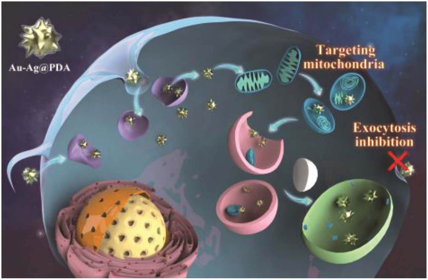
Recently, mitochondria-targeting nanomaterials have gained major attention as mitochondria are cells’ powerhouse, controls various signaling pathways including apoptosis and necrosis and produce reactive oxygen species (ROS). It is important to mention that increases ROS can cause the proliferation of cancer cells and drug resistance. In this present work, researchers used a mitochondria-targeted and exocytosis inhibition strategy. They used polydopamine-coated gold-silver alloy nanoparticles (Au-Ag@PDA NPs) to target papillary thyroid cancer cells (TPC-1 cells). In order to understand the nano-bio interactions between the nanoparticles and the cancer cells, the authors systematically studied the endocytosis pathway, the subcellular localization, and the cellular responses to the nanoparticles.

The results showed that:

(i)Au-Ag@PDA NPs were internalized through a caveolae-mediated and macropinocytosis pathway, localized in mitochondria and block exocytosis pathway

(ii) This lead to cell cycle arrest in S-phase and this inhibited the cell proliferation

(iii) The TPC-1 cells can survive by an autophagy-mediated method to escape the apoptosis or necrosis

The researchers, using the mitrochondria targeting behavior of the nanoparticles, then carried out photothermal therapy for the enhanced treatment of the papillary thyroid cancer cells. These findings indicate that PDA-coated inorganic nanoparticles have potential in mitrochondria-targeted cancer treatments and, one day, these could be provide an alternative to surgery for patients suffering from papillary thyroid cancer.


Read the full paper for free until the 13th May

Targeting mitochondria with Au–Ag@Polydopamine nanoparticles for papillary thyroid cancer therapy Biomater. Sci., 2019, 7, 1052-1063


About the Web/Blog writer:

Dr Sudip MukherjeeDr. Sudip Mukherjee is a Web Writer for Biomaterials Science. He is currently a Postdoctoral Research Associate at the Rice University. His research is involved in the development of advanced nanomaterials for drug/gene delivery in cancer theranostics, immunomodulatory applications & angiogenesis. He published a total of ~35 research articles/patents. He serves as International Advisory Board Member for ‘Materials Research Express‘, IOP Sciences. He is an associate member (AMRSC) of RSC, UK. He serves as reviewer for several international journals like ChemComm, J Mater Chem A, J Mater Chem B, Journal of Biomedical Nanotechnology, RSC Advances, IOP Nanotechnology, Biofabrication etc.

Contact Email: sudip.mukherjee@rice.edu
Twitter: https://twitter.com/sudip_88


Digg This
Reddit This
Stumble Now!
Share on Facebook
Bookmark this on Delicious
Share on LinkedIn
Bookmark this on Technorati
Post on Twitter
Google Buzz (aka. Google Reader)

Sericin hydrogels promote skin wound healing

Severe skin injuries affect millions of patients each year worldwide. These lead to serious issues, such as the formation of nonfunctional large scar tissue and the loss of skin appendages (such as hair follicles and sebaceous glands; etc). This can cause the patients insufferable bodily discomfort and a poor quality of life. Current available treatments include autologous skin grafting, allotransplantation and artificial skin substitutes. However, it remains very challenging to functionally regenerate skin tissue after severe loss of the epidermis and dermis. Other limitations include the lack of donor skin, costly medical expenses, the chance of immune-rejection and unsatisfactory skin regeneration. Hence, the development of an efficient alternative skin substitute is highly desired.

10.1039/C8BM00934A

Sericin is a natural biomaterial derived from silk cocoons has been used previously for a variety of types of injury repair. Previously, Wang and coworkers utilized sericin made hydrogels or scaffolds for transected sciatic nerve regeneration, repair of ischemic stroke, and cartilage regeneration. In this present work, a photo-crosslinkable sericin hydrogel (SMH) for the repair of scarless skin and sebaceous glands regeneration is reported. The sericin hydrogel promotes such regeneration by the following mechanisms: (a) effective inhibition of inflammation; (b) promotion of angiogenesis by stimulating the growth factors like VEGF and EGF; (c) reduction of scar formation through regulating the expressions of TGF-β1 and TGF-β3; and (d) effective conscription of stem cells to injury sites, where they differentiate and regenerate into skin appendages. Overall, these results showcase the potential of this innovative bimodal tool for the development of new artificial skin substitutes for the clinical treatment of severe skin injuries.

Read the full article for free until 19th November

Sericin hydrogels promote skin wound healing with effective regeneration of hair follicles and sebaceous glands after complete loss of epidermis and dermis  Biomater. Sci., 2018, Advance Article DOI: 10.1039/C8BM00934A.


About the webwriter
Dr Sudip MukherjeeDr. Sudip Mukherjee is a Web Writer for Biomaterials Science. He is currently a Postdoctoral Research Associate at the Rice University. His research is involved in the development of advanced nanomaterials for drug/gene delivery in cancer theranostics, immunomodulatory applications & angiogenesis. He published a total of ~35 research articles/patents. He serves as International Advisory Board Member for ‘Materials Research Express‘, IOP Sciences. He is an associate member (AMRSC) of RSC, UK. He serves as reviewer for several international journals like ChemComm, J Mater Chem A, J Mater Chem B, Journal of Biomedical Nanotechnology, RSC Advances, IOP Nanotechnology, Biofabrication etc.

Contact Email: sudip.mukherjee@rice.edu

Twitter: https://twitter.com/sudip_88

Digg This
Reddit This
Stumble Now!
Share on Facebook
Bookmark this on Delicious
Share on LinkedIn
Bookmark this on Technorati
Post on Twitter
Google Buzz (aka. Google Reader)

New hybrid nanoparticles for enhanced drug accumulation in glioblastoma cells

Glioblastoma is the most frequent and aggressive primary malignant tumor of the central nervous system. The efficacy of antineoplastic drugs that are able to cross the blood-brain barrier is limited mainly by various resistance mechanisms. Hence, local strategies have been developed to improve the therapeutic efficacy. Platinum derivatives and one among them, cisplatin, demonstrated promising results when locally administered into the brain of glioblastoma-bearing rats. A more specific vectorization of the active substance may further promote its accumulation within cancer cells, therefore improving its bioavailability. In this context, biocompatible and biodegradable copolymers that are approved for medical applications can be synthesized without any toxic organic solvents or excipients. Their amphiphilicity, namely the combination of both a hydrophilic and a hydrophobic sequence, is responsible for their spontaneous self-assembly in water. Such a formulation process is simple and flexible. The versatile structure of these drug delivery systems allows imaging moieties to be grafted onto their surface while encapsulating a drug within their core. The diagnosis of glioblastoma relies on magnetic resonance imaging (MRI) after the injection of gadolinium-based contrast agents. Thus, the injection of hybrid nanoplatforms that combine an MRI contrast agent and a drug would enable to non-invasively monitor the biodistribution of the treatment by analyzing the tumor response in real time. Personalized medicine and theranostic applications require similar strategies for the purposes of adjusting the treatment regimen to the patient response.


Such smart drug delivery systems were designed by Lajous and coworkers based on amphiphilic block copolymers. Gadolinium complexes were grafted at the end of the hydrophilic chain while a chemical modification of the other block allowed for cisplatin cross-linking with the copolymer backbone. The self-assembly of these functionalized copolymers in water resulted in stable cisplatin cross-linked nanoparticles with a mean size of 100.63 ± 12.04 nm consistent with biological investigations. High field MRI confirmed the intrinsic potential of these hybrid nanoparticles as alternative MRI contrast agents compared to conventional low molar mass Gd-DTPA complexes. Their infusion within the striatum of glioblastoma-bearing mice resulted in a signal that persisted over time. The accumulation of platinum compounds in human glioblastoma cells when treated with these drug delivery systems and the subsequent formation of Pt-DNA adducts was significantly increased in comparison with free cisplatin by up to 50-fold and 32-fold respectively. These results support the potential of this innovative bimodal tool for further applications.

Hybrid Gd3+/cisplatin cross-linked polymer nanoparticles enhance platinum accumulation and formation of DNA adducts in glioblastoma cell lines  Biomater. Sci., 2018, 6, 2386-2409


Read the full article now for free until 11 October


About the web writer

Dr. Sudip MukherjeeDr. Sudip Mukherjee is a Web Writer for Biomaterials Science. He is currently a Postdoctoral Research Associate at the Rice University. His research is involved in the development of advanced nanomaterials for drug/gene delivery in cancer theranostics, immunomodulatory applications & angiogenesis. He published a total of ~35 research articles/patents. He serves as International Advisory Board Member for ‘Materials Research Express‘, IOP Sciences. He is an Associate Member (AMRSC) of RSC, UK. He serves as reviewer for several international journals like ChemComm, J Mater Chem A, J Mater Chem B, Journal of Biomedical Nanotechnology, RSC Advances, IOP Nanotechnology, Biofabrication etc.


Digg This
Reddit This
Stumble Now!
Share on Facebook
Bookmark this on Delicious
Share on LinkedIn
Bookmark this on Technorati
Post on Twitter
Google Buzz (aka. Google Reader)

Chemically modified nanogels for superior bioavailability

The pharmacokinetics and pharmacodynamics of any drug or nanoparticles plays an important role towards the therapeutic activity, half-life of the therapeutic agent and frequency of dosing of the biomaterial in various diseases. Recently, the development of biopolymer nanogels has received tremendous attention due to their effective delivery of therapeutics, size uniformity, high drug encapsulation capacity, easy preparation, high biocompatibility and size tunability. However, the tendency of these nanogels to disassemble in the bloodstream is cause for concern, due to the interactions with serum proteins and excessive dilution volume that decreases the tumor targeting efficiency through EPR effects. In this regard, substantial research is needed to develop novel platform technologies for bioactive nanogels with enhanced bioavailability.

c8bm00396c

In this work, Auzély-Velty and co-workers, developed a novel and easy method to synthesize stable self-assembled hyaluronic acid (HA) nanogels, modified with a thermoresponsive ketone-functional copolymer by hydrazone formation. The cross-linking density played a crucial role in the nanogel stability and pharmacokinetics, and it was easily tuned by varying dihydrazide crosslinker to ketone ratio. Several physicochemical characterizations including cryo-transmission, dynamic light scattering and scanning electron microscopy were performed to analyze the size, morphology and stability of the nanogels. The authors showed the in vitro cellular uptake of the nanogels by CD44 receptor mediated pathway further confirmed the effectiveness of the cross-linking strategy. The modified nanogels demonstrated superior bioavailability in tumors, with enhanced blood circulation for over 24 hours, demonstrated in vivo in biodistribution studies with mouse tumor models. Overall, these nanogels are inexpensive, stable, easily tunable and biocompatible, thus holding promise for the discovery of a new class of molecules for cancer therapy application in near future.


This article is free to read and download until 13th August.

A versatile method for the selective core-crosslinking of hyaluronic acid nanogels via ketone-hydrazide chemistry: from chemical characterization to in vivo biodistribution Biomater. Sci., 2018, 6, 1754-1763


About the web writer

Dr. Sudip MukherjeeDr. Sudip Mukherjee is a Web Writer for Biomaterials Science. He is currently a Postdoctoral Research Associate at the Rice University. His research is involved in the development of advanced nanomaterials for drug/gene delivery in cancer theranostics, immunomodulatory applications & angiogenesis. He published a total of ~35 research articles/patents. He serves as International Advisory Board Member for ‘Materials Research Express‘, IOP Sciences. He is an associate member (AMRSC) of RSC, UK. He serves as reviewer for several international journals like ChemComm, J Mater Chem A, J Mater Chem B, Journal of Biomedical Nanotechnology, RSC Advances, IOP Nanotechnology, Biofabrication etc.

Contact Email: sudip.mukherjee@rice.edu
Twitter: https://twitter.com/sudip_88

Digg This
Reddit This
Stumble Now!
Share on Facebook
Bookmark this on Delicious
Share on LinkedIn
Bookmark this on Technorati
Post on Twitter
Google Buzz (aka. Google Reader)

Cancer theranostics applications of Graphene Oxides

Graphene oxide (GO) has actively been used in various biomedical applications, including biomedical sensors, electronic sensors, functional composites and so on. In addition to having a large surface area, large-scale manufacturability and being dispersible in water, GO shows strong fluorescence and conjugation properties, owing to the abundance of oxygen functional groups on its surface.

Numerous methods have been developed to enable photon emission from GO sheets, such as reduction, labeling with fluorescent protein, and cleaving GO sheets into smaller fragments to produce graphene quantum dots (QDs), although this method does remove oxygen from the carbon lattice. This is a problem because reducing the oxygen content prevents the further functionalization of GO structures with biomolecules. Furthermore, such methods have been reported to cause cytotoxicity.

Cancer theranostics applications of Graphene Oxides

In this work by the Chen and co-workers, a blue fluorescence was induced in a GO suspension by triggering a phase transformation in GO through treatment with a simple, one-step mild annealing. Previously, it had been difficult to facilitate light emission from GO while preserving the oxygen content and maintaining low cytotoxicity. However, this work suggests that by providing a nano-bio interface for reactions with biomolecules the physical difficulties can be overcome. In this case, GO acts as a bio-imaging agent as well as a functionalization platform for biomolecules. Material characterization and biocompatibility tests were performed to examine the purity, inherent property and non-toxicity of the system. The mechanism for enhanced blue fluorescence upon mild annealing was discovered and modeled through atomistic simulations. Most importantly, GO shows an appealing capability in drug delivery and cellular imaging simultaneously. Overall, this method is expected to be inexpensive, rapid and straightforward, thus holding promise for the development of a whole new class of GO-based nanomaterials for cancer theranostics application in near future.


This article is free to read until 30 May


Simultaneous drug delivery and cellular imaging using graphene oxide Biomater. Sci., 2018, 6, 813-819


About the webwriter

Dr. Sudip MukherjeeDr. Sudip Mukherjee is a Web Writer for Biomaterials Science. He is currently a Postdoctoral Research Associate at the Rice University. His research is involved in the development of advanced nanomaterials for drug/gene delivery in cancer theranostics, immunomodulatory applications & angiogenesis. He published a total of ~30 research articles/patents. He serves as International Advisory Board Member for ‘Materials Research Express‘, IOP Sciences. He is an associate member (AMRSC) of RSC, UK. He serves as reviewer for several international journals like Chem Comm, J Mater Chem A, J Mater Chem B, Journal of Biomedical Nanotechnology, RSC Advances, IOP Nanotechnology etc.

Contact Email: sudip.mukherjee@rice.edu

Twitter: https://twitter.com/sudip_88

Digg This
Reddit This
Stumble Now!
Share on Facebook
Bookmark this on Delicious
Share on LinkedIn
Bookmark this on Technorati
Post on Twitter
Google Buzz (aka. Google Reader)

On-demand chemotherapy using photo-activated micelles

Preventing or minimizing the adverse effects of anti-cancer chemotherapy has been a great challenge, due to the non-specific distribution of chemodrugs in healthy organs. Many previous works have attempted to address this issue by improving the accumulation of cytotoxic drugs in the tumor, with various targeting strategies. All the drug targeting methods enhance efficacy and reduce side-effects to a certain extent, but they cannot completely avoid the unfavourable adverse effects. Site-specific activation of chemotherapeutic drugs via a photo trigger has been a promising means of reducing side-effects.

On-demand chemotherapy using photo-activated micelles


The Zhao group at Tianjin University report the use of cyclodextrin-bearing polymer micelles for the on-demand delivery of a photoswitchable microtubule inhibitor, to achieve precision chemotherapy. The tailored inhibitor displays conformation-dependent cytotoxicity with a low potency; the “trans” isomer is thermodynamically stable and inactive, whereas the “cis” isomer is active, but thermodynamically unstable. Light irradiation activates the drug from the “trans” to “cis” form, which then instantly induces rapid drug release. Such simultaneous drug activation and release could compromise the low drug potency to a certain extent, showing improved anti-cancer efficacy in vitro and in vivo. Therefore, photo-triggered nanosystems could open new avenues of on-demand precision chemotherapy, without the risk of adverse effects on healthy organs and tissues.

This article is free to read until 30 April 18

Photo-triggered micelles: simultaneous activation and release of microtubule inhibitors for on-demand chemotherapy Biomater. Sci., 2018, 6, 511-518


About the Web/Blog writer:

Dr. Sudip MukherjeeDr. Sudip Mukherjee is a Web Writer for Biomaterials Science. He is currently a Postdoctoral Research Associate at the Rice University. His research is involved in the development of advanced nanomaterials for drug/gene delivery in cancer theranostics, immunomodulatory applications & angiogenesis. He published a total of ~30 research articles/patents. He serves as International Advisory Board Member for ‘Materials Research Express‘, IOP Sciences. He is an associate member (AMRSC) of RSC, UK. He serves as reviewer for several international journals like ChemComm, J Mater Chem A, J Mater Chem B, Journal of Biomedical Nanotechnology, RSC Advances, IOP Nanotechnology etc.

Contact Email: sudip.mukherjee@rice.edu

Twitter: https://twitter.com/sudip_88


Digg This
Reddit This
Stumble Now!
Share on Facebook
Bookmark this on Delicious
Share on LinkedIn
Bookmark this on Technorati
Post on Twitter
Google Buzz (aka. Google Reader)

3D Multiscale Fiber Matrices for Bioartificial Livers

The liver is the largest internal organ and gland in the human body and it performs numerous vital functions including metabolism regulation, synthesis, detoxification, excretion, and body homeostasis. Many factors, such as viral hepatitis, drugs, toxins, or cardiac-related hepatic ischemia, can cause liver failure. According to the World Health Organisation (WHO) around 10% of the world’s population is suffering from chronic liver disease. The eventual treatment for chronic liver diseases is liver transplantation; however, an inadequate supply of donors is a major limitation for this therapy. With the large number of patients awaiting a liver transplant, there is urgent need for the development of a temporary liver support system, which could be used until a transplant liver is available, or until the patient’s own liver regains its function. Temporary liver supports can be artificial or bioartificial. Artificial support devices are used for detoxification only, whereas bioartificial support devices are more promising, and have biomaterial and cellular components, allowing for both detoxification and synthetic functions. The aim of this study is to prepare a hollow fiber membrane (HFM) based three-dimension matrix, which is the most crucial part of bioartificial livers, and acts as cell growth promoting material. The microenvironment of the three-dimensional matrix influences the cellular behavior and function, exhibiting proliferation, metabolism and interaction of cells with each other and their environment.

3D multiscale fiber matrices

In this study, the authors developed a three-dimensional liver cell (HepG2) compatible bio-matrix which supports the liver cells, allowing them to grow and multiply robustly. The outer surface of indigenously prepared HFM was modified and made accommodating for the growth of the liver cells. Nanofibers of biocompatible compounds including polycaprolactone, chitosan, and gelatin, which mimic the native cell attachment surface, were deposited on the HFMs. The developed material exhibited excellent hemocompatibility with human blood. Minimal induction of inflammatory response and negligible cytotoxicity was observed. Further evaluation of the liver cell functional activity showed that cells exhibited the key characterstics of typical liver cells by secreting urea and albumin in the medium. The specific activity of cytochrome P450 2C9 (detoxifying enzyme) was found to have increased by 2.78-fold. Hence, these results significantly indicate that the three-dimensional fiber matrix developed in this study open up the possibility to use it for the various applications including (1) a membrane material for bio-artificial liver development, (2) cell metabolic studies and (3) drug testing bioreactor.

Three-dimensional multiscale fiber matrices: development and characterization for increased HepG2 functional maintenance for bio-artificial liver application Biomater. Sci., 2018,6, 280-291.

This article is free to read until the 16 April 2018


About the Web/Blog writer:

Dr. Sudip Mukherjee Dr. Sudip Mukherjee is a Web Writer for Biomaterials Science. He is currently a Postdoctoral Research Associate at the Rice University. His research is involved in the development of advanced nanomaterials for drug/gene delivery in cancer theranostics, immunomodulatory applications & angiogenesis. He published a total of ~30 research articles/patents. He serves as International Advisory Board Member for ‘Materials Research Express‘, IOP Sciences. He is an associate member (AMRSC) of The Royal Society of Chemistry, UK. He serves as reviewer for several international journals like Chem Comm, J Mater Chem A, J Mater Chem B, Journal of Biomedical Nanotechnology, RSC Advances, IOP Nanotechnology etc.

Contact Email: sudip.mukherjee@rice.edu
Twitter: https://twitter.com/sudip_88

Digg This
Reddit This
Stumble Now!
Share on Facebook
Bookmark this on Delicious
Share on LinkedIn
Bookmark this on Technorati
Post on Twitter
Google Buzz (aka. Google Reader)

Bio-mimicking melanin-manganese nanoparticles for tumor targeting MRI agent

Good quality MRI contrast agents require profound tumor-targeting ability, well relaxivity, along with rapid clearance properties. In this context, endogenous biological mimicking biomaterials, with high biodegradability and biocompatibility hold enormous potential for the development of clinically translatable nanotheranostics platforms. Manganese plays a crucial role in mitochondrial and cellular function and recently, manganese (Mn)-based contrast agents have been receiving significant attention, due to improved biosafety and superior contrast abilities. However, the long term toxicity and non-biodegradability of these inorganic nanoplatforms have significantly halted their clinical progress. In contrast to this, melanin, an asymmetrical natural biopolymer, has garnered enormous attention due to good biocompatibility, biodegradability and MRI contrast imaging abilities. Hence, exploring endogenous natural materials with high contrast properties seems promising as clinically translatable in vivo MRI imaging contrast agent.

melanin-manganese nanoparticles for tumor targeting MRI agent

The Wang group developed an ultra small and water soluble Mn2+ chelating pegylated melanin nanoparticles (MNP-PEG-Mn) demonstrating excellent tumor-targeting Magnetic Resonance Imaging (MRI) ability. The MNP-PEG-Mn nanoparticles show a size of 5.6 nm displaying high chelating stability and low cytotoxicity. Interestingly, the MNP-PEG-Mn nanoparticles show improved longitudinal relaxivity compared to clinically approved MRI contrast agent Gadodiamide. In vivo studies further showcased excellent tumor targeting abilities upon intravenous administration of MNP-PEG-Mn nanoparticles in mouse model. The author further showed that the MNP-PEG-Mn nanoparticles could be excreted via hepatobiliary and renal routes. In this process negligible toxicity was generated to body tissues that indicate high biocompatibility. Altogether, these results clinically validate the tumor targeted T1 MRI contrast properties of bio-mimicking melanin conjugated manganese nanoparticles.

Melanin-manganese nanoparticles with ultrahigh efficient clearance in vivo for tumor-targeting T1 magnetic resonance imaging contrast agent . Biomater. Sci., 2018, 6, 207-215.

This article is free to read until 28 February!


About the Web writer:

Dr. Sudip MukherjeeDr. Sudip Mukherjee is a Web Writer for Biomaterials Science. He is currently a Postdoctoral Research Associate working at the Department of Bioengineering at the Rice University. His research is involved in the development of advanced nanomaterials for drug/gene delivery in cancer theranostics, immunotherapy, immunomodulatory applications & angiogenesis. He published a total of ~30 research articles/patents. He serves as International Advisory Board Member for ‘Materials Research Express‘, IOP Sciences. He is an associate member (AMRSC) of The Royal Society of Chemistry, UK. He serves as reviewer for several international journals like Chem Comm, J Mater Chem A, J Mater Chem B, Journal of Biomedical Nanotechnology, RSC Advances, IOP Nanotechnology etc. He can be contacted by email at sudip.mukherjee@rice.edu or on Twitter.

Digg This
Reddit This
Stumble Now!
Share on Facebook
Bookmark this on Delicious
Share on LinkedIn
Bookmark this on Technorati
Post on Twitter
Google Buzz (aka. Google Reader)

Biocompatible CuS-based nanoplatforms for multifunctional theranostics

The rationale for combination therapy is to employ various therapeutic methods which work using different mechanisms, thereby decreasing the chance of developing resistant cancer cells. Photothermal therapy (PTT) is the use of electromagnetic radiation to eradicate cancer cells which can also be utilized to increase the effectiveness of chemotherapy or radiation therapy. The application of targeted and functional nanoparticles can be used to overcome the existing limitations of nonspecific toxicity which is associated with PTT. Therefore, functional nanomaterials with near-infrared (NIR) PTT, high biocompatibility and excellent photothermal conversion efficiency can be dynamic tools for cancer ablation without affecting normal healthy tissue.
Biocompatible CuS-based nanoplatforms for multifunctional theranostics

The Chen group used core-shell water-soluble copper sulphide nanoparticles (CuSNPs) coated with mesoporous silica nanoshells (MSNs) for effective delivery of anti-cancer drug doxorubicin (DOX) towards H22 liver cancer. Cleverly, the hollow cavity of MSN was utilized for the loading of anti-cancer drug DOX. CuS@MSN-DOX demonstrated good water dispersibility, high stability, excellent biocompatibility and strong NIR absorption. Its excellent photothermal and NIR thermal imaging properties are due its strong NIR photothermal conversion efficiency. The anti-tumor activity of CuS@MSN-DOX was extensively studied in both in vitro and in vivo therapeutic models which supports excellent chemotherapeutic activity. Complete eradication of the liver tumor was observed by combination therapy of PTT and chemotherapy using CuS@MSN-DOX. Infrared thermal imaging was used to monitor the photothermal treatment. These results clinically validate the multifunctional cancer theranostics property of CuS@MSN-DOX that has enormous potential for clinically translatable thermochemotherapy and enhanced drug delivery in the future.


Biocompatible CuS-based nanoplatforms for efficient photothermal therapy and chemotherapy in vivo Biomater. Sci., 2017, 5, 475 – 484


About the WebwriterDr. Sudip Mukherjee

Dr. Sudip Mukherjee is a Web Writer for Biomaterials Science. He is currently a Postdoctoral Research Associate working alongside Dr. Omid Veiseh at the Department of Bioengineering at the Rice University. His research is involved in the development of advanced nanomaterials for drug/gene delivery in cancer theranostics, immunomodulatory applications & angiogenesis. He published a total of ~30 research articles/patents. He serves as International Advisory Board Member for‘Materials Research Express‘, IOP Sciences. He is an associate member (AMRSC) of RSC, UK. He serves as reviewer for several international journals like Chem Comm, J Mater Chem A, J Mater Chem B, Journal of Biomedical Nanotechnology, RSC Advances, IOP Nanotechnology etc.
Contact Email: sudip.mukherjee@rice.edu

Digg This
Reddit This
Stumble Now!
Share on Facebook
Bookmark this on Delicious
Share on LinkedIn
Bookmark this on Technorati
Post on Twitter
Google Buzz (aka. Google Reader)