Author Archive

Graphene Hybrids Join the Hunt for Mercury

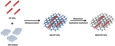
Graphene-polyfuran hybrids for Hg sensing

Graphene-polyfuran hybrids for Hg sensing

Commonly associated with dental fillings, mercury has a wide range of industrial and domestic applications. However, it can be toxic to humans and the environment even in small concentrations, and has been linked to a number of fatal diseases including pulmonary edema and cyanosis.

Previously developed methods of mercury detection include photochemical, colorimetric and oligonucleotide-based approaches. However, these methods can be slow, expensive and associated with low sensitivity and selectivity for the analyte. Graphene composite materials, with their unusual and enhanced conductivity properties, are an attractive prospect for metal ion analysis.

Researchers led by Professor Jyongsik Jang at the Seoul National University, Korea, have developed a new class of graphene oxide-polyfuran nanotube hybrids. In this “HOT” Analyst paper, the authors test the potential of their graphene-based materials as high performance sensors for mercury ion detection. This method has proved to be highly selective for Hg2+ over other metal ions and has excellent sensitivity even at low mercury concentrations.

This article will be free to read until 30th June.

High-performance Hg2+ FET-type sensors based on reduced graphene oxide–polyfuran nanohybrids
Jin Wook Park, Seon Joo Park, Oh Seok Kwon, Choonghyen Lee and Jyongsik Jang
Analyst, 2014, Advance Article
DOI: 10.1039/C4AN00403E

Digg This
Reddit This
Stumble Now!
Share on Facebook
Bookmark this on Delicious
Share on LinkedIn
Bookmark this on Technorati
Post on Twitter
Google Buzz (aka. Google Reader)

Enhancing transmission Raman spectroscopy (TRS) for pharmaceutical analysis

Data sub-selection in transmission Raman spectroscopy

Data sub-selection in transmission Raman spectroscopy

The analysis of compound mixtures in powder and tablet form has a range of purposes, from monitoring the stability of a formulation over time and quality control of a product, to the forensic analysis of illicit substances. Transmission Raman spectroscopy is a promising candidate for this type of analysis. TRS is fast and non-destructive, it produces data that is easy to interpret, and has good penetration depth for opaque samples such as powders.

Researchers led by Jonathan Burley at the University of Nottingham (UK) have investigated ways to improve the accuracy of TRS for quantitative analysis. In this HOT Analyst paper, they report the first detailed analysis of data sub-selection for a set of transmission Raman data obtained from a model pharmaceutical formulation. Burley and co-workers also focus on the utility of low-wavenumber data, which has only become accessible in recent years. The authors anticipate that their findings may shape the future development of Raman instrumentation.

To read more about this work, please access the link below. This paper will be free to read until 6 January 2014.

Quantification of pharmaceuticals via transmission Raman spectroscopy: data sub-selection
Jonathan C. Burley, Adeyinka Aina, Pavel Matousek and Christopher Brignell
Analyst, 2014,139, 74-78
DOI: 10.1039/C3AN01293J

Digg This
Reddit This
Stumble Now!
Share on Facebook
Bookmark this on Delicious
Share on LinkedIn
Bookmark this on Technorati
Post on Twitter
Google Buzz (aka. Google Reader)

LEDs Overcome the “Enemy” of Raman Spectroscopy

LED-SERDS in Raman Spectroscopy

LED-SERDS in Raman Spectroscopy

Raman spectroscopy finds a range of uses in analytical chemistry. Like infra-red (IR) spectroscopy it can provide a vibrational fingerprint by which compounds may be identified. Unlike IR, Raman is a scattering technique that does not require complex sample preparation, and water can be used as a solvent. In some samples, however, the Raman spectrum is rendered useless by intense fluorescence signals.

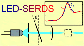
Chemical engineers Renata Adami (University of Salerno, Italy) and Johannes Kiefer (University of Aberdeen, UK) have developed a new approach to shifted-excitation Raman difference spectroscopy (SERDS) for suppression of fluorescence. SERDS experiments conventionally require expensive diode lasers, but in this Analyst paper Kiefer and Adami demonstrate the use of light-emitting diodes (LEDs) as an inexpensive light source. A dielectric bandpass filter narrows and stabilises the broad LED spectrum to make it usable for SERDS. While the resulting spectral resolution is low, different compounds and functional groups can be clearly distinguished using this method.

To read the full access, please click on the link below. This paper will be free to read for the next three weeks:

Light-emitting diode based shifted-excitation Raman difference spectroscopy (LED-SERDS)
Renata Adami and Johannes Kiefer
Analyst, 2013,138, 6258-6261
DOI: 10.1039/C3AN01367G
Digg This
Reddit This
Stumble Now!
Share on Facebook
Bookmark this on Delicious
Share on LinkedIn
Bookmark this on Technorati
Post on Twitter
Google Buzz (aka. Google Reader)

Use Your Eyes: A simple test for E. Coli

Detection of E. coli in Ringer's solution

Detection of E. coli in Ringer's solution

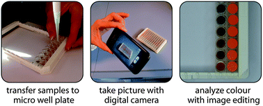
Sophisticated analytical tests producing a readout which can be interpreted by the naked eye, or by image editing software, are becoming increasingly popular.  They offer a low cost alternative to tests requiring expensive equipment and highly trained analysts. Jürgen Popp and colleagues from the Friedrich Schiller University and the Institute of Photonic Technology in Jena, Germany, have developed a new detection method for DNA which provides a distinctive colour change in the case of a positive result.

The bacterial contamination of intravenous fluids in hospitals is a significant concern in modern medicine, so Popp and his team chose the detection of E. Coli in Ringer’s solution as the first test for their assay. The technique uses surface-modified magnetic beads which can bind complementary target DNA by hybridisation. A silver deposition kit provides the visual colour change: for a positive result, silver nanoparticles are generated by enzymatic silver deposition, turning the sample black. As well as being an inexpensive and convenient qualitative test, a quantitative result can be found by analysing a digital photograph of the colour change in standard image editing software.

To read the full article, please cick on the link below. The paper will be free to read for the next three weeks.

Convenient detection of E. coli in Ringer’s solution
Martha Schwarz, Susanne Pahlow, Thomas Bocklitz, Carolin Steinbrücker, Dana Cialla, Karina Weber and Jürgen Popp
Analyst, 2013, Advance Article
DOI: 10.1039/C3AN01240A

Digg This
Reddit This
Stumble Now!
Share on Facebook
Bookmark this on Delicious
Share on LinkedIn
Bookmark this on Technorati
Post on Twitter
Google Buzz (aka. Google Reader)

In the Blood: “Smelling” Bacteria in Cases of Sepsis

Rapid detection and identification of bacteria in sepsis

Rapid detection and identification of bacteria in sepsis

Infections of the blood are a major cause of death, even in countries where antibiotics are widely available. A large hurdle to successful treatment of this type of infection is the identification of the responsible bacteria, which can take several days. Clinicians often resort to the use of broad spectrum antibiotics, which encourages the rise of antibiotic resistant bacteria in the population.

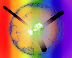
In this collaborative work, researchers led by James Carey at the National University of Kaohsiung, Taiwan, and David Engebretson of Oklahoma City University in the United States, report the design and testing of a colorimetric sensor for bacterial identification. The process simultaneously cultures the bacteria and analyses the unique fingerprint of gases and metabolites produced. The single-step testing kit is automated and disposable, unlike current methods which require skilled technicians, and is so far able to distinguish between eight of the most common pathogens.

By combining the detection and identification of bacteria, the authors are confident that this new technology will prove to be a major step forward in the treatment and outcome of sepsis in patients.

To read the full paper, free for you for the next three weeks, please access the link below:

Single step, rapid identification of pathogenic microorganisms in a culture bottle
Yu W. Chu, Bo Y. Wang, David A. Engebretson and   James R. Carey
Analyst, 2013, Advance Article
DOI: 10.1039/C3AN01175E

Digg This
Reddit This
Stumble Now!
Share on Facebook
Bookmark this on Delicious
Share on LinkedIn
Bookmark this on Technorati
Post on Twitter
Google Buzz (aka. Google Reader)

Shining a Light on Palladium

CyN1 chemosensor for detection of palladium

CyN1 chemosensor for detection of palladium

Palladium is all around us, from the catalytic converters in our cars to watches, jewellery and mobile phones. However, palladium ions can bind to DNA and proteins, leading to concerns that accumulation of the metal in human tissue may pose a health risk.

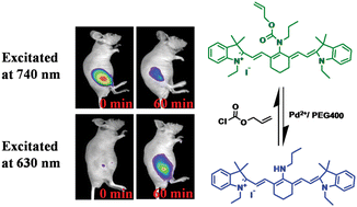
Fengling Song and Xiaojun Peng from the Dalian University of Technology, China, have developed a new near-infrared ratiometric fluorescent chemosensor for palladium. The CyN1 molecule is based on the structure of a cyanine dye and undergoes a Tsuji-Trost reaction in the presence of catalytic amounts of palladium.

The CyN1 sensor offers significant advantages over traditional methods of palladium detection such as atomic absorption spectroscopy (AAS) and inductively coupled plasma mass spectrometry (ICP-MS), which require expensive instrumentation and highly skilled analysts.

Demonstrating the potential bioapplications of the chemosensor, the authors used fluorescence imaging to conduct the first in vivo visualisation of palladium in living mice.

A near-infrared and ratiometric fluorescent chemosensor for palladium
Junyu Wang, Fengling Song,* Jingyun Wang and Xiaojun Peng*
Analyst, 2013, Advance Article
DOI: 10.1039/C3AN00616F

Digg This
Reddit This
Stumble Now!
Share on Facebook
Bookmark this on Delicious
Share on LinkedIn
Bookmark this on Technorati
Post on Twitter
Google Buzz (aka. Google Reader)

Diagnose and Deliver: Hybrid Nanoparticles to Detect and Treat Disease

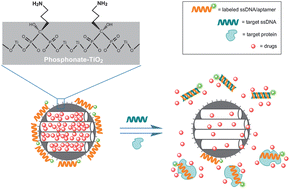
Using nanoparticles for diagnosis and drug release

A vast array of illnesses can be diagnosed by detecting specific proteins or fragments of DNA in the body. But is it possible to build a 2-in-1 system which both detects a disease and provides treatment?

Researchers at Nankai University, China, have created the first functioning system for simultaneous diagnosis and controlled drug release, a so-called “diagnospy” carrier.   To demonstrate the concept, De-Ming Kong and co-workers constructed a mesoporous phosphonate-TiO2 nanoparticle scaffold loaded with ibuprofen. The amino groups on the phosphonates hold fluorescein-labelled single strands of DNA in place over the porous surface, trapping the drug molecules inside. When a complementary target strand is present, a displacement reaction uncaps the pores and the drug is released.

This mechanism relies on the highly specific affinity between complementary strands of DNA, or between proteins and their aptamers, resulting in a very sensitive detection and drug delivery system. The simultaneous sense-and-release approach is hoped to make treatments more efficient and convenient for patients in the future.

To find out more, please access the full article below. It will be free to read for the next two weeks.

Mesoporous phosphonate-TiO2 nanoparticles for simultaneous bioresponsive sensing and controlled drug release
Hui Li,  Tian-Yi Ma,  De-Ming Kong and Zhong-Yong Yuan
Analyst, 2013, 138, 1084-1090
DOI: 10.1039/C2AN36631B

Digg This
Reddit This
Stumble Now!
Share on Facebook
Bookmark this on Delicious
Share on LinkedIn
Bookmark this on Technorati
Post on Twitter
Google Buzz (aka. Google Reader)