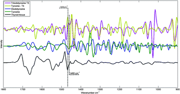
FTIR spectra of hormones triiodothyronine, thyroxine, diiodotyrosine, tyrosine and thyroid tissue
The combination of vibrational spectroscopy with mapping and imaging techniques to address important biochemical questions is an area of active and expanding research. By subjecting adjacent tissue sections to standard histopathological screening and spectroscopic imaging, a greater understanding of the biochemical processes underlying tissue form and function can be achieved.
Researchers from Northeastern University, USA, and the Institute of Energy and Nuclear Research (IPEN), Sao Paolo, have used Fourier transform infrared (FTIR) microspectroscopy to examine healthy thyroid tissues. FTIR allows spatially resolved images, or maps, to be obtained by use of focal plane array detectors and spectral processing of the data then reveals chemical images. In this HOT Analyst paper, Denise Zezell and coworkers present an example from their investigation of 80 different patient samples, which were analysed by transflection-mode FTIR mapping. The approach was applied to the study of healthy thyroid tissue and, with reference to thyroid specific hormones, iodination state.
To know more about this research, click on the links below. This paper will be free to read for the next three weeks:
The characterization of normal thyroid tissue by micro-FTIR spectroscopy
Thiago M. Pereira, Denise M. Zezell, Benjamin Bird, Milos Miljković and Max Diem
Analyst, 2013,138, 7094-7100
DOI: 10.1039/C3AN00296A


